

Case Reports
doi: 10.4251/wjgo.v8.i3.326.
Rare case of entero-enteric intussusception caused by small bowel metastasis from a cardiac liposarcoma
Affiliations
- PMID: 26989469
- PMCID: PMC4789619
- DOI: 10.4251/wjgo.v8.i3.326
Item in Clipboard
Case Reports
Rare case of entero-enteric intussusception caused by small bowel metastasis from a cardiac liposarcoma
World J Gastrointest Oncol.
.
Abstract
Primary cardiac liposarcoma is exceedingly rare and its metastatic potential varies based on the actual tumor subclass. Intestinal intussusception is also an uncommon cause of abdominal pain and bowel obstruction in adults and it usually generates at a malignant lead point in this age group. We report a case of a primary cardiac dedifferentiated liposarcoma in a pregnant woman causing small bowel seeding leading to bowel intussusception.
Keywords: Cardiac; Enteroenteric; Intussusception; Liposarcoma; Small bowel metastasis.
Figures
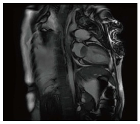
Cardiac magnetic resonance imaging showing a large mass in the left atrium.
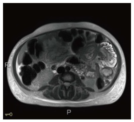
Magnetic resonance imaging of abdomen showing entero-enteric intussusception in axial cut.

Magnetic resonance imaging of the abdomen showing an entero-enteric intussusception in the left upper quadrant.

Normal small bowel mucosa (left hand corner) in contrast to infiltrative area of increased cellularity (right lower hand corner).

High power: Population of poorly differentiated malignant cells that are high grade (pleomorphic, hyperchromatic and contain increased mitotic activity) and are similar to the intracardiac dedifferentiated liposarcoma.

Poorly differentiated malignant neoplasm composed of variably spindled polygonal or histiocytoid cells and irregular vesicular nuclei consistent with a dedifferentiated liposarcoma.
References
-
- Harvey WP. Clinical aspects of cardiac tumors. Am J Cardiol. 1968;21:328–343. - PubMed
-
- Pino PG, Zampi G, Pergolini A, Pero G, Polizzi V, Sbaraglia F, Minardi G, Musumeci F. Metastatic liposarcoma of the heart. Case series and brief literature review. Herz. 2013;38:938–942. - PubMed
-
- Barreiro M, Renilla A, Jimenez JM, Martin M, Al Musa T, Garcia L, Barriales V. Primary cardiac tumors: 32 years of experience from a Spanish tertiary surgical center. Cardiovasc Pathol. 2013;22:424–427. - PubMed
Publication types
LinkOut - more resources
Full Text Sources
Other Literature Sources
