. 2016:2016:3764372.
doi: 10.1155/2016/3764372. Epub 2016 Feb 16.

Activation of the Extracellular Signal-Regulated Kinase Signaling Is Critical for Human Umbilical Cord Mesenchymal Stem Cell Osteogenic Differentiation

Affiliations

Activation of the Extracellular Signal-Regulated Kinase Signaling Is Critical for Human Umbilical Cord Mesenchymal Stem Cell Osteogenic Differentiation

Chen-Shuang Li et al. Biomed Res Int. 2016.

Abstract

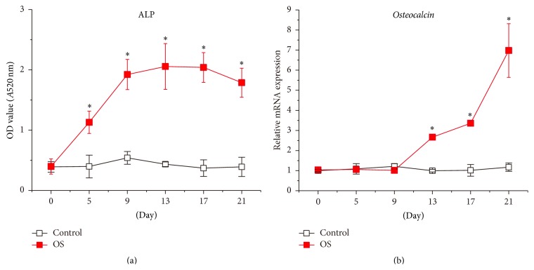
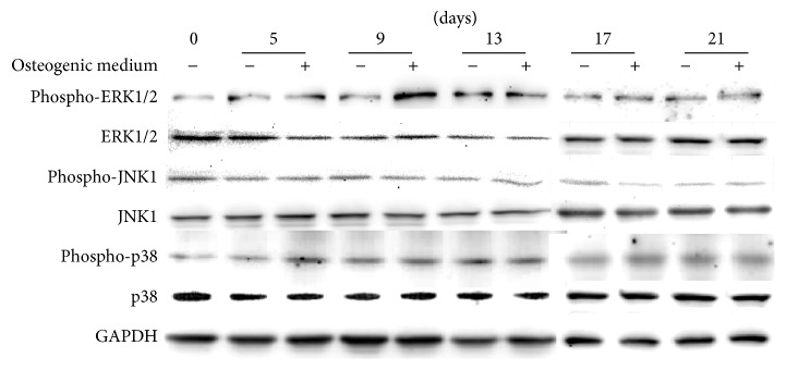
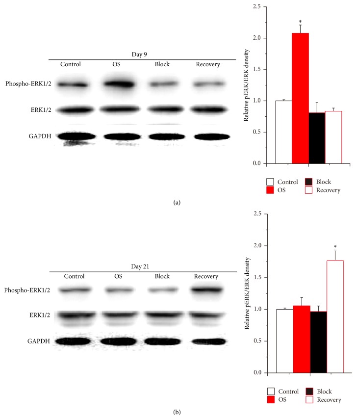
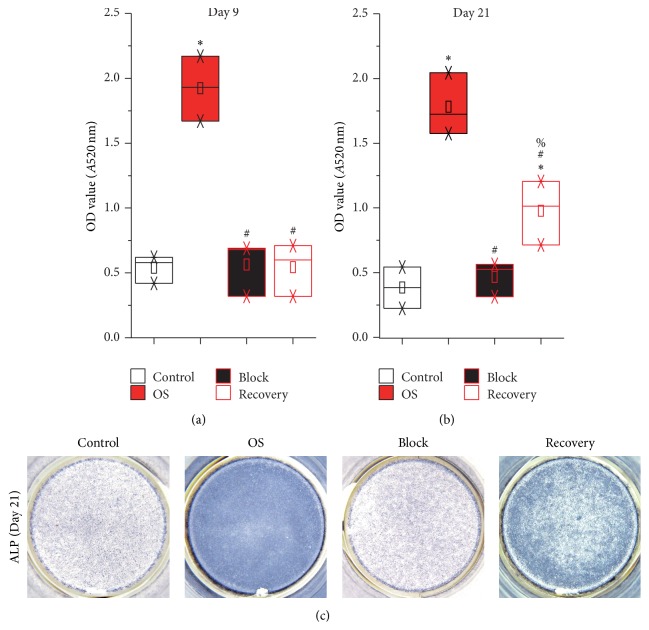
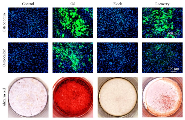
Human umbilical cord mesenchymal stem cells (hUCMSCs) are recognized as candidate progenitor cells for bone regeneration. However, the mechanism of hUCMSC osteogenesis remains unclear. In this study, we revealed that mitogen-activated protein kinases (MAPKs) signaling is involved in hUCMSC osteogenic differentiation in vitro. Particularly, the activation of c-Jun N-terminal kinases (JNK) and p38 signaling pathways maintained a consistent level in hUCMSCs through the entire 21-day osteogenic differentiation period. At the same time, the activation of extracellular signal-regulated kinases (ERK) signaling significantly increased from day 5, peaked at day 9, and declined thereafter. Moreover, gene profiling of osteogenic markers, alkaline phosphatase (ALP) activity measurement, and alizarin red staining demonstrated that the application of U0126, a specific inhibitor for ERK activation, completely prohibited hUCMSC osteogenic differentiation. However, when U0126 was removed from the culture at day 9, ERK activation and osteogenic differentiation of hUCMSCs were partially recovered. Together, these findings demonstrate that the activation of ERK signaling is essential for hUCMSC osteogenic differentiation, which points out the significance of ERK signaling pathway to regulate the osteogenic differentiation of hUCMSCs as an alternative cell source for bone tissue engineering.


Figures

Figure 1
Figure 1
The osteogenic differentiation medium stimulates hUCMSC osteogenic differentiation. ALP activity (a) and transcription of Osteocalcin (b) were monitored in hUCMSCs during the 21-day cultivation in either the maintenance medium (Control) or the osteogenic differentiation medium (OS). Data were presented as Mean + SD; p < 0.05 (N = 6).
Figure 2
Figure 2
Western blotting revealed that the activation of MAPK signaling pathways was different during hUCMSC osteogenic differentiation. hUCMSCs were cultured in either the maintenance medium (−) or the osteogenic differentiation medium (+).
Figure 3
Figure 3
The activation of ERK signaling in hUCMSCs was blocked by U0126. Treated hUCMSCs with U0126 for 9 days completely prohibited the osteogenic differentiation medium-induced increase of ERK activation (a). After the inhibitor was removed, ERK activation of hUCMSCs of Recovery group was upregulated at day 21 (b). The relative densities were normalized to the Control group. Data were presented as Mean + SD; p < 0.05 (N = 6).
Figure 4
Figure 4
ALP activity was partially rescued in the Recovery group hUCMSCs at day 21. The secreted ALP activities in the hUCMSC culture media were analyzed at days 9 (a) and 21 (b). In addition, ALP staining of hUCMSCs was documented at day 21 (c). p < 0.05 compared with the Control group; # p < 0.05 compared with the OS group; % p < 0.05 compared with the Block group (N = 6).
Figure 5
Figure 5
Expression of osteogenic marker genes in hUCMSCs was partially recovered by removing the ERK activation inhibitor U0126 at day 21. Data were presented as Mean + SD; p < 0.05 compared with the Control group; # p < 0.05 compared with the OS group; % p < 0.05 compared with the Block group (N = 6).
Figure 6
Figure 6
Immunocytochemistry staining of Osteopontin and Osteocalcin as well as the alizarin red staining confirmed the osteogenic differentiation of hUCMSCs in the Recovery group at day 21.

Similar articles

Cited by

References

    1. Rahaman M. N., Mao J. J. Stem cell-based composite tissue constructs for regenerative medicine. Biotechnology and Bioengineering. 2005;91(3):261–284. doi: 10.1002/bit.20292. - DOI - PubMed
    1. Wieser K., Zingg P., Dora C. Trochanteric osteotomy in primary and revision total hip arthroplasty: risk factors for non-union. Archives of Orthopaedic and Trauma Surgery. 2012;132(5):711–717. doi: 10.1007/s00402-011-1457-4. - DOI - PubMed
    1. Wang X., Wang Y., Gou W., Lu Q., Peng J., Lu S. Role of mesenchymal stem cells in bone regeneration and fracture repair: a review. International Orthopaedics. 2013;37(12):2491–2498. doi: 10.1007/s00264-013-2059-2. - DOI - PMC - PubMed
    1. Amini A. R., Laurencin C. T., Nukavarapu S. P. Bone tissue engineering: recent advances and challenges. Critical Reviews in Biomedical Engineering. 2012;40(5):363–408. doi: 10.1615/critrevbiomedeng.v40.i5.10. - DOI - PMC - PubMed
    1. Ohishi M., Schipani E. Bone marrow mesenchymal stem cells. Journal of Cellular Biochemistry. 2010;109(2):277–282. doi: 10.1002/jcb.22399. - DOI - PubMed

Publication types

MeSH terms

Substances

LinkOut - more resources