. 2016:2016:8432759.
doi: 10.1155/2016/8432759. Epub 2016 Feb 18.

Antiobesity and Antidiabetes Effects of a Cudrania tricuspidata Hydrophilic Extract Presenting PTP1B Inhibitory Potential

Affiliations

Antiobesity and Antidiabetes Effects of a Cudrania tricuspidata Hydrophilic Extract Presenting PTP1B Inhibitory Potential

Dae Hoon Kim et al. Biomed Res Int. 2016.

Abstract

Diabetes and obesity represent the major health problems and the most age-related metabolic diseases. Protein-tyrosine phosphatase 1B (PTP1B) has emerged as an important regulator of insulin signal transduction and is regarded as a pharmaceutical target for metabolic disorders. To find novel natural materials presenting therapeutic activities against diabetes and obesity, we screened various herb extracts using a chip screening allowing the determination of PTP1B inhibitory effects of the tested compounds using insulin receptor (IR) as the substrate. Cudrania tricuspidata leaves (CTe) had a strong inhibitory effect on PTP1B activity and substantially inhibited fat accumulation in 3T3-L1 cells. CTe was orally administrated to diet-induced obesity (DIO) mice once daily for 3 weeks after which changes in glucose, insulin metabolism, and fat accumulation were examined. Hepatic enzyme markers (aspartate aminotransferase, AST, and alanine aminotransferase, ALT) and total fat mass and triglyceride levels decreased in CTe-treated mice, whereas body weight and total cholesterol concentration slightly decreased. CTe increased the phosphorylation of IRS-1 and Akt in liver tissue. Furthermore, CTe treatment significantly lowered blood glucose levels and improved insulin secretion in DIO mice. Our results strongly suggest that CTe may represent a promising therapeutic substance against diabetes and obesity.


Figures

Figure 1
Figure 1
Effect of CTe on PTP1B activity and lipid droplets. (a) The substrate (IR; insulin receptor) was subjected to autophosphorylation for 1 h, followed by spotting on a chip slide. Reaction mixture was incubated for 24 h in the presence of PTP1B (100 mg/mL) with sodium orthovanadate (Na3VO4; 800 μM), CTe (50, 100, or 200 μg/mL), or 10% PEG alone. The fluorescent signal corresponding to the phosphorylated IR incubated with an anti-phospho-IR, followed by incubation with an anti-Cy5 antibody, was measured using a microarray scanner system. (b) CTe inhibition of fat droplet formation in 3T3-L1 cells. 3T3-L1 cells were stained with Oil Red O dye and examined using a light microscope. (c) Quantification of lipid accumulation in differentiated 3T3-L1 cells by measuring the absorbance of the cell extract at 520 nm. Statistical significance was determined by Student's t-test. p < 0.05 and ∗∗∗ p < 0.001 versus MDI treated cells.
Figure 2
Figure 2
Time line of the experimental procedures using the diet-induced obesity (DIO) mouse model, and effects of CTe on the body weight of DIO mice. (a) C57BL/6 mice were treated daily with vehicle or 60% high fat diet (HFD) by oral gavage for 12 weeks. Week 0 is the starting point beginning on the 1st week with normal chow. The mice were treated with a placebo for 3 weeks prior to CTe treatment. The vehicle group received the placebo treatment for 6 weeks without oral administration of CTe (upper panel). The HFD-fed mice were orally administered CTe or a positive control (PC; sibutramine or sitagliptin) for 1 month (lower panel). (b) Body weights of DIO mice treated with vehicle or CTe (20 or 100 mg/kg) for 3 weeks. Sibutramine was used as a positive control. Data are presented as the mean ± SD for each group (n = 7). p < 0.05 and ∗∗∗ p < 0.001 versus vehicle administered DIO mice.
Figure 3
Figure 3
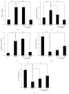
Effects of CTe on fat mass and insulin signaling in DIO mice. The HFD-fed mice were orally administered vehicle or CTe (20 or 100 mg/kg) for 3 weeks. At 28 h after the final CTe treatment, fat was collected from DIO mice. (a) Total fat weight. (b) Inguinal fat weight. (c) Epididymal fat weight. (d) p-IRS-1 and (e) p-Akt detection was estimated by Pathscan sandwich ELISA assay. Data are presented as the mean ± SD for each group (n = 7). Statistical significance was determined by Student's t-test. ∗∗ p < 0.01 and ∗∗∗ p < 0.001 versus lean mice; p < 0.05, †† p < 0.01, and ††† p < 0.001 versus vehicle administered DIO mice.
Figure 4
Figure 4
The HFD-fed mice were orally administered CTe (20 or 100 mg/kg) for 3 weeks. Blood was collected from the abdominal vein. (a) Levels of total cholesterol (CHOL), (b) low-density lipoprotein (LDL), (c) high-density lipoprotein (HDL), and (d) triglycerides (TRIG) were analyzed in triplicate using a serum biochemical analyzer. Liver tissue was collected for toxicity test. (e) Aspartate aminotransferase (AST) and (f) alanine aminotransferase (ALT) levels were monitored by clinical chemistry analyzer. Data are presented as the mean ± SD for each group (n = 7). Statistical significance was determined by Student's t-test. ∗∗∗ p < 0.001 versus lean mice; p < 0.05 and ††† p < 0.001 versus vehicle administered DIO mice.
Figure 5
Figure 5
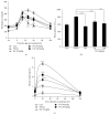
Effect of CTe on blood glucose and insulin levels in DIO mice. The HFD-fed mice were orally administered CTe (20 or 100 mg/kg) for 3 weeks. (a) The effect of CTe on plasma glucose levels in DIO mice. (b) The area under the plasma glucose concentration-time curve for 2 h (AUC0–2 h) in an OGTT. (c) The effect of CTe on plasma insulin in DIO mice. Sitagliptin was used as a positive control. Data are presented as the mean ± SD for each group (n = 7). Statistical significance was determined by Student's t-test. ∗∗∗ p < 0.001 versus vehicle administered DIO mice.

References

    1. Requejo O. H., Rodriguez M. C. R. Nutrition and cancer. Nutrición Hospitalaria. 2015;32(supplement 1):67–72. - PubMed
    1. Kotsis V., Stabouli S., Papakatsika S., Rizos Z., Parati G. Mechanisms of obesity-induced hypertension. Hypertension Research. 2010;33(5):386–393. doi: 10.1038/hr.2010.9. - DOI - PubMed
    1. Lavie C. J., Milani R. V., Ventura H. O. Obesity and cardiovascular disease: risk factor, paradox, and impact of weight loss. Journal of the American College of Cardiology. 2009;53(21):1925–1932. doi: 10.1016/j.jacc.2008.12.068. - DOI - PubMed
    1. Toda N., Ayajiki K., Okamura T. Obesity-induced cerebral hypoperfusion derived from endothelial dysfunction: one of the risk factors for Alzheimer's disease. Current Alzheimer Research. 2014;11(8):733–744. doi: 10.2174/156720501108140910120456. - DOI - PubMed
    1. Kaur J. A comprehensive review on metabolic syndrome. Cardiology Research and Practice. 2014;2014:21. doi: 10.1155/2014/943162.943162 - DOI - PMC - PubMed

Publication types

MeSH terms

LinkOut - more resources