Clinical Trial
. 2016:2016:9672082.
doi: 10.1155/2016/9672082. Epub 2016 Feb 17.

Quiescent and Active Tear Protein Profiles to Predict Vernal Keratoconjunctivitis Reactivation

Affiliations
Clinical Trial

Quiescent and Active Tear Protein Profiles to Predict Vernal Keratoconjunctivitis Reactivation

Alessandra Micera et al. Biomed Res Int. 2016.

Abstract

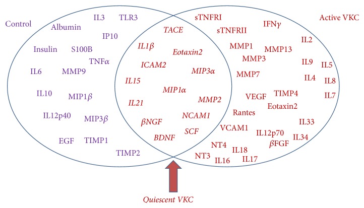
Objective: Vernal keratoconjunctivitis (VKC) is a chronic recurrent bilateral inflammation of the conjunctiva associated with atopy. Several inflammatory and tissue remodeling factors contribute to VKC disease. The aim is to provide a chip-based protein analysis in tears from patients suffering from quiescent or active VKC.

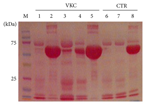
Methods: This study cohort included 16 consecutive patients with VKC and 10 controls. Participants were subjected to clinical assessment of ocular surface and tear sampling. Total protein quantification, total protein sketch, and protein array (sixty protein candidates) were evaluated.

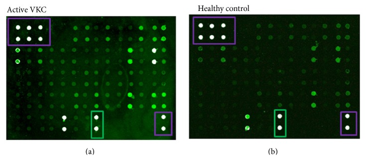
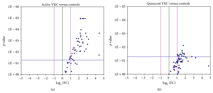
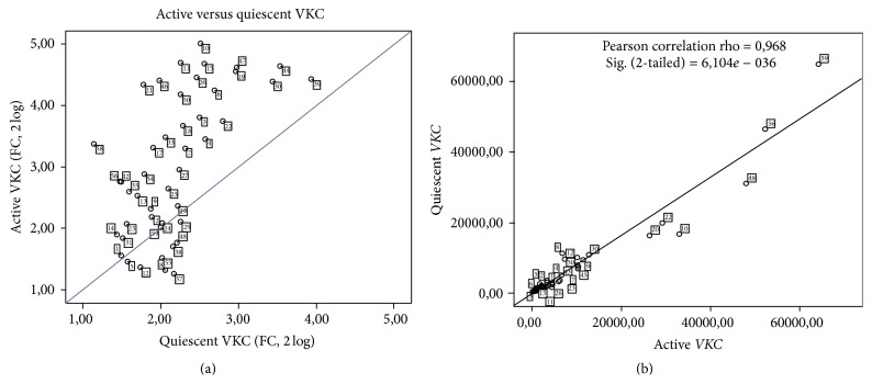
Results: An overall increased Fluorescent Intensity expression was observed in VKC arrays. Particularly, IL1β, IL15, IL21, Eotaxin2, TACE, MIP1α, MIP3α, NCAM1, ICAM2, βNGF, NT4, BDNF, βFGF, SCF, MMP1, and MMP2 were increased in quiescent VKC. Of those candidates, only IL1β, IL15, IL21, βNGF, SCF, MMP2, Eotaxin2, TACE, MIP1α, MIP3α, NCAM1, and ICAM2 were increased in both active and quiescent VKC. Finally, NT4, βFGF, and MMP1 were highly increased in active VKC.

Conclusion: A distinct "protein tear-print" characterizes VKC activity, confirming some previously reported factors and highlighting some new candidates common to quiescent and active states. Those candidates expressed in quiescent VKC might be considered as predictive indicators of VKC reactivation and/or exacerbation out-of-season.


Figures

Figure 1
Figure 1
Tear protein profile. Equal protein amounts (20 µg/lane/sample) were subjected to electrophoretic separation (SDS-PAGE) and membranes were stained with Ponceau S before image acquisition (see M&M). Note the presence of albumin (60 kDa), low/high IgG bands (40/100 kDa), and fibronectin (200 kDa) in some tear samples (1–5, VKC; 6–8 healthy controls). To retrieve low-expressed antigens, samples showing high albumin/IgGs were treated with a specific preclearing kit (see M&M).
Figure 2
Figure 2
Overview of the protein array. Equal protein amounts were loaded for each subarray and the presence of an equal number of VKC: control samples were guaranteed for each array-chip (14 subarrays). (a), (b) Representative active VKC (a) and control (b) arrays, as provided by the GenePix scanner (with no color adjustment). White spots framed violet are positive controls, and black spots are negative referring controls and black spots framed green are albumin specific signals.
Figure 3
Figure 3
Plot graphs of the tear expression profile in active and quiescent VKC. Fold changes (Log2 (FC); x-axis) are ranked in Volcano plots according to the statistical significance (p values as negative Log10; y-axis). For each marker, FC between case and control values were calculated from mean of Fluorescent Intensity values provided by the software, as described in M&M. The two-sided unpaired t-test comparisons of active (a) and quiescent (b) samples were carried out versus controls. Both ±2 FC and p ≤ 0.05 were used as initial cut-offs. Red lines indicate differences of ±1 FC (log2) and blue line shows the initial significance level. Those candidates, having ≥2 FC and p ≤ 0.05 initial cut-offs, are localized in the upper left quadrant.
Figure 4
Figure 4
Comparison between quiescent and active subgroups. The scatter plot in (a) shows the candidate fold changes between quiescent and active tears, as calculated from mean of FI values provided by the software (see M&M). Those candidates, having ≥2 FC and p ≤ 0.05 initial cut-offs, are localized in the lower left quadrant. Correlation between quiescent and active biomarkers in VKC tears is shown in MFI values (b). The Pearson correlation analysis is reported in the panel. Note the close association of quiescent and active VKC candidates in the lower left region of the slope.
Figure 5
Figure 5
Venn diagram of predicted candidate biomarkers. Venn diagram showing the partial overlap between quiescent and active VKC groups. As predicted by this experimental approach, overlapping biomarkers are highlighted by the red arrow. At least in this study, all candidates showed at least ≥2-fold differences and a p value ≤ 0.05 or 0.00085, according to the Bonferroni correction.

References

    1. Bonini S., Bonini S., Lambiase A., et al. Vernal keratoconjunctivitis revisited: a case series of 195 patients with long-term followup. Ophthalmology. 2000;107(6):1157–1163. doi: 10.1016/s0161-6420(00)00092-0. - DOI - PubMed
    1. Bonini S., Lambiase A., Sgrulletta R., Bonini S. Allergic chronic inflammation of the ocular surface in vernal keratoconjunctivitis. Current Opinion in Allergy and Clinical Immunology. 2003;3(5):381–387. doi: 10.1097/00130832-200310000-00011. - DOI - PubMed
    1. Leonardi A., Lazzarini D., Motterle L., et al. Vernal keratoconjunctivitis-like disease in adults. American Journal of Ophthalmology. 2013;155(5):796–803. doi: 10.1016/j.ajo.2012.11.018. - DOI - PubMed
    1. Bonini S., Sacchetti M., Mantelli F., Lambiase A. Clinical grading of vernal keratoconjunctivitis. Current Opinion in Allergy and Clinical Immunology. 2007;7(5):436–441. doi: 10.1097/ACI.0b013e3282efb726. - DOI - PubMed
    1. Uchio E., Ono S. Y., Ikezawa Z., et al. Tear levels of IFN-gamma, interleukin (IL) 2, IL4 and IL5 in patients with vernal keratoconjunctivitis, atopic keratitis and allergic conjunctivitis. Clinical & Experimental Allergy. 2000;30(1):103–109. - PubMed

Publication types