. 2016 Jul;20(7):1381-91.
doi: 10.1111/jcmm.12828. Epub 2016 Mar 17.

Effects of short-chain acyl-CoA dehydrogenase on cardiomyocyte apoptosis

Affiliations

Effects of short-chain acyl-CoA dehydrogenase on cardiomyocyte apoptosis

Zhenhua Zeng et al. J Cell Mol Med. 2016 Jul.

Abstract

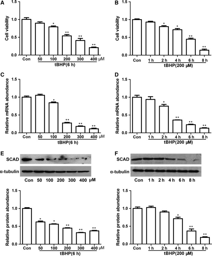
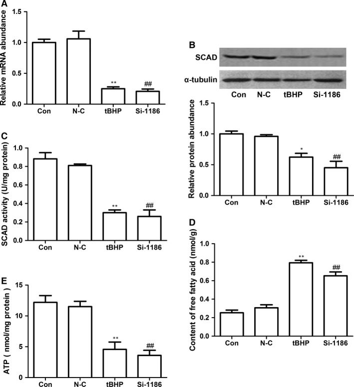
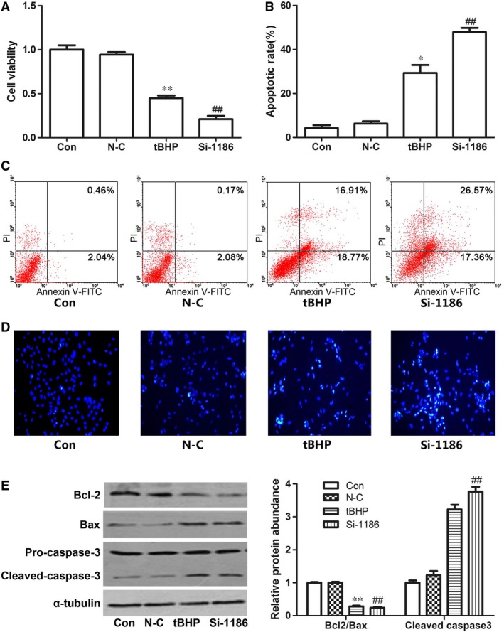
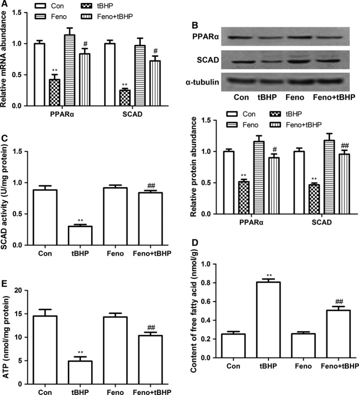
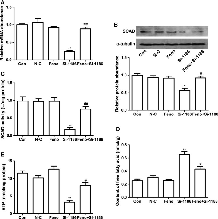
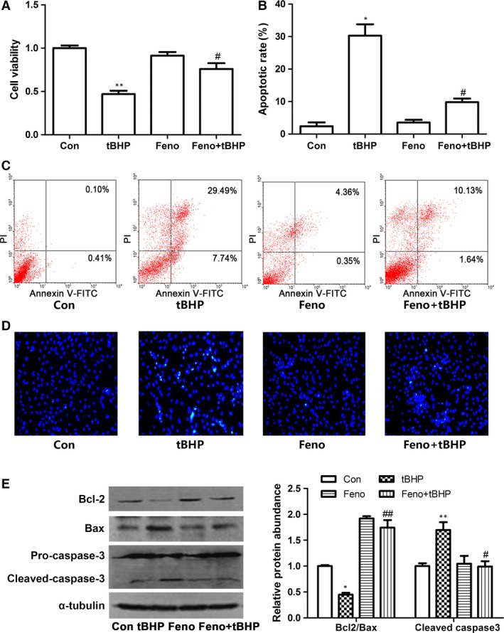
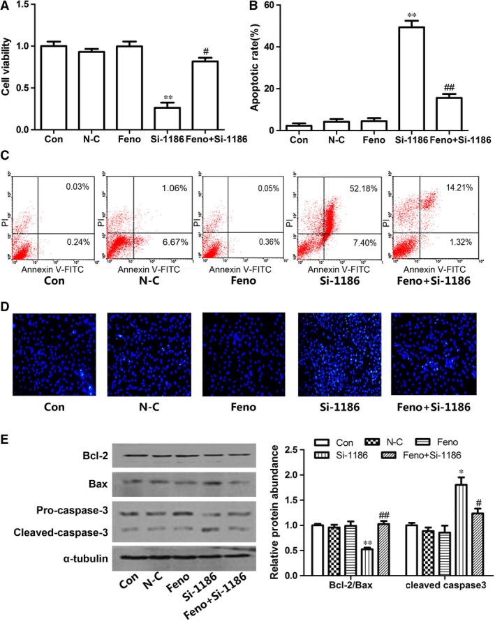
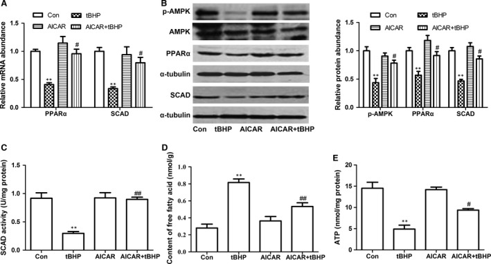
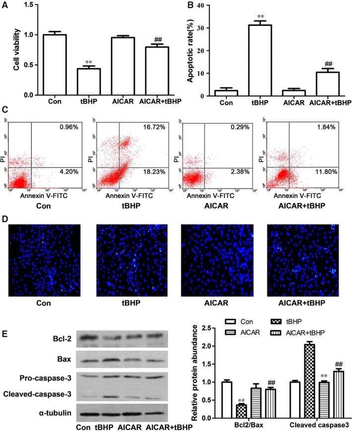
Short-chain acyl-CoA dehydrogenase (SCAD), a key enzyme of fatty acid β-oxidation, plays an important role in cardiac hypertrophy. However, its effect on the cardiomyocyte apoptosis remains unknown. We aimed to determine the role of SCAD in tert-butyl hydroperoxide (tBHP)-induced cardiomyocyte apoptosis. The mRNA and protein expression of SCAD were significantly down-regulated in the cardiomyocyte apoptosis model. Inhibition of SCAD with siRNA-1186 significantly decreased SCAD expression, enzyme activity and ATP content, but obviously increased the content of free fatty acids. Meanwhile, SCAD siRNA treatment triggered the same apoptosis as cardiomyocytes treated with tBHP, such as the increase in cell apoptotic rate, the activation of caspase3 and the decrease in the Bcl-2/Bax ratio, which showed that SCAD may play an important role in primary cardiomyocyte apoptosis. The changes of phosphonate AMP-activated protein kinase α (p-AMPKα) and Peroxisome proliferator-activated receptor α (PPARα) in cardiomyocyte apoptosis were consistent with that of SCAD. Furthermore, PPARα activator fenofibrate and AMPKα activator AICAR treatment significantly increased the expression of SCAD and inhibited cardiomyocyte apoptosis. In conclusion, for the first time our findings directly demonstrated that SCAD may be as a new target to prevent cardiomyocyte apoptosis through the AMPK/PPARα/SCAD signal pathways.

Keywords: AMP-activated protein kinase; cardiomyocyte apoptosis; energy metabolism•; peroxisome proliferator-activated receptor α; short-chain acyl-CoA dehydrogenase; tert-butyl hydroperoxide.


Figures

Figure 1
Figure 1
Cell viability and SCAD expression were decreased by tBHP in cardiomyocyte. (A) Cardiomyocytes were treated with various concentrations of tBHP for 6 hrs and assessed for cell viability. (B) Cardiomyocytes were treated with 200 μM tBHP for different incubation times. The levels of mRNA (C and D) and protein of SCAD (E and F) were mearused by real‐time PCR and Western blots. Cardiomyocytes were treated with different concentrations of tBHP for 6 hrs and 200 μM tBHP for different incubation time. Mean ± S.D., n = 3, *P < 0.05; **P < 0.01 versus control.
Figure 2
Figure 2
The expression and enzyme activity of SCAD in the cardiomyocytes treated with tBHP (200 μM, 6 hrs) or siRNA‐1186 (72 hrs). (A) The relative mRNA level of SCAD was significantly inhibited by siRNA‐1186 and tBHP. (B) The relative protein level of SCAD was significantly inhibited by siRNA‐1186 and tBHP. (C) The enzyme activity of SCAD was significantly inhibited by siRNA‐1186 and tBHP. (D) Content of free fatty acid was significantly increased in cardiomyocytes treated with siRNA‐1186 or tBHP. (E) The ATP level was significantly decreased in cardiomyocytes treated with siRNA‐1186 or tBHP. N‐C: negative control siRNA. Mean ± S.D., n = 3, *P < 0.05, **P < 0.01 versus control. ##P < 0.01 versus N‐C.
Figure 3
Figure 3
The cell apoptotic in the cardiomyocytes treated with tBHP (200 μM, 6 hrs) or siRNA‐1186 (72 hrs). The changes of cell viability (A) and cell apoptotic rate (B and C) in the cardiomyocytes treated with tBHP (200 μM, 6 hrs) or siRNA‐1186 (72 hrs). (D) Cell apoptosis was determined with Hoechst 33258 staining. 200× magnification. (E) The expression of apoptosis‐related proteins in tBHP‐ or siRNA‐1186‐induced cardiomyocytes. N‐C: negative control siRNA. Mean ± S.D., n = 3, *P < 0.05; **P < 0.01 versus control. ##P < 0.01 versus N‐C.
Figure 4
Figure 4
Effects of Feno on cardiomyocyte apoptosis in tBHP‐treated cardiomyocytes. (A) The PPARα and SCAD mRNA expression was significantly decreased in cardiomyocytes treated with tBHP, however, increased in cardiomyocytes pre‐treated with Feno. (B) The PPARα and SCAD protein expression was significantly decreased in cardiomyocytes treated with tBHP, however, increased in cardiomyocytes pre‐treated with Feno. (C) The SCAD enzyme activity was significantly decreased in cardiomyocytes treated with tBHP, however, increased in cardiomyocytes pre‐treated with Feno. (D) The content of free fatty acid was significantly increased in cardiomyocytes treated with tBHP, however, decreased in cardiomyocytes pre‐treated with Feno. (E) The ATP level was significantly decreased in cardiomyocytes treated with tBHP, however, increased in cardiomyocytes pre‐treated with Feno. Mean ± S.D., n = 3, *P < 0.05, **P < 0.01 versus control, #P < 0.05, ##P < 0.01 versus tBHP.
Figure 5
Figure 5
Effects of Feno on cardiomyocyte apoptosis in siRNA‐1186‐treated cardiomyocytes. (A) The SCAD mRNA expression was significantly decreased in cardiomyocytes treated with siRNA‐1186, however, increased in cardiomyocytes pre‐treated with Feno. (B) The SCAD protein expression was significantly decreased in cardiomyocytes treated with siRNA‐1186, however, increased in cardiomyocytes pre‐treated with Feno. (C) The SCAD enzyme activity was significantly decreased in cardiomyocytes treated with siRNA‐1186, however, increased in cardiomyocytes pre‐treated with Feno. (D) The content of free fatty acid was significantly increased in cardiomyocytes treated with siRNA‐1186, however, decreased in cardiomyocytes pre‐treated with Feno. (E) The ATP level was significantly decreased in cardiomyocytes treated with siRNA‐1186, however, increased in cardiomyocytes pre‐treated with Feno N‐C: negative control siRNA. Mean ± S.D., n = 3, *P < 0.05, **P < 0.01 versus control, #P < 0.05, ##P < 0.01 versus si‐1186.
Figure 6
Figure 6
The protective effects of Feno on cardiomyocyte apoptosis induced by tBHP. (A) The decrease in cell viability were abrogated in tBHP group pre‐treated with Feno. (B and C) The cell apoptotic rate was increased significantly in tBHP group, however, decreased in cardiomyocytes pre‐treated with Feno. (D) Cell apoptosis was determined with Hoechst 33258 staining. 200× magnification. (E) Effects of Feno on the expression of apoptosis‐related proteins in tBHP‐induced cardiomyocytes. Mean ± S.D., n = 3, *P < 0.05, **P < 0.01 versus control, #P < 0.05, ##P < 0.01 versus tBHP.
Figure 7
Figure 7
The protective effects of Feno on cardiomyocyte apoptosis induced by siRNA‐1186. (A) The derease of cell viability were abrogated in siRNA‐1186 group pre‐treated with Feno. (B and C) The cell apoptotic rate was increased significantly in siRNA‐1186 group, however, decreased in cardiomyocytes pre‐treated with Feno. (D) Cell apoptosis was determined with Hoechst 33258 staining. 200× magnification. (E) Effects of Feno on the expression of apoptosis‐related proteins in siRNA‐1186‐induced cardiomyocytes. N‐C: negative control siRNA. Mean ± S.D., n = 3, *P < 0.05, **P < 0.01 versus control, #P < 0.05, ##P < 0.01 versus si‐1186.
Figure 8
Figure 8
Effects of AICAR on cardiomyocyte apoptosis in tBHP‐treated cardiomyocytes. (A) The PPARα and SCAD mRNA expression was significantly decreased in cardiomyocytes treated with tBHP, however, increased in cardiomyocytes pre‐treated with AICAR. (B) The p‐AMPK, PPARα and SCAD protein expression was significantly decreased in cardiomyocytes treated with tBHP, however, increased in cardiomyocytes pre‐treated with AICAR. (C) The SCAD enzyme activity was significantly decreased in cardiomyocytes treated with tBHP, however, increased in cardiomyocytes pre‐treated with AICAR. (D) The content of free fatty acid was significantly increased in cardiomyocytes treated with tBHP, however, decreased in cardiomyocytes pre‐treated with AICAR. (E) The ATP level was significantly decreased in cardiomyocytes treated with tBHP, however, increased in cardiomyocytes pre‐treated with AICAR. Mean ± S.D., n = 3, *P < 0.05, **P < 0.01 versus control, #P < 0.05, ##P < 0.01 versus tBHP.
Figure 9
Figure 9
The protective effects of AICAR on cardiomyocyte apoptosis induced by tBHP. (A) The derease of cell viability were abrogated in tBHP group pre‐treated with AICAR. (B and C) The cell apoptotic rate was increased significantly in tBHP group, however, decreased in cardiomyocytes pre‐treated with AICAR. (D) Cell apoptosis was determined with Hoechst 33258 staining. 200× magnification. (E) Effects of AICAR on the expression of apoptosis‐related proteins in tBHP‐induced cardiomyocytes. Mean ± S.D., n = 3, *P < 0.05, **P < 0.01 versus control, #P < 0.05, ##P < 0.01 versus tBHP.

Similar articles

Cited by

References

    1. Whelan RS, Kaplinskiy V, Kitsis RN. Cell death in the pathogenesis of heart disease: mechanisms and significance. Annu Rev Physiol. 2010; 72: 19–44. - PubMed
    1. Zhang Y, Herman B. Apoptosis and successful aging. Mech Ageing Dev. 2002; 123: 563–5. - PubMed
    1. Lee Y, Gustafsson AB. Role of apoptosis in cardiovascular disease. Apoptosis. 2009; 14: 536–48. - PubMed
    1. Nishida K, Otsu K. Cell death in heart failure. Circ J. 2008; 72 (Suppl. A): A17–21. - PubMed
    1. Lu FH, Fu SB, Leng X, et al Role of the calcium‐sensing receptor in cardiomyocyte apoptosis via the sarcoplasmic reticulum and mitochondrial death pathway in cardiac hypertrophy and heart failure. Cell Physiol Biochem. 2013; 31: 728–43. - PubMed

Publication types

MeSH terms