. 2016 Mar 18;11(3):e0151635.
doi: 10.1371/journal.pone.0151635. eCollection 2016.

Pyruvate Kinase M2 and Lactate Dehydrogenase A Are Overexpressed in Pancreatic Cancer and Correlate with Poor Outcome

Affiliations

Pyruvate Kinase M2 and Lactate Dehydrogenase A Are Overexpressed in Pancreatic Cancer and Correlate with Poor Outcome

Goran Hamid Mohammad et al. PLoS One. .

Abstract

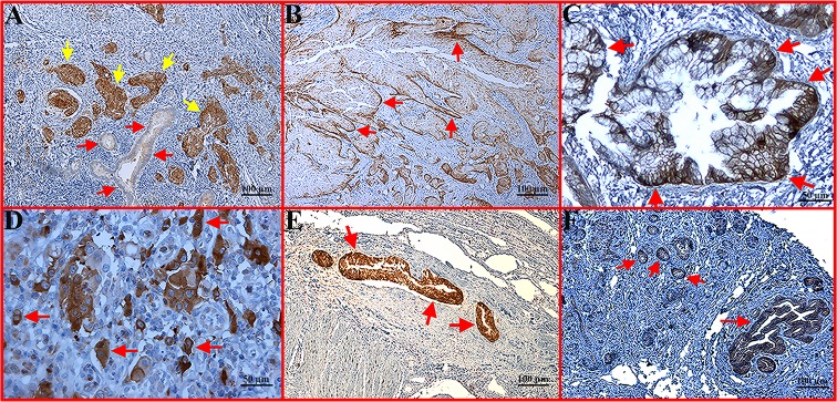
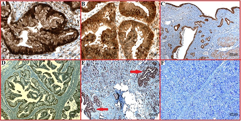
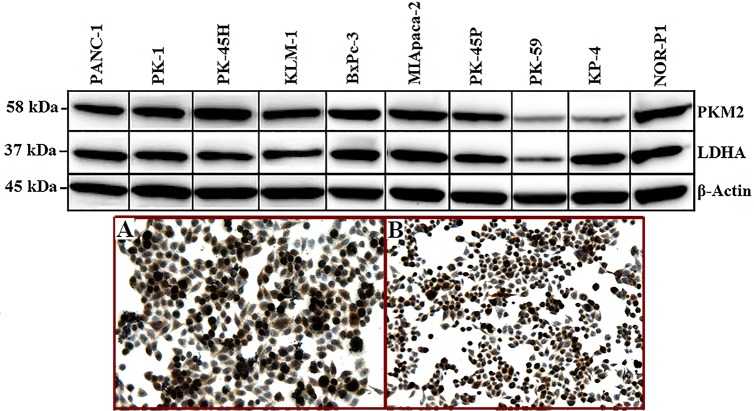
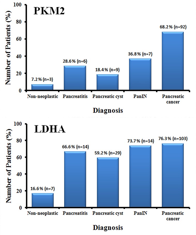
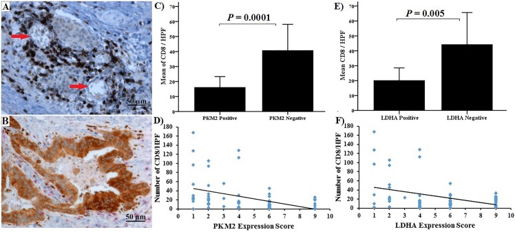
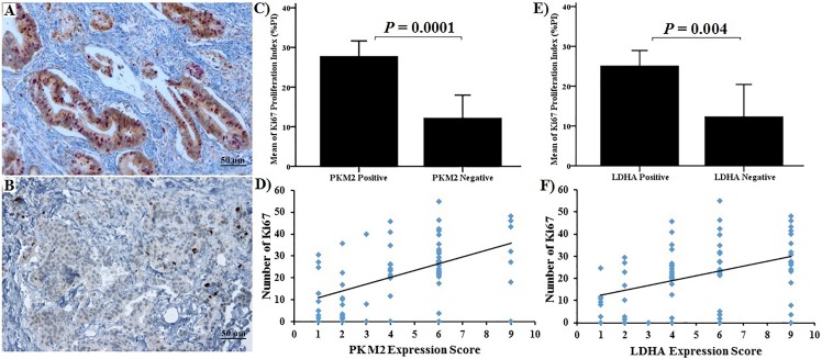
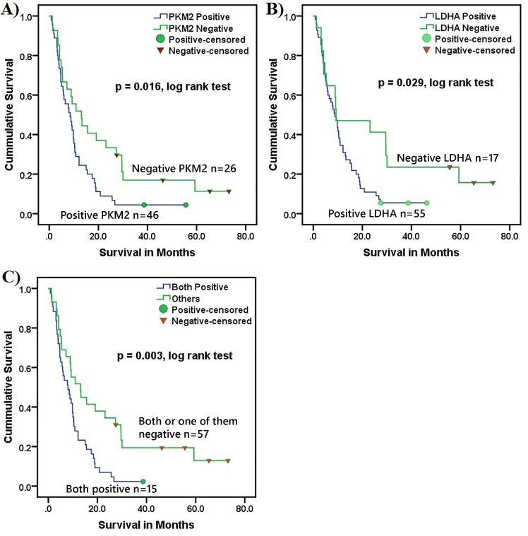
Pancreatic cancer has a 5-year survival rate of less than 4%. Despite advances in diagnostic technology, pancreatic cancer continues to be diagnosed at a late and incurable stage. Accurate biomarkers for early diagnosis and to predict treatment response are urgently needed. Since alteration of glucose metabolism is one of the hallmarks of cancer cells, we proposed that pyruvate kinase type M2 (M2PK) and lactate dehydrogenase A (LDHA) enzymes could represent novel diagnostic markers and potential therapeutic targets in pancreatic cancer. In 266 tissue sections from normal pancreas, pancreatic cystic neoplasms, pancreatic intraepithelial neoplasia (PanIN) and cancer, we evaluated the expression of PKM2, LDHA, Ki-67 and CD8+ by immunohistochemistry and correlated these markers with clinicopathological characteristics and patient survival. PKM2 and LDHA expression was also assessed by Western blot in 10 human pancreatic cancer cell lines. PKM2 expression increased progressively from cyst through PanIN to cancer, whereas LDHA was overexpressed throughout the carcinogenic process. All but one cell line showed high expression of both proteins. Patients with strong PKM2 and LDHA expression had significantly worse survival than those with weak PKM2 and/or LDHA expression (7.0 months vs. 27.9 months, respectively, p = 0.003, log rank test). The expression of both PKM2 and LDHA correlated directly with Ki-67 expression, and inversely with intratumoral CD8+ cell count. PKM2 was significantly overexpressed in poorly differentiated tumours and both PKM2 and LDHA were overexpressed in larger tumours. Multivariable analysis showed that combined expression of PKM2 and LDHA was an independent poor prognostic marker for survival. In conclusion, our results demonstrate a high expression pattern of two major glycolytic enzymes during pancreatic carcinogenesis, with increased expression in aggressive tumours and a significant adverse effect on survival.


Conflict of interest statement

Competing Interests: The authors have declared that no competing interests exist.

Figures

Fig 1
Fig 1. Immunohistochemical staining of PKM2 expression in representative pancreatic tumour sections.
(A) Well differentiated area of tumour showing weak PKM2 expression (red arrow), with high expression in poorly differentiated areas (brown colour, yellow arrow) (x100 magnification). (B) Growing margin of tumour nodules with strong expression of PKM2 (red arrow) (x100 magnification). (C) Membranous expression of PKM2 (red arrow) (x200 magnification). (D) Heterogeneous expression of PKM2 with predominant expression in the proliferating cells (red arrow) (x200 magnification). (E) Strongly positive tumour expression of PKM2 in vascular invasion (red arrow) (x100 magnification). (F) Strongly positive tumour expression of PKM2 with muscular invasion (red arrow) (x100 magnification).
Fig 2
Fig 2. LDHA expression pattern in pancreatic cancer, benign and normal tissue sections.
(A) Membranous expression of LDHA (x200 magnification). (B) Cytoplasmic and nuclear expression (x200 magnification). (C) Strong expression in pancreatic cyst with mild expression in the surrounding normal pancreatic tissue (x50 magnification). (D) Nuclear expression of LDHA in pancreatic cancer (x100 magnification). (E) Strong expression in PanIN lesion (x100 magnification). (F) Negative staining in normal pancreas (x100 magnification).
Fig 3
Fig 3. Expression of PKM2 and LDHA in pancreatic cancer cell lines as detected by Western blot analysis (upper panel).
Immunostaining of Miapaca-2 cells with PKM2 (A) and LDHA (B) is shown in the lower panel. Strong cytoplasmic and nuclear staining is noted in proliferating cells.
Fig 4
Fig 4. Percentages of PKM2 and LDHA expression in different tissue types.
PKM2 was strongly expressed by pancreatic cancer tissue specimens and was significantly higher than in normal, pancreatitis, pancreatic cyst and PanIN tissues (P<0.001). Expression of LDHA was significantly higher in pancreatic cancer than in normal pancreas (P<0.001), whereas, there was no significant differences in LDHA expression between pancreatic cancer, PanIN, pancreatic cysts and pancreatitis.
Fig 5
Fig 5. Immunohistochemical staining and correlation between PKM2 and LDHA and CD8+TIL.
(A) Well differentiated tumours with negative PKM2 expression had strong infiltration by CD8+ positive T-lymphocytes (x200 magnification). (B) Poorly differentiated tumours strongly positive for PKM2 had sparse infiltration by CD8+ positive T-lymphocytes (x200 magnification). There was a significant negative correlation between CD8+ positive cells and both PKM2 (C) & (D) and LDHA staining (E) & (F).
Fig 6
Fig 6. Immunohistochemical staining and correlation between PKM2, LDHA and Ki-67.
(A) Tumours with strongly positive PKM2 expression had most of the nucleus stained for Ki-67 (x100 magnification). (B) Tumours weakly positive for PKM2 had scant Ki67 staining (x100 magnification). (C) & (D) Correlation between PKM2 staining and Ki-67 positive cells. (E) & (F) Correlation between LDHA staining and Ki-67 positive cells. There was a significant correlation for both PKM2 and LDHA.
Fig 7
Fig 7. Overall patient survival in relation to PKM2 and LDHA expression.
Both PKM2 (A) and LDHA (B) had a significant prognostic impact on patient survival (p = 0.016, 0.029, log rank test, respectively) and the combined expression for both markers further stratified the patients (C) (p = 0.003, log rank test). As expected, patients who underwent surgical resection had a longer survival than unresected patients.

Similar articles

Cited by

References

    1. American Cancer Society. Cancer Fact and Figures 2014. Accessed 16 Dec 2014. Available: http://www.cancer.org/research/cancerfactsfigures/cancerfactsfigures/can.... 2014.
    1. Cancer Research UK. Pancreatic Cancer Statistics. Accessed 20 Feb 2015. Available: http://www.cancerresearchuk.org/cancer-info/cancerstats/types/pancreas/. 2014.
    1. Conroy T, Desseigne F, Ychou M, Bouché O, Guimbaud R, Bécouarn Y, et al. FOLFIRINOX versus gemcitabine for metastatic pancreatic cancer. N Engl J Med. 2011;364(19):1817–25. 10.1056/NEJMoa1011923 - DOI - PubMed
    1. Gourgou-Bourgade S, Bascoul-Mollevi C, Desseigne F, Ychou M, Bouché O, Guimbaud R, et al. Impact of FOLFIRINOX compared with gemcitabine on quality of life in patients with metastatic pancreatic cancer: Results from the PRODIGE 4/ACCORD 11 randomized trial. J Clin Oncol. 2013;31(1):23–9. 10.1200/JCO.2012.44.4869 - DOI - PubMed
    1. Wong N, Ojo D, Yan J, Tang D. PKM2 contributes to cancer metabolism. Cancer Letters. 2015;656 (2): 184–191. - PubMed

Publication types

MeSH terms