Case Reports
. 2016 Mar 18;11(3):e0151429.
doi: 10.1371/journal.pone.0151429. eCollection 2016.

ATP1A3 Mutation in Adult Rapid-Onset Ataxia

Affiliations
Case Reports

ATP1A3 Mutation in Adult Rapid-Onset Ataxia

Kathleen J Sweadner et al. PLoS One. .

Abstract

A 21-year old male presented with ataxia and dysarthria that had appeared over a period of months. Exome sequencing identified a de novo missense variant in ATP1A3, the gene encoding the α3 subunit of Na,K-ATPase. Several lines of evidence suggest that the variant is causative. ATP1A3 mutations can cause rapid-onset dystonia-parkinsonism (RDP) with a similar age and speed of onset, as well as severe diseases of infancy. The patient's ATP1A3 p.Gly316Ser mutation was validated in the laboratory by the impaired ability of the expressed protein to support the growth of cultured cells. In a crystal structure of Na,K-ATPase, the mutated amino acid was directly apposed to a different amino acid mutated in RDP. Clinical evaluation showed that the patient had many characteristics of RDP, however he had minimal fixed dystonia, a defining symptom of RDP. Successive magnetic resonance imaging (MRI) revealed progressive cerebellar atrophy, explaining the ataxia. The absence of dystonia in the presence of other RDP symptoms corroborates other evidence that the cerebellum contributes importantly to dystonia pathophysiology. We discuss the possibility that a second de novo variant, in ubiquilin 4 (UBQLN4), a ubiquitin pathway component, contributed to the cerebellar neurodegenerative phenotype and differentiated the disease from other manifestations of ATP1A3 mutations. We also show that a homozygous variant in GPRIN1 (G protein-regulated inducer of neurite outgrowth 1) deletes a motif with multiple copies and is unlikely to be causative.


Conflict of interest statement

Competing Interests: KJS, CT, CTW, BMS, JFC, LJO, and TCM have declared that no competing interests exist. AB is a consultant to Allergan, Ipsen, Concert and Revance. Her conflict of interest was managed by Wake Forest School of Medicine. This did not alter the authors' adherence to PLOS ONE policies on sharing data and materials.

Figures

Fig 1
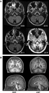
Fig 1. Sequential and comparative MRI.
(A) Axial T1-weighted images from successive MRI scans acquired from 2008 (year of onset) to 2014 demonstrate the progressive development of cerebellar atrophy. (B) Coronal (top row) and sagittal (bottom row) T1-weighted images from MRI demonstrate relatively normal cerebellar structure and volume of a representative RDP patient (left images) in contrast to marked cerebellar atrophy of the patient in 2014 (right images).
Fig 2
Fig 2. Stereo images of Gly316 and Phe780 in the K+ and Na+ conformations.
The structures can be viewed in stereo by focusing the eyes behind the plane of the top or bottom image pair and letting the eyes drift separately until a central image fuses. On the top, the transmembrane domain of Na,K-ATPase is shown from a crystal structure in the E2 (K+-bound) conformation. Gold spheres are K+ ions in the ion binding pocket. The yellow space-fill residue is Gly316. Opposite it, the blue-green residue is Phe780, mutated to leucine in RDP. On the bottom, red spheres are Na+ ions in the ion binding pocket of a crystal structure in the E1 (Na+ bound) conformation. The rearrangement of transmembrane helices has separated Gly316 and Phe780 significantly.
Fig 3
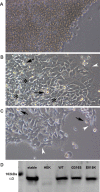
Fig 3. Impairment of Na,K-ATPase function.
(A-C) Ouabain is a specific Na,K-ATPase inhibitor. HEK293 cells are shown 5 d after they were transfected and 4 d after 0.5 μM ouabain was added. In A, mock-transfected HEK cells had all died. In B, transfection was with unmutated ATP1A3. Cells survived and divided, becoming crowded (asterisks), medium acidified overnight, and some death due to overcrowding was apparent (arrows). Cells at the edge of open spaces were flattened (white arrowheads). In C, the p.Gly316Ser mutation was transfected. Many cells survived, but were unable to divide, and the medium did not acidify. Over the subsequent two weeks there was a slow attrition of cells and no detectable division of the remaining living p.Gly316Ser cells. (D) Na,K-ATPase α3 western blot of equal amounts of total protein from cell homogenates demonstrating that the mutant protein is expressed at levels comparable to controls. A stable WT ATP1A3-transfected cell line was a positive control for optimal ATP1A3 expression. The visible molecular weight marker (Amersham Rainbow) was 102 kDa, and α3 migrates at ~93 kDa (faster than its molecular mass). HEK cells express only ATP1A1, which is not detected by the specific antibody (Santa Cruz Biotechnology sc-16052). The other lanes show transient expression of WT ATP1A3, p.Gly316Ser, and p.Glu815Lys, an inactive mutation found in AHC [28]. The expression results and the failure to support cell growth are representative of three experiments.
Fig 4
Fig 4. Repeated motifs in GPRIN1.
GPRIN1 (formerly known as GRIN1 until the name conflicted with the official gene name of an NMDA glutamate receptor) has sequence characteristics of a relatively unstructured protein, and in human it has 23 copies, of differing fidelity, of a six amino acid motif typified by KEDPGS. The diagram shows the distribution of motifs (red bars) and the location of an abrupt change of the degree of conservation between human and mouse. The less-conserved segment is likely to be relatively unstructured, and the conserved segment is likely to be compactly folded. One motif copy (green bar) is homozygously deleted in the patient.

References

    1. Gahl WA, Markello TC, Toro C, Fuentes Fajardo K, Sincan M, Gill F, et al. The National Institutes of Health Undiagnosed Diseases Program: insights into rare diseases. Genet Med. 2012;14: 51–59. 10.1038/gim.0b013e318232a005 - DOI - PMC - PubMed
    1. McGrail KM, Phillips JM, Sweadner KJ. Immunofluorescent localization of three Na,K-ATPase isozymes in the rat central nervous system: both neurons and glia can express more than one Na,K-ATPase. J Neurosci. 1991;11: 381–391. - PMC - PubMed
    1. Brashear A, Sweadner KJ, Cook JF, Swoboda KJ, Ozelius LJ (2014) ATP1A3-related neurologic disorders In: Pagon RA, Adam MP, Ardinger HH, et al. , editors. GeneReviews. NCBI Bookshelf.
    1. Rosewich H, Ohlenbusch A, Huppke P, Schlotawa L, Baethmann M, Carrilho I et al. The expanding clinical and genetic spectrum of ATP1A3-related disorders. Neurology. 2014;82: 945–955. 10.1212/WNL.0000000000000212 - DOI - PubMed
    1. Sweney MT, Newcomb TM, Swoboda KJ. The expanding spectrum of neurological phenotypes in children with ATP1A3 mutations, Alternating Hemiplegia of Childhood, Rapid-onset Dystonia-Parkinsonism, CAPOS and beyond. Pediatr Neurol. 2015;52: 56–64. 10.1016/j.pediatrneurol.2014.09.015 - DOI - PMC - PubMed

Publication types

MeSH terms

Supplementary concepts