Review
. 2016 Aug 1;594(15):4099-116.
doi: 10.1113/JP271919. Epub 2016 Jun 9.

Insights into the mechanisms underlying colonic motor patterns

Affiliations
Review

Insights into the mechanisms underlying colonic motor patterns

Nick J Spencer et al. J Physiol. .

Abstract

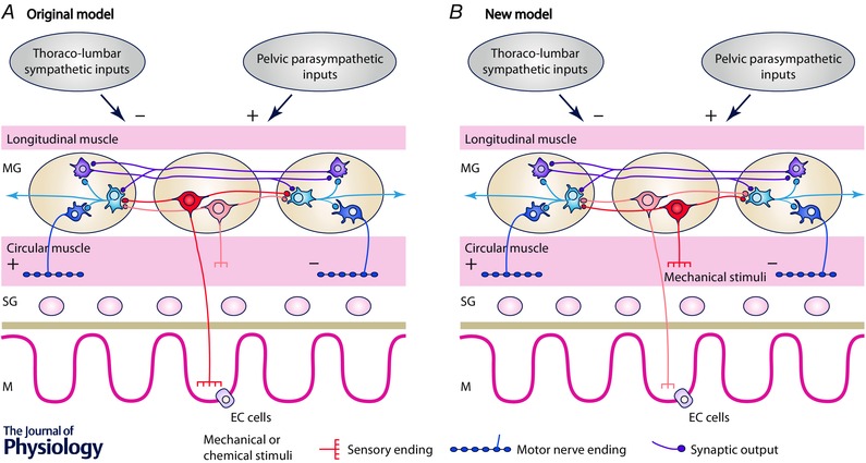
In recent years there have been significant technical and methodological advances in our ability to record the movements of the gastrointestinal tract. This has led to significant changes in our understanding of the different types of motor patterns that exist in the gastrointestinal tract (particularly the large intestine) and in our understanding of the mechanisms underlying their generation. Compared with other tubular smooth muscle organs, a rich variety of motor patterns occurs in the large intestine. This reflects a relatively autonomous nervous system in the gut wall, which has its own unique population of sensory neurons. Although the enteric nervous system can function independently of central neural inputs, under physiological conditions bowel motility is influenced by the CNS: if spinal pathways are disrupted, deficits in motility occur. The combination of high resolution manometry and video imaging has improved our knowledge of the range of motor patterns and provided some insight into the neural and mechanical factors underlying propulsion of contents. The neural circuits responsible for the generation of peristalsis and colonic migrating motor complexes have now been identified to lie within the myenteric plexus and do not require inputs from the mucosa or submucosal ganglia for their generation, but can be modified by their activity. This review will discuss the recent advances in our understanding of the different patterns of propagating motor activity in the large intestine of mammals and how latest technologies have led to major changes in our understanding of the mechanisms underlying their generation.


Figures

Figure 1
Figure 1. Originally proposed enteric neural pathway underlying distension‐evoked reflexes (A) compared with latest understanding of this neural pathway (see B)
A, Originally proposed arrangement of enteric neural pathways in the intestine. Mucosally projecting sensory neurons (in red) respond to chemical and mechanical stimuli and are responsible for initiating the peristaltic reflex. Mechanosensory interneurons respond to circumferential stretch and have transduction sites in the circular muscle. They can initiate polarized neural reflexes to the circular and longitudinal muscle in the distal colon. Possible other enteric circuits of interneurons generate migrating motor complexes. Extrinsic inputs from sympathetic and parasympathetic pathways inhibit and excite motility by acting on the enteric circuits. Current knowledge of submucosal neurons and longitudinal muscle have been deliberately omitted to maintain simplicity. B, the new model describes how our understanding of these pathways has changed in recent years. We propose a refinement of the enteric circuits involved in colonic motility. The mechanosensory enteric neurons (located in the myenteric plexus) have essential mechanosensitive nerve endings located in the circular muscle (see Spencer et al. 2006) (now shown in red) which initiate polarized neural pathways that result in oral contraction and anal relaxation. These pathways do not require mucosal inputs and represent the bases of the neuromechanical loop responsible for propulsion adapted to the consistency of contents. However, stimulation of mucosal sensory nerve endings can initiate and modulate enteric neural activity. The polarized enteric circuits involved in the neuromechanical loop are modulated by underlying cyclic neural activity initiated by any maintained distension and providing the bases of the migrating motor complexes. Extrinsic excitatory inputs from both pelvic and vagal sources are involved in the greater or lesser permissive role depending on the degree of central control of colonic movements. EC cells, enterochromaffin cells, with permission.
Figure 2
Figure 2. Propulsion of luminal contents is modified by differences in the consistency of luminal contents
In the upper half of the figure, five different images are shown where boluses of different sizes and shapes (pellets) are present in the distal colon. Natural pellets are represented in purple, distal colonic migrating motor complexes (DCMMCs) in red, fluid pellets in blue, viscous pellets in grey, and peristaltic contractions in green. It was noted that peristaltic contractions often started approximately half‐way along the preparation (white arrow). A–C shows the relationships between speed of propulsion and the bolus size; speed correlates positively with bolus length (A); speed in relation to bolus surface area (B); speed in relation to average bolus diameter (C). In the upper quadrant of each graph (A–C) the probability density is represented on a log scale. The distributions of means are shaded within their 95% highest density interval (HDI, see text) and the gaps between shaded 95% HDIs represent significant differences between the various measures. Note that for the average diameter (C) there is a clear separation between all of the shaded 95% HDIs, indicating that the diameters of different boluses are significantly different from one another. However, a poor correlation exists between the speed of propulsion and average diameter. For surface areas (B) from about 1 cm2, the speeds of all bolus types (except DCMMCs) differ significantly, as shown by the gap between their shaded 95% HDIs (see centre square), with natural, viscous, liquid and peristaltic liquid boluses moving at increasing speeds. The inset in B depicts the distributions of the steepest slope of the sigmoid curve for each bolus type. A shows that a significant overlap exists between the length of the natural, viscous and fluid pellets. DCMMCs (shown in red) are shorter than all other bolus types and peristaltic contractions (shown in green) are significantly longer than all other bolus types. Figure reproduced from Costa et al. (2015), with permission.
Figure 3
Figure 3. Spatio‐temporal map of the steady states of linear and quiescent orbits
This was constructed from the composite DPMaps (shown in A) and the extracted orbits that were formed from the independent variables (diameter and pressure) (see B). In B each of the 12 possible mechanical states are mapped in different colours. The map portrays the periods of quiescence either when the intestine remains passively dilated (light green) or passively occluded (dark green). Red, orange and yellow areas represent active contractions and mark the propagating area of contraction during neural peristalsis. Active relaxation (aqua and light blue) precedes both in time and space the propagating contraction. Myogenic activity in this particular example consists of isometric relaxations and contractions. The last map (C) represents a simplification of the full composite map of states by clustering all areas undergoing active contraction (red) and active relaxation (blue). All other passive states are in black. The spatio‐temporal nature of active contractions and relaxations during neurogenic activity and myogenic activity is made very distinct. Figure reproduced from Costa et al. (2013 b).
Figure 4
Figure 4. The frequency of colonic migrating motor complexes (CMMCs) is significantly increased by the presence of faecal contents
A, schematic diagram showing an isolated whole mouse colon containing multiple faecal pellets. B and C show spatio‐temporal maps from the whole colon of different mice in vivo. CMMCs occur frequently and propagate over significant lengths of colon. D and E, when the colon has expelled all contents, the same segments of colon shown in B and C, respectively, rarely generate CMMCs. When CMMCs do occur, their velocity of propagation is significantly slower. This shows that the presence of multiple pellets in the lumen enhances the velocity and frequency of CMMCs. Figure reproduced from Barnes et al. (2014).
Figure 5
Figure 5. Myogenic ‘ripples’ recorded from isolated rabbit distal colon
In the presence of hexamethonium and tetrodotoxin, random chaotic ripple contractions can be visualized, with initiation sites that vary and an irregular direction of propagation. Figure reproduced from Dinning et al. (2012 a).
Figure 6
Figure 6. Colonic manometry recordings made using high resolution fibre optic technology taken from healthy adult human colon
The left‐hand images are displayed as spatio‐temporal colour plots and on the right is the same image displayed as a conventional line plot. A shows the well‐described high amplitude propagating sequences. In B, three long single propagating motor patterns can be seen (black arrows). These motor patterns rapidly propagate across the transverse and descending colon and the component pressure waves have a lower amplitude than the pressure wave shown in A. In C, an example of a slowly propagating retrograde motor pattern is shown (solid white arrow). These originate in the sigmoid colon and over several minutes propagate into the transverse colon. These motor patterns appear during an unstimulated period of recording (i.e. before a meal). Preceding the slow retrograde motor patterns is a short single motor pattern (short white arrow). In D, the cyclic propagating motor pattern is shown. This is the most common postprandial propagating motor pattern. It is largely confined to the distal regions of the colon and propagates predominantly in a retrograde direction, although in this instance both antegrade and retrograde propagation can be seen. The cyclic propagating motor patterns occur at the colonic slow wave frequency of 2–4 cycles min−1. Figure constructed from data published in Dinning et al. (2014 a).
Figure 7
Figure 7. Colonic motor patterns recorded with a fibre optic manometry catheter in the sigmoid colon and rectum
A shows the low‐resolution recording (10 cm spaced sensors) used for most colonic manometry studies. In B, the complete data set is shown. With the high resolution recording (1 cm spaced sensors) four propagating motor patterns can be seen (blue arrows indicate the actual direction of propagation). These propagating contractions may represent the colonic motor complex. Figure constructed from data published in Dinning et al. (2013).
Figure 8
Figure 8. Examples of pan‐colonic pressurizations recorded from descending colon of a patient with slow transit constipation, using a fibre optic manometry catheter
Figure constructed from data published in Dinning et al. (2015).
Figure 9
Figure 9. Schematic interpretation of the neuro‐mechanical loop mechanisms for propulsion of intestinal content initiated and sustained by a bolus
Three separate images have been taken of a bolus (yellow dashed line) moving along the rabbit distal colon at 2 s intervals. The inferred state of enteric motor neuron activity was determined from the relationships that exist between changes in diameter (video image) and the corresponding changes in intraluminal pressure (manometry). These relationships allow for the calculation of the mechanical state of the muscle (Costa et al. 2013 b; Dinning et al. 2014 b). The red regions indicate activation of excitatory motor neurons and the blue regions indicate the activation of enteric inhibitory motor neurons. The liquid bolus is propelled by oral excitation and anal inhibition, as proposed by Bayliss & Starling (1899), and predicted by the neuro‐mechanical loop mechanisms. The bolus distends the gut and activates the polarized ascending excitatory reflex pathways resulting in oral active contraction of the muscle (red) and anal active relaxation (blue). This results in a mechanical event of propulsion of the bolus in the anal direction distending a new area of intestine, then initiating neural activity of polarized enteric pathways, resulting in further propulsion of the bolus, which becomes self‐sustained.

References

    1. Adler HF, Atkinson AJ & Ivy AC (1941). A study of the motility of the human colon: an explanation of dysynergia of the colon, or of the ‘unstable colon’. Am J Dig Dis 8, 197–202.
    1. Anderson KD (2004). Targeting recovery: priorities of the spinal cord‐injured population. J Neurotrauma 21, 1371–1383. - PubMed
    1. Arkwright JW, Underhill ID, Maunder SA, Blenman N, Szczesniak MM, Wiklendt L, Cook IJ, Lubowski DZ & Dinning PG (2009). Design of a high‐sensor count fibre optic manometry catheter for in‐vivo colonic diagnostics. Opt Express 17, 22423–22431. - PubMed
    1. Auli M, Martínez E, Gallego D, Opazo A, Espín F, Marti‐Gallostra M, Jiménez M & Clavé P (2008). Effects of excitatory and inhibitory neurotransmission on motor patterns of human sigmoid colon in vitro . Br J Pharmacol 155, 1043–1055. - PMC - PubMed
    1. Bampton PA, Dinning PG, Kennedy ML, Lubowski DZ & Cook IJ (2001). Prolonged multi‐point recording of colonic manometry in the unprepared human colon: Providing insight into potentially relevant pressure wave parameters. Am J Gastroenterol 96, 1838–1848. - PubMed

Publication types

LinkOut - more resources