. 2016 Jun;245(6):653-66.
doi: 10.1002/dvdy.24404. Epub 2016 Apr 13.

Vertebrate Claudin/PMP22/EMP22/MP20 family protein TMEM47 regulates epithelial cell junction maturation and morphogenesis

Affiliations

Vertebrate Claudin/PMP22/EMP22/MP20 family protein TMEM47 regulates epithelial cell junction maturation and morphogenesis

Yi Dong et al. Dev Dyn. 2016 Jun.

Abstract

Background: TMEM47 is the vertebrate orthologue of C. elegans VAB-9, a tetraspan adherens junction protein in the PMP22/EMP/Claudin family of proteins. VAB-9 regulates cell morphology and adhesion in C. elegans and TMEM47 is expressed during kidney development and regulates the activity of Fyn. The conserved functions of VAB-9 and TMEM47 are not well understood.

Results: expression of TMEM47 in C. elegans functionally rescues vab-9 mutations. Unlike Claudins, expression of TMEM47 in L fibroblasts does not generate tight junction strands; instead, membrane localization requires E-cadherin expression. Temporally, TMEM47 localizes at cell junctions first with E-cadherin before ZO-1 colocalization and in polarized epithelia, TMEM47 colocalizes with adherens junction proteins. By immunoprecipitation, TMEM47 associates with classical adherens junction proteins, but also with tight junction proteins Par6B and aPKCλ. Over-expression of TMEM47 in MDCK cells decreases apical surface area, increases activated myosin light chain at cell-cell contacts, disrupts cell polarity and morphology, delays cell junction reassembly following calcium switch, and selectively interferes with tight junction assembly. Reduced TMEM47 expression results in opposite phenotypes.

Conclusions: TMEM47 regulates the localization of a subset of tight junction proteins, associated actomyosin structures, cell morphology, and participates in developmental transitions from adherens to tight junctions. Developmental Dynamics 245:653-666, 2016. © 2016 Wiley Periodicals, Inc.

Keywords: Par6B; aPKCλ; actomyosin; adherens junction; cellular junctions; epithelial morphogenesis; tight junction.


Figures

Figure 1
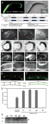
Figure 1. Murine TMEM47 functionally rescues the C. elegans vab-9 mutation
TMEM47-GFP was expressed under control of the ajm-1 promoter in vab-9 mutant animals. (A) Cell junction localization of TMEM47-GFP in epidermis and expression in the pharynx. (B) Matching DIC image of panel A showing vab-9 body and tail shape defect rescue (inset: control second larval stage vab-9 mutant). (C) Schematic of murine TMEM47 and C. elegans VAB-9. TM indicates transmembrane spanning domains. The Amino terminus (N-term) and Carboxy terminus (C-term) are predicted to be intracellular. Below are shown the predicted cytoplasmic N-term and C-term amino acids. TMEM47 and VAB-9 contain 181 and 211 residues, respectively. TMEM47 canine and murine cytoplasmic domains are identical. (D-V) TMEM47 rescues vab-9 defects. Expression of TMEM47-GFP variants in embryonic seam cells (D-F) and pharyngeal (K-N) epithelia. Full length (“FL”) TMEM47-GFP and amino terminal deletion “ΔN” (E) localize strongly, but Y180/181A “YA” (F) is mis-localized basolaterally in seam cells. (G-J). Phenotypes of the variants in vab-9(ju6) null mutants is shown. All constructs except the C-terminal deletion “ΔC” rescue all vab-9 phenotypes (tail, body shape, and egg-laying defects), scored as adults. (K-N) Expression of variants in the pharynx during embryonic elongation “FL” localizes normally, while “ΔN” and “YA” are not exclusively concentrated at junctions. TMEM47-GFP carboxy terminal “ΔC” was completely mislocalized from pharyngeal cell junctions (N). (O-R) Expression of Full length “FL” “N-term” and “YA” but not “ΔC” TMEM47-GFP variants restores the regular circumferential F-actin filaments in vab-9 mutant epidermis. Anterior is to the left and the strong white band(s) indicate F-actin in muscle quadrants that run anterior to posterior just below and attached to the epidermis. The bands orthogonal to the muscle actomyosin are regularly spaced in rescued animals and are discontinuous in “ΔC” epidermis. (S, T) Lateral projections of TMEM47 FL and YA expression in dorsal epidermal cells; drawings shown below represent the TMEM47 localization observed in panels S and T, respectively. (U) Quantification of vab-9 phenotypic rescue by untagged (“wt”) TMEM47, TMEM47-GFP (FL), and TMEM47-GFP variants YA, ΔN, and ΔC. Animals were scored as adults and were only scored as rescued if all vab-9 phenotypes were absent and animals were indistinguishable from wild type. (V) Western blot of extracts from TMEM47-GFP and variant containing strains probed with anti-GFP antibodies. Arrow indicates TMEM47-GFP fusion proteins. Scale, 10um.
Figure 2
Figure 2. TMEM47 associates with adherens junctions in MDCK cells
(A,B) Immunolocalization of endogenous TMEM47 (“anti-TM4”) with E-Cadherin and ZO-1 in the apical domain of MDCK cells. The merged top focal planes (3μm) are shown. (C, D) Lateral views (Z axis) showing localization of E-Cadherin and ZO-1, respectively, relative to TMEM47-GFP. (E) Co-localization of β-Catenin with TMEM47-GFP. (F) Co-localization of endogenous TMEM47 (“anti-TM4”) with F-actin at new cell contacts in MCK cells. (G) Expression of C. elegans VAB9-GFP in MDCK cells shows a pattern similar to TMEM47. Gamma adjustments were made to merged images.
Figure 3
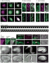
Figure 3. TMEM47 expression during junctional maturation and dependence on E-Cadherin
(A) Co-localization of TMEM47-GFP with β-Catenin, E-Cadherin, and Occludin in isolated, non-contacting MDCK cells. TMEM47-GFP is observed in cytoplasmic structures and at membranes, co-localizing with β-Catenin and E-Cadherin. Less overlap is observed with Occludin. (B, C) Co-localization of E-Cadherin and ZO-1 with TMEM47 in (B) new cell contacts (<1 hour after plating) or (C) more established cell contacts (>4 hours after plating). Arrows indicate ZO-1-free cell contacts containing TMEM47-GFP. (D) Kymograph of video recording showing similar movement of TMEM47-tagRFP (top) and β-catenin-GFP (bottom). Total run time is 16 minutes with 24 of 58 total frames shown (E) Images of first frame from video, indicating region shown in (D). The merged image of the two tagged proteins demonstrates co-localization in a remodeling cell-cell contact. Arrows indicate the point of cell-cell contact. . (F) In L cells, which lack E-Cadherin, there was no placement of TMEM47-GFP at cell-cell contacts. (G) In L cells transfected with E-Cadherin, TMEM47-GFP now was present at cell-cell contacts and co-localized with E-Cadherin. (H-L) ZOO-1-GFP (H-J) and ZOO-1 (K,L) localize the same in wild type animals (H and K) and vab-9(ju6) mutants (I and J, L). Nomarski images in Columns H, I, and J match the fluorescent images above. ZOO-1-GFP localizes normally prior to development of Vab phenotypes (I) and after phenotypes are visible (fourth larval stage) (J). Scale=10um. Gamma adjustments were made to images in A, F, and G.
Figure 4
Figure 4. TMEM47 over-expression affects tight junction protein localization and F-actin organization, but not adherens junction protein localization
TMEM47-GFP over-expression in confluent MDCK cells (A,D,G) and immunolocalization of Occludin (B), Claudin 1 (E), and β-Catenin (H). Regions of TMEM47-GFP over-expression correlated with mislocalization of Occludin (B,C) and Claudin 1 (E,F), but not β-Catenin (H,I). Loss of cell membrane staining in TMEM47-GFP positive cells indicated with arrows. Scale=10um. Gamma adjustments were made to merged images (C,F,I).
Figure 5
Figure 5. Association of TMEM47 with catenins and tight junction proteins Par6B and aPKCλ
(A-C) endogenous α-catenin and β-catenin (A), myc-tagged aPKCλ (B), and endogenous Par6B (C) co-immunoprecipitate with TMEM47-GFP. (D-G) Over-expression of TMEM47-GFP disrupts localization of Par6B and aPKCλ at cell junctions. (D) Normal Par6B localization in top 3um of the cell (top) compared to ZO-1 control (bottom). (E) Similar focal planes as (D) showing Par6B displaced from membrane (top) in TMEM-47-GFP expressing cells. TMEM47-GFP expression is shown (bottom). (F) aPKCλ and Par6B localization in MDCK cells. (G) mislocalized aPKCλ (top) in TMEM47-GFP over-expressing cells (bottom). Arrows indicate similar cytoplasmic aPKCλ and TMEM47-GFP localization. Thin panels show projected lateral (Z-axis) views. (H,I) TMEM47 shRNA in MDCK cells stained with Par6B and β-catenin, respectively. TMEM47 RNAi is shown on the left panel and scrambled oligo in pGSuper is on the right lys = lysate, T47-GFP = TMEM47-GFP, HC = heavy chain.
Figure 6
Figure 6. TMEM47 regulates cell junction reassembly following calcium switch
Confluent monolayers of MDCK cells were placed in low calcium medium to disrupt existing cell-cell junctions and then returned to normal calcium media to allow reassembly of cell junctions. (A-D) E-Cadherin expression pattern at 3 and 7 hours following low calcium incubation. Compared to normal MDCK cells (A, B), re-assembly of E-Cadherin-containing junctions were delayed in TMEM47-GFP over-expressing cells 3 hours following calcium switch (C); however junctions were re-established after 7 hours (D) but with obvious morphological defects. (E-H) Similar delays and morphological defects in ZO-1 junctional reassembly were observed in TMEM47-GFP cells (G,H) compared to control cells (E,F). (I-K) Reducing TMEM47 expression accelerates cell junction reassembly. MDCK cells transfected with scrambed oligo shRNA control vectors (I) or cells transfected with TMEM47 shRNA (J) were subjected to calcium switch and stained for ZO-1 1 hour after re-addition of calcium. TMEM47 shRNA expressing cells displayed more rapid junctional reformation after 1 hour compared to controls, quantified in (K). *P<0.027 and **P<0.015. Scale=10um.
Figure 7
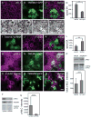
Figure 7. TMEM47 regulates apical surface area and p-MLC
(A-C) MDCK cells and MDCK cells stably expressing TMEM47-GFP were mixed and grown in confluent monolayers. (A) ZO-1 staining delineating the apical surface area. (B) TMEM47-GFP cells expressing cells (C) merged image. (D) Quantification of apical surface areas. *P<0.0001. (E-H) MDCK control cells (E,F) and TMEM47-GFP cells (G,H) phalloidin stained for F-actin reveal ventral stress fibers (E,G), and cell borders (F,H). The number of F-actin bundles/cell were not statistically different, but cell area for TMEM47-GFP was half that of MDCK cells (see text). (I-L) TMEM47 shRNA knock down cells were immunostained for F-actin to reveal cell borders and apical surface area of expressing and non-expressing cells were measured; note that F-actin appeared reduced in shRNA cells. (L) Quantification of the apical surface area of control shRNA and TMEM47 shRNA cells. **P<0.0001. (M-O) Phosphorylated myosin light chain (p-MLC) expression (M) was increased at cell borders in TMEM47-GFP cells (N); merged in (O). (P) Despite increased concentration of p-MLC at cell junctions in TMEM47-GFP overexpressing cells, overall p-MLC levels in TMEM47 cells were not significantly greater than in MDCK cells. A representative blot of three experiments is shown in the upper panel and quantified in the third panel (*** P=0.45). (Q-S) TMEM47-GFP cells also displayed increased apical F-actin accumulation (T,U) p-MLC is reduced in Tmem47 shRNA cells. A Representative blot (T, upper panel) of three averaged experiments (U) is shown. **** P= 0.0372. (T, second panel) Knockdown efficiency of TMEM47 culture was determined by Western blotting. SA = Surface Area. Scale=10um., Gamma adjustments were made to merged images C,K, and O.

References

    1. Adams CL, Chen YT, Smith SJ, Nelson WJ. Mechanisms of epithelial cell-cell adhesion and cell compaction revealed by high-resolution tracking of E-cadherin-green fluorescent protein. J Cell Biol. 1998;142:1105–1119. - PMC - PubMed
    1. Armenti ST, Nance J. Adherens junctions in C. elegans embryonic morphogenesis. Subcell Biochem. 2012;60:279–299. - PMC - PubMed
    1. Azhibekov TA, Wu Z, Padiyar A, Bruggeman LA, Simske JS. TM4SF10 and ADAP interaction in podocytes: Role in Fyn activity and Nephrin phosphorylation. Am J Physiol Cell Physiol 2011 - PMC - PubMed
    1. Baum B, Georgiou M. Dynamics of adherens junctions in epithelial establishment, maintenance, and remodeling. J Cell Biol. 2011;192:907–917. - PMC - PubMed
    1. Bruggeman LA, Martinka S, Simske JS. Expression of TM4SF10, a Claudin/EMP/PMP22 family cell junction protein, during mouse kidney development and podocyte differentiation. Dev Dyn. 2007;236:596–605. - PubMed

Publication types

MeSH terms

LinkOut - more resources