. 2016 Mar 18;11(3):e0150320.
doi: 10.1371/journal.pone.0150320. eCollection 2016.

Streptococcus pneumoniae Cell-Wall-Localized Phosphoenolpyruvate Protein Phosphotransferase Can Function as an Adhesin: Identification of Its Host Target Molecules and Evaluation of Its Potential as a Vaccine

Affiliations

Streptococcus pneumoniae Cell-Wall-Localized Phosphoenolpyruvate Protein Phosphotransferase Can Function as an Adhesin: Identification of Its Host Target Molecules and Evaluation of Its Potential as a Vaccine

Yaffa Mizrachi Nebenzahl et al. PLoS One. .

Abstract

In Streptococcus pneumonia, phosphoenolpyruvate protein phosphotransferase (PtsA) is an intracellular protein of the monosaccharide phosphotransferase systems. Biochemical and immunostaining methods were applied to show that PtsA also localizes to the bacterial cell-wall. Thus, it was suspected that PtsA has functions other than its main cytoplasmic enzymatic role. Indeed, recombinant PtsA and anti-rPtsA antiserum were shown to inhibit adhesion of S. pneumoniae to cultured human lung adenocarcinoma A549 cells. Screening of a combinatorial peptide library expressed in a filamentous phage with rPtsA identified epitopes that were capable of inhibiting S. pneumoniae adhesion to A549 cells. The insert peptides in the phages were sequenced, and homologous sequences were found in human BMPER, multimerin1, protocadherin19, integrinβ4, epsin1 and collagen type VIIα1 proteins, all of which can be found in A549 cells except the latter. Six peptides, synthesized according to the homologous sequences in the human proteins, specifically bound rPtsA in the micromolar range and significantly inhibited pneumococcal adhesion in vitro to lung- and tracheal-derived cell lines. In addition, the tested peptides inhibited lung colonization after intranasal inoculation of mice with S. pneumoniae. Immunization with rPtsA protected the mice against a sublethal intranasal and a lethal intravenous pneumococcal challenge. In addition, mouse anti rPtsA antiserum reduced bacterial virulence in the intravenous inoculation mouse model. These findings showed that the surface-localized PtsA functions as an adhesin, PtsA binding peptides derived from its putative target molecules can be considered for future development of therapeutics, and rPtsA should be regarded as a candidate for vaccine development.


Conflict of interest statement

Competing Interests: This study was partly funded by BG Negev Technologies. The study and part of the authors were partially funded by a commercial company (Nasvax Ltd./Protea Vaccine Technologies Ltd.) through the BGNegev Biotechnologies which is the BGU industrial liaison. Nasvax Ltd./Protea Vaccine Technologies Ltd. is the employer of authors Shahar Dotan, Michael Tal and Ronald Ellis. The PtsA protein discussed in the enclosed manuscript is protected by several pending patents: US 7,504,110; EP 2275128; US 8,691,243 B2. The protein vaccine is at an early stage of preclinical studies. There are no further patents, products in development or marketed products to declare. This does not alter the authors' adherence to all the PLOS ONE policies on sharing data and materials.

Figures

Fig 1
Fig 1. Surface expression of PtsA.
A) Immunoblot of untagged rPtsA: total S. pneumoniae extract (Total), total cell-wall proteins (CW), cytoplasmic proteins (Cyto) and membrane extract (mem) were probed with rabbit anti-rPtsA antiserum. B) Immunoblot of recombinant pneumolysin (rPly): total S. pneumoniae extract, total cell-wall proteins, cytoplasmic proteins and membrane extract were probed with rabbit anti-rPly antiserum. C) Immunoblot of tagged rFabD: total S. pneumoniae extract, total cell-wall proteins, cytoplasmic proteins and membrane extract were probed with rabbit anti-rFabD antiserum. D) FACS analysis of PtsA surface expression using anti-rPtsA monoclonal antibody (mAb) and isotype control IgG. Red line–preimmune control serum; green line–anti rPtsA mAb diluted 1:50; blue line–anti rPtsA mAb diluted 1:20. M = molecular weight markers. E) SIM orthogonal view image demonstrating the surface staining with mouse anti rPtsA antibody detected with donkey anti-mouse IgG (green). Three planes XZ, XY and YZ are shown. PtsA is visible on the perimeter of the bacterium in green, surrounding the white bacterial genome.
Fig 2
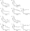
Fig 2. PtsA mediates S. pneumoniae adhesion to target host cells.
A–E. A549 cells were grown to confluence and then blocked with 0,5% gelatin for 1 h. Excess gelatin was removed, and the A549 cells were incubated for 1 h with rPtsA at the denoted concentrations. Excess protein was then removed. S. pneumoniae was added for 1 h to the cells, non-adherent bacteria were removed, and cells were detached with trypsin and plated onto blood agar plates for counting. rPtsA inhibited the adhesion to A549 cells of: A) strain WU2 (p < 0.0001; r = −0.943), B) strain 3.8DW (p < 0.0001; r = −0.371), C) strain D39 (p < 0.0001; r = −1), and D) strain R6 (p < 0.0006; r = −1). E) rKLH, a protein used as a negative control, did not inhibit D39 adhesion to A549 cells (p = 0.8; r = −0.2). F–J. S. pneumoniae cells (WU2 and 3.8DW) were treated for 1 h with rabbit anti-rPtsA antiserum and added to gelatin-blocked A549 cells for 1 h; non-adherent bacteria were removed; and cells were detached with trypsin and plated onto blood agar plates for counting. Rabbit anti-rPtsA inhibited adhesion to A549 cells of: F) strain WU2 (p < 0.0001; r = −0.359); G) strain 3.8DW (p < 0.0001; r = −0.886); H) strain D39 (p < 0.0001; r = −0.943); and I) strain R6 (p < 0.0001, r = −0.406); J) Serum obtained from a rabbit prior to immunization did not inhibit the adhesion of strain WU2 to A549 (J p>0.005; r = +0.667). (p > 0.005; r = +0.667). Experiments were performed in triplicate and repeated at least 3 times. Values are means±SD. *Student's t-test p<0.05.
Fig 3
Fig 3. Putative target molecules in A549 cells.
A549 cells were fixed with 4% para-formaldehyde and stained with a combination of a mouse anti Eps 1 and one of the following: rabbit anti Int β4, PCDH19, MMRN1, or BMPER. The secondary antibodies used were either Alexa Fluor 405-conjugated AffiniPure Goat Anti-Mouse IgG (blue) or Alexa Fluor 594-conjugated AffiniPure Goat Anti-Rabbit IgG (red) in accordance with the primary antibody used. 3D SIM images were taken using the Elyra SIM imaging system.
Fig 4
Fig 4. Immunostaining of S. pneumoniae adhesion to A549 cells.
CFDA-stained S. pneumoniae strain WU2 cells were incubated with A549 cells for 1 h. Excess bacteria were removed, and the culture was fixed with 4% para-formaldehyde, stained, and viewed with a FluoView FV1000 confocal system (Olympus, Japan). The following stains were used: A) mouse anti-Eps 1 antiserum and rabbit anti-integrin β4 antiserum; C) mouse anti-epsin 1 antiserum and rabbit anti-BMPER antiserum; E) mouse anti-Eps 1 antiserum and rabbit anti-MMRN1 antiserum; and G) mouse anti-Eps 1 antiserum and anti-PCDH19 antiserum. The same cells viewed by Nomarsky microscopy overlaid with the CFDA-stained bacteria are shown, respectively, in B), D), F) and H). Secondary antibodies used were Alexa Fluor 405-conjugated AffiniPure Goat Anti-Mouse IgG (H+L) antiserum and Alexa Fluor 594-conjugated AffiniPure Goat Anti-Rabbit IgG (H+L (antiserum, in accordance with the primary antiserum used.
Fig 5
Fig 5. SIM images of CFDA-stained bacteria with Int β4 on A549 cells.
CFDA-stained S. pneumoniae strain WU2 cells were incubated with A549 cells for 1 h. Excess bacteria were removed, and the culture was fixed with 4% para-formaldehyde and stained with a rabbit anti Int β4. Secondary antibody used was Alexa Fluor 594 (red) conjugated AffiniPure Goat Anti-Rabbit IgG (H+L). 3D SIM images were obtained with the Elyra SIM imaging system with a 63x oil objective (NA = 1.4); actual magnification of the image is as indicated in the scale bar. A) S. pneumoniae (CFDA 488 nm—green) adhering to A549 epithelial cells are seen coated by Int β4 (red). B) Higher magnification of bacteria (green) enveloped with Int β4 (red). C) Intensity alignment profile. D) Pedestal-like structure formed at the site of adherence of S. pneumoniae (green) to A549 cells recruits Int β4 (red). E) Higher magnification demonstrates the recruitment of Int β4 (red) to the pedestal-like structure underneath the adhered S. pneumoniae (green). F) Intensity alignment profile.
Fig 6
Fig 6. MicroScale thermophoresis (MST) analysis.
In all cases, 16 serial dilutions of each putative receptor-derived peptide (~90 nM–3.175 mM) were mixed with a constant concentration of 335 nM labeled rPtsA protein. The normalized change in the fluorescence of the labeled rPtsA protein was plotted against the peptide concentration, and a fit was computed by the NTP program. The experiments were performed in triplicate. Presented here are the affinities of rPtsA binding to: the Eps1-derived peptide Kd = 43.900 ± 9.35 μM; the BMPER-derived peptide Kd = 21.00± 5.49μM; the MMRN1-derived peptide Kd = 391 ± 72.7 μM; the PCDH19-derived peptide Kd = 58.00 ± 13.4 μM; and the Int β4-derived peptide Kd = 115±20.5μM. For the control, namely, the Nox putative receptor (contactin 4)-derived peptide, a fit could not be generated due to a very low signal-to-noise ratio.
Fig 7
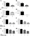
Fig 7. Inhibition of S. pneumoniae adhesion to A549 cells by Eps 1-derived peptide.
S. pneumoniae cells were treated for 1 h with Eps 1-derived peptide and added to A549 cells for 1 h; non-adherent bacteria were removed; and cells were detached with trypsin and plated onto blood agar plates for counting. A) Strain WU2 (p < 0.0001; r = −0.943); B) Strain 3.8DW (p < 0.0001; r = −0.056); C) Strain D39 (p < 0.0001; r = −0.943); D) Strain R6 (p <0.0001; r = −0.406; *t-test p < 0.05). Experiments were performed in 3–6 replicates and repeated at least 3 times. Values are means±SD. *Student's t-test p < 0.05.
Fig 8
Fig 8. Inhibition of adhesion of S. pneumoniae cells to A549 cells by target-derived peptides.
S. pneumoniae cells were treated for 1 h with peptide and added to A549 cells for 1 h; non-adherent bacteria were removed; and cells were detached with trypsin and plated onto blood agar plates for counting. A) BMPER, WU2 (P < 0.0001, r = −0.887); B) BMPER, 3.8DW (P < 0.0001, r = −883); C) Col VIIα WU2 (P < 0.0001, r = −1); D) Col VIIα, 3.8DW (P < 0.0001, r = −0.886); E) MMRN1, WU2 (P < 0.0001, r = −1); F) MMRN1, 3.8DW (P < 0.0001, r = −0.821); G) PCDH 19, WU2 (P < 0.0001, r = −0.829); H) PCDH 19, 3.8DW (P < 0.0001, r = −0.754); I) Int β4, WU2 (P < 0.0001, r = −0.5619); J) Int β4, 3.8DW (P < 0.0001, r = −0.967); K) No significant inhibition was observed with a scrambled peptide (K scrambled peptide, P = 0.9981). Experiments were performed in triplicates and repeated at least 3 times. Values are means±SD. *Student's t-test p<0.05.
Fig 9
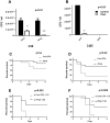
Fig 9. Effects of target-derived peptides on nasopharyngeal and lung colonization and survival in mice.
Strain WU2 cells were pretreated with peptide and then inoculated IN into adult CBA/Nxid mice. As a control, another group of mice was injected with bacteria treated ex vivo for 1 h with PBS alone. Mice were euthanized 3, 24 or 48 h later. The nasopharynx and lungs were excised, homogenized and plated onto blood agar plates for counting. A) Eps 1 (14.7 μM), nasopharynx, 3 h (P < 0.001); B) Eps 1 (14.7 μm), lung, 3 h (P < 0.0001); C) Eps 1 (14.7 μm), nasopharynx, 24 h (P < 0.0001); D) Eps 1 (14.7 μm) lung, 24 h (P < 0.0001); E) BMPER, nasopharynx, 48 h at 7 and 14 μM (P < 0.0001); F) BMPER, lung, 48 h at 7 and 14 μM (P < 0.0001); G) PCDH19, nasopharynx, 48 h at 14.7 and 17.4 μM (P < 0.001); H) PCDH19, lung, 48 h (P < 0.0001).
Fig 10
Fig 10. Vaccine potential of rPtsA.
BALB/c mice were immunized subcutaneously (SC) with 25 μg of rPtsA emulsified with CFA and boosted (days 14 and 28) with IFA. Mice were challenged IN on day 42 with a sublethal dose (5 × 10⁷) of strain WU2. Mice were sacrificed 3 and 24 h (strain WU2) following inoculation, and the nasopharynx and right lobe lung were excised, homogenized and plated onto blood agar plates for bacterial colony counting. Control mice were immunized with the adjuvant alone. A) Bacterial load in the nasopharynx and the lungs 3 h post inoculation of BALB/c mice with strain WU2 (Student's t-test; P < 0.01 and P < 0.05, respectively). B. Bacterial load in the nasopharynx (P < 0.05) and lung of BALB/c mice 24 h post inoculation with WU2 strain. C) After a similar immunization regimen, BALB/c mice were challenged IN with a lethal dose (108 CFU) of strain WU2; control mice were immunized with the adjuvant alone. Mortality was monitored daily. Each group contained 10 mice (p = ns). D) After a similar immunization regimen, CBA/Nxid mice were challenged intravenously (IV) with a lethal dose (104 CFU) of strain WU2. Control mice were immunized with the adjuvant alone. Mortality was monitored continuously (P<0.02). Each group contained 15 mice. E) S. pneumoniae serotype 3 (104 CFU) was incubated with 1:10 dilution of serum obtained from adjuvant or 1:50 dilution of serum obtained from rPtsA + adjuvant immunized BALB/c mice and inoculated IV into BALB/c mice. Survival was monitored continuously (P < 0.002). Each group contained 10 mice. F) S. pneumoniae serotype 3 strain WU2 was incubated with 1:10 dilution of serum obtained from adjuvant or rPtsA + adjuvant (1:50) immunized CBA/Nxid mice. Then, 104 CFU of WU2 were inoculated IV into CBA/Nxid mice, and survival was monitored continuously (P < 0.006). Each group contained 10 mice.*Log-rank (Mantel-Cox) test.

References

    1. Dagan R, Poolman J, Siegrist CA. Glycoconjugate vaccines and immune interference: A review. Vaccine. 2010;28(34):5513–23. Epub 2010/07/06. 10.1016/j.vaccine.2010.06.026 . - DOI - PubMed
    1. Mond JJ, Lees A, Snapper CM. T cell-independent antigens type 2. Annual review of immunology. 1995;13:655–92. Epub 1995/01/01. 10.1146/annurev.iy.13.040195.003255 . - DOI - PubMed
    1. Shapiro ED, Berg AT, Austrian R, Schroeder D, Parcells V, Margolis A, et al. The protective efficacy of polyvalent pneumococcal polysaccharide vaccine. The New England journal of medicine. 1991;325(21):1453–60. Epub 1991/11/21. 10.1056/NEJM199111213252101 . - DOI - PubMed
    1. van der Poll T, Opal SM. Pathogenesis, treatment, and prevention of pneumococcal pneumonia. Lancet. 2009;374(9700):1543–56. Epub 2009/11/03. 10.1016/S0140-6736(09)61114-4 . - DOI - PubMed
    1. Hammitt LL, Bulkow LR, Singleton RJ, Nuorti JP, Hummel KB, Miernyk KM, et al. Repeat revaccination with 23-valent pneumococcal polysaccharide vaccine among adults aged 55–74 years living in Alaska: no evidence of hyporesponsiveness. Vaccine. 2011;29(12):2287–95. Epub 2011/01/25. 10.1016/j.vaccine.2011.01.029 . - DOI - PubMed

Publication types

MeSH terms

Substances

LinkOut - more resources