Review
. 2016 Jun;23(6):915-26.
doi: 10.1038/cdd.2015.172. Epub 2016 Mar 18.

The clearance of dying cells: table for two

Affiliations
Review

The clearance of dying cells: table for two

D R Green et al. Cell Death Differ. 2016 Jun.

Abstract

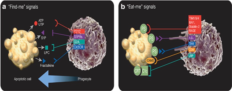
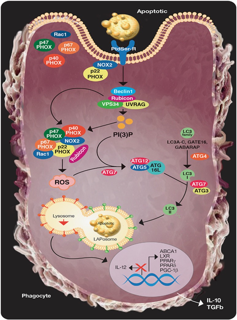
Phagocytic cells of the immune system must constantly survey for, recognize, and efficiently clear the billions of cellular corpses that arise as a result of development, stress, infection, or normal homeostasis. This process, termed efferocytosis, is critical for the prevention of autoimmune and inflammatory disorders, and persistence of dead cells in tissue is characteristic of many human autoimmune diseases, notably systemic lupus erythematosus. The most notable characteristic of the efferocytosis of apoptotic cells is its 'immunologically silent' response. Although the mechanisms by which phagocytes facilitate engulfment of dead cells has been a well-studied area, the pathways that coordinate to process the ingested corpse and direct the subsequent immune response is an area of growing interest. The recently described pathway of LC3 (microtubule-associated protein 1A/1B-light chain 3)-associated phagocytosis (LAP) has shed some light on this issue. LAP is triggered when an extracellular particle, such as a dead cell, engages an extracellular receptor during phagocytosis, induces the translocation of autophagy machinery, and ultimately LC3 to the cargo-containing phagosome, termed the LAPosome. In this review, we will examine efferocytosis and the impact of LAP on efferocytosis, allowing us to reimagine the impact of the autophagy machinery on innate host defense mechanisms.


Figures

Figure 1
Figure 1
The recruitment of phagocytes and recognition of dying cells by phagocytes. (a) Dying cells release ‘find-me' signals, such as ATP, UTP, S1P, lysophosphatidylcholine (LPC), or fractalkine, that recruit phagocytes to sites of cell death. Phagocytes sense these ‘find-me' signals via cognate receptors (P2Y2, S1PRs, G2A, and CXCR3, respectively). (b) Phagocytes express a variety of receptors and bridging molecules that recognize and engage dying cells via ‘eat-me' signals exposed on apoptotic cell surfaces. The most common ‘eat-me' signal, phosphatidylserine (PtdSer or PS), engages the PtdSer-specific receptors, TIM1, TIM3, TIM4, BAI1, stabilin-2, and RAGE, as well as the PS-specific bridging molecules MFG-E8, Gas6, and protein S. These bridging molecules engage other surface engulfment receptors (αvβ3 or TAM) to facilitate uptake. Other ‘eat-me' signals, such as calreticulin (CRT) and ICAM3, exist and mediate recognition and engulfment via the receptors LRP (via C1q) and CD14, respectively
Figure 2
Figure 2
The processing of engulfed dying cells requires LC3-associated phagocytosis (LAP) and promotes an anti-inflammatory response. Upon engulfment of dying cells, components of the LAP pathway are recruited to dead cell-containing phagosome (or LAPosome). The class III PI3 K complex, comprised of Beclin 1, VPS34, UVRAG, and Rubicon, is critical to the sustained and localized production of PI(3)P at the LAPosome. PI(3)P serves two roles – the recruitment of the downstream LAP machinery (such as ATG5, ATG12, ATG16L, and ATG7) and stabilization of the NOX2 complex for the production of ROS. Rubicon is also required for the stabilization of the NOX2 complex. Both ROS and PI(3)P are required for successful LC3-II decoration of the LAPosome, and LC3-II is required for fusion to the lysosome and maturation of LAPosome. The anti-inflammatory effects of efferocytosis are mediated by the activity of lipid and cholesterol sensors, such as ABCA1, LXR, PPARγ/δ, and PGC-1β, leading to the production of IL-10 and TGFβ, whereas pro-inflammatory mediators, such as IL-12, are actively repressed

References

    1. Green DR. Means To An End: Apoptosis And Other Cell Death Mechanisms. Cold Spring Harbor Laboratory Press: Cold Spring Harbor, NY, USA, 2011, p 220.
    1. Kinchen JM. A model to die for: signaling to apoptotic cell removal in worm, fly and mouse. Apoptosis 2010; 15: 998–1006. - PubMed
    1. Penaloza C, Lin L, Lockshin RA, Zakeri Z. Cell death in development: shaping the embryo. Histochem Cell Biol 2006; 126: 149–158. - PubMed
    1. McIlwain DR, Berger T, Mak TW. Caspase functions in cell death and disease. Cold Spring Harb Perspect Biol 2013; 5: a008656. - PMC - PubMed
    1. Oberst A, Dillon CP, Weinlich R, McCormick LL, Fitzgerald P, Pop C et al. Catalytic activity of the caspase-8-FLIP(L) complex inhibits RIPK3-dependent necrosis. Nature 2011; 471: 363–367. - PMC - PubMed