. 2016 Mar 17;61(6):809-20.
doi: 10.1016/j.molcel.2016.02.032.

Glutamine Triggers Acetylation-Dependent Degradation of Glutamine Synthetase via the Thalidomide Receptor Cereblon

Affiliations

Glutamine Triggers Acetylation-Dependent Degradation of Glutamine Synthetase via the Thalidomide Receptor Cereblon

T Van Nguyen et al. Mol Cell. .

Abstract

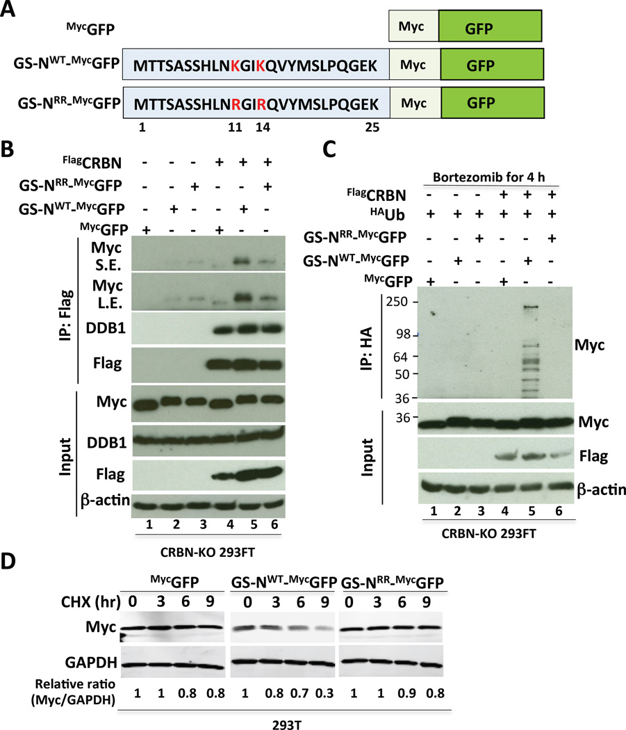
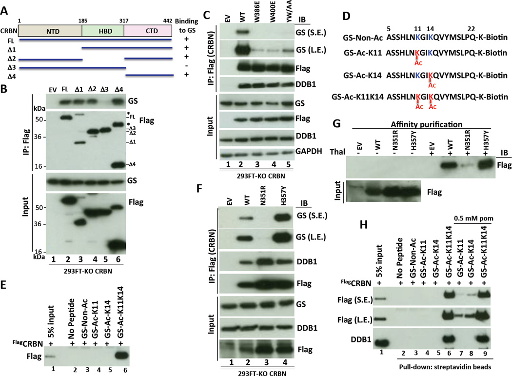
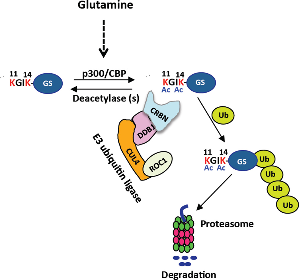
Cereblon (CRBN), a substrate receptor for the cullin-RING ubiquitin ligase 4 (CRL4) complex, is a direct protein target for thalidomide teratogenicity and antitumor activity of immunomodulatory drugs (IMiDs). Here we report that glutamine synthetase (GS) is an endogenous substrate of CRL4(CRBN). Upon exposing cells to high glutamine concentration, GS is acetylated at lysines 11 and 14, yielding a degron that is necessary and sufficient for binding and ubiquitylation by CRL4(CRBN) and degradation by the proteasome. Binding of acetylated degron peptides to CRBN depends on an intact thalidomide-binding pocket but is not competitive with IMiDs. These findings reveal a feedback loop involving CRL4(CRBN) that adjusts GS protein levels in response to glutamine and uncover a new function for lysine acetylation.


Figures

Figure 1
Figure 1. GS is an endogenous substrate of CRL4CRBN
(A) Identification of GS as a CRBN-interacting protein. 293T cells stably expressing FlagCRBN and grown in either ‘heavy’ or ‘light’ SILAC medium were treated with DMSO (light) or 50 µM thalidomide (heavy) for 4 h prior to lysis and immunoprecipitation (IP) with anti-Flag followed by mass spectrometry. The heavy:light ratios for GS and subunits of CRL4 and CSN are shown. The asterisk indicates a ratio that differs significantly from 1 (p-value 1×10⁻¹⁹). The data are an average of two experiments. Error bars indicate ± SD. (B) GS binds CRBN. 293T cells stably expressing empty vector or wild-type FlagCRBN were treated with or without lenalidomide (10 µM) for 3 h. Protein extracts were immunoprecipitated with Flag antibody followed by Western blot analysis with the indicated antibodies. The ratio of GS bound to CRBN normalized to input GS is shown. (C) Endogenous CRBN and GS interact. MM.1S cells were supplemented with DMSO or 1 µM lenalidomide 2 h prior to lysis and IP with mouse IgG control or CRBN antibodies. IP and input samples were fractionated by SDS-PAGE and immunoblotted with the indicated antibodies. Quantification was as described in (B). (D–E) CRBN promotes GS ubiquitylation in cells (D) and in vitro (E). (D) 293T cells were transiently transfected with plasmids expressing GSFlag and HAubiquitin (HAUb). After 30h, cells were treated with 10 µM MG132 for 4 h, followed by cell lysis and Flag IP under denaturing conditions. The input and bound fractions were evaluated by immunoblotting with HA and Flag antibodies. Ubiquitin conjugates in the input are shown in Figure S1C. (E) 293T cells stably expressing FlagCRBN were treated with proteasome inhibitor (1 µM bortezomib) for 6 h. After IP with Flag antibody, in vitro ubiquitylation of endogenous, co-precipitated GS was carried out for 1 h at 30°C in the presence or absence of E1+E2 and HAUb. Where indicated, methylated ubiquitin (Me-Ub) or recombinant (r) CUL4A-RBX1 was added. Reactions were analyzed by SDS-PAGE and immunoblotting with GS antibody. (Ub)n indicates polyubiquitylation. S.E., L.E.: short and long exposures.
Figure 2
Figure 2. CRBN is required for glutamine-induced degradation of GS
(A) Glutamine regulates GS protein abundance. Hep3B cells were maintained in DMEM 10% FCS without glutamine for 48 h. The cells were then treated with glutamine (4 mM) for the indicated times. Equal amounts of protein extracts were analyzed by SDS-PAGE and immunoblotting with the indicated antibodies. GAPDH served as a loading control. (B) Glutamine-induced GS degradation is blocked by the proteasome inhibitor bortezomib or the Nedd8-activating enzyme inhibitor MLN4924. Hep3B cells were starved of glutamine for 36 h, and then pretreated with or without bortezomib (200 nM) or MLN4924 (2 µM) for 30 min, followed by 4 mM glutamine treatment for 7 h. Cell lysates were analyzed by SDS-PAGE and immunoblotting with antibodies against GS, CRBN, and GAPDH. The relative ratio of GS:GAPDH, normalized to lane 1, is shown. (C) Glutamine-induced GS degradation is promoted by CRBN. Wild-type (WT) and CRISPR/Cas9-derived CRBN-knockout (KO) Hep3B cells were starved of glutamine for 36 h, followed by addition of 4 mM glutamine for 0, 12 and 16 h. The relative ratio of GS:GAPDH protein level, normalized to that at 0-time, is shown. Note that this experiment was done with a pool of KO cells (i.e. non-clonal) and there appears to be a small amount of residual CRBN in the population. (D–F) GS protein levels are elevated in the kidneys, skeletal muscles and lungs of Crbn−/− mice. Left panels: Tissue extracts prepared from total kidneys and skeletal muscles of wild-type (WT) and Crbn−/− (KO) mice were analyzed by SDS-PAGE and Western blotting, using GS, CRBN and GAPDH antibodies. n = 3–4 mice per group. Right panels: densitometric quantification of relative band intensities. Error bars represent the SEM. (G) Crbn−/− mice exhibit an increased glutamine/glutamate ratio in serum. Glutamine and glutamate levels in serum of wild-type (WT) and homozygous mutant Crbn−/− mice (KO) were quantified by mass spectrometry. Glutamine/glutamate ratio was calculated and represented as mean ± SD; n = 6 mice per group (P = 0.02615 by t-test).
Figure 3
Figure 3. The N-terminal extension of GS and its KxxK motif promote binding to CRBN, ubiquitylation, and degradation
(A–B) The N-terminal extension of GS is required to bind CRBN. (A) Schematic diagram of full-length (FL) human GS protein structure and deletion constructs used in (B). The GS degron (amino acids 1–24) recognized by CRBN is highlighted. (B) 293T cells stably expressing FlagCRBN were transfected with the indicated plasmids. After 36 h, cells were treated with 10 µM MG132 for 4 h. Cellular extracts were immunoprecipitated with Flag antibody, fractionated by SDS-PAGE and immunoblotted with Myc and Flag antibodies. *, indicates a non-specific band. A band ∼25 kDa represents IgG light chains (IgG-LC). (C) The N-terminal extension of GS is required for degradation. 293T cells stably expressing Flag-HA-tagged GS (FHGS) or GS with deletion of the N-terminal 24 amino acids (d1FHGS) were cultured in complete DMEM with 2 mM glutamine, and treated with cycloheximide (CHX; 100 µg/ml) for 0, 2, 4, and 6 h. Cell lysates were analyzed by SDS-PAGE and immunoblotting with Flag and GAPDH antibodies. The relative ratio of GS:GAPDH, normalized to that of zero-time, is shown. (D) The N-terminal KxxK motif modulates binding of GS to CRBN. 293T cells stably expressing FlagCRBN were transfected with empty vector (EV) or plasmids encoding the indicated GS mutants. After 36 h, cells were treated with 10 µM MG132 for 4 h. Cell extracts were immunoprecipitated with Flag antibody and the precipitated and input fractions were analyzed by SDS-PAGE and immunoblotting with DDB1, Myc, and Flag antibodies. WT: wild type. RR: K11R, K14R. AA: K11A, K14A. (E) The N-terminal KxxK motif modulates ubiquitylation of GS. 293T cells were transfected with plasmids encoding HAUb and the indicated Myc-tagged GS mutants. After 24 h, the cells were treated with 10 µM MG132 for 4 h, followed by cell lysis, denaturation of the lysate proteins, and IP with anti-Myc. The input lysates and bound fractions were evaluated by SDS-PAGE and immunoblotting with HA and Myc antibodies. (F) The N-terminal KxxK motif modulates degradation of GS. 293T cells were transfected with plasmids encoding wild type GSMyc or the RR and AA mutants. After 24–30 h, the cells growing in medium containing 2 mM glutamine were treated with 100 µg/ml cycloheximide (CHX). At the indicated times following addition of CHX, cells were harvested, and their content of GS and GAPDH was evaluated by immunoblotting. GSRR: K11R, K14R GS. GSAA: K11A, K14A GS.
Figure 4
Figure 4. The N-terminal extension of GS comprises a sufficient, KxxK-dependent ubiquitylation and degradation signal
(A) Schematic of GS Degron-GFP fusion proteins. Wild type (GS-NWT) or mutant (GS-NRR) versions of the N-terminal extension (amino acids 1–25) of GS were fused to Myc-tagged GFP. RR refers to the double mutant in which K11 and K14 were changed to R. (B) The N-terminal extension of GS is sufficient to bind CRBN in a manner that depends on an intact KxxK motif. CRBN-KO 293FT cells stably expressing MycGFP, GS-NWT_MycGFP, or GS-NRR_MycGFP fusion proteins were transfected with empty plasmid (lanes 1–3) or plasmid expressing FlagCRBN (lanes 4–6). After 36 h, cell extracts were immunoprecipitated with Flag antibody, fractionated by SDS-PAGE and immunoblotted with the indicated antibodies. (C) The N-terminal extension of GS is sufficient to confer CRBN- and KxxK-dependent ubiquitylation. CRBN-KO 293FT cells stably expressing MycGFP, GS-NWT_MycGFP, and GS-NRR_MycGFP fusion proteins were transfected with plasmid expressing HAUb (lanes 1–6) and empty plasmid (lanes 1–3) or plasmid expressing FlagCRBN (lanes 4–6). After 48 h, cells were treated with bortezomib (1 µM) for 4 h prior to lysis and IP with HA antibody. Immunoprecipitates and input samples were fractionated by SDS-PAGE and immunoblotted with the indicated antibodies. The anti-HA blots are in Figure S4B. (D) The N-terminal region of wild type GS is sufficient to confer degradation. 293T cells, stably expressing MycGFP, GS-NWT_MycGFP, and GS-NRR_MycGFP fusion proteins, grown in 2 mM glutamine were treated with 100 µg/ml cycloheximide (CHX) for the indicated times. Extracts were evaluated by SDS-PAGE and immunoblotting with Myc and GAPDH antibodies. The relative ratio of test protein:GAPDH, normalized to that of 0-time, is shown.
Figure 5
Figure 5. p300-mediated acetylation promotes the degradation of GS
(A) p300 promotes GS acetylation in cells. CRBN-KO 293FT cells were transfected with GSFlag and HA-tagged p300 (p300HA) plasmids. After 36 h, the cells were treated with or without HDAC inhibitors (1 µM TSA and 10 mM NAM) for 12 h. Cell lysates were immunoprecipitated with anti-Flag and precipitated and input fractions were analyzed by SDS-PAGE and immunoblotting with the indicated antibodies. Ac-Lys refers to antibody that recognizes acetylated lysine. (B) Lysines 11 and/or 14 are acetylation sites. Lysates from CRBN-KO 293FT cells transfected with plasmids expressing Myc-tagged wild type or RR (K11R/K14R) mutant GS were immunoprecipitated with anti-Myc, eluted with Myc peptide, and then analyzed by SDS-PAGE and immunoblotting with the indicated antibodies. S.E., L.E.: short and long exposures. (C) Glutamine induces p300-mediated acetylation of GS. CRBN-KO 293FT cells were transiently transfected with plasmids expressing wild type or RR mutant GSFlag. After 24 h, cells were starved of glutamine for 24 h, then pre-treated with or without 10 µM p300/CBP inhibitor C646 in fetal bovine serum-free DMEM medium for 2 h, followed by treatment with 4 mM glutamine for 2 h. The cell lysates were immunoprecipitated with anti-Flag, and then analyzed by SDS-PAGE and immunoblotting (IB) with the indicated antibodies. The relative ratio of acetylated GSFlag to total GSFlag protein (Ac-Lys/Flag ratio), normalized to that of untreated cells, is shown. (D) HDAC inhibitors enhance glutamine-induced GS degradation. Hep3B cells were starved of glutamine for 24 h, and then supplemented (or not) with 4 mM glutamine for 12 h in the presence or absence of HDAC inhibitors SAHA (1 µM) and NAM (10 mM). Equal amounts of cell extracts were analyzed by SDS-PAGE and immunoblotting with antibodies against GS, CRBN, and GAPDH. The relative ratio of GS:GAPDH, normalized to that of untreated cells, is shown. S.E., L.E.: short and long exposures. (E) Inhibition of the acetyltransferases p300 and CBP by C646 counteracts HDAC inhibitor-induced GS degradation. 293T cells were starved of glutamine for 24 h and then pretreated (or not) with 10 µM C646 in fetal bovine serum-free medium for 1 h. Afterwards, cells were treated with 4 mM glutamine in the presence or absence of HDAC inhibitors (1 µM TSA and 10 mM NAM) for 4 h. Equal amounts of cell extracts were analyzed by SDS-PAGE and immunoblotting with the indicated antibodies. The relative ratio of GS:GAPDH, normalized to that of untreated cells, is shown. (F) HDAC inhibitor-induced GS degradation requires CRBN. Hep3B cells stably expressing control shRNA or different CRBN shRNAs were starved of glutamine for 48 h. Starved cells were mock-treated or supplemented with 4 mM glutamine and 1 µM TSA plus 10 mM NAM, as indicated, for 7 h. Cell lysates were analyzed by SDS-PAGE and immunoblotting with the indicated antibodies. The relative ratio of GS:GAPDH, normalized to that of untreated cells, is shown. S.E., L.E.: short and long exposures. (G) CRBN interacts with acetylated endogenous GS. Cell extracts were prepared from 293T cells stably expressing FlagCRBN or empty vector. Immunoprecipitation (1st IP) was performed with anti-Flag antibody. One twenty-fifth of the unbound fractions was precipitated with anti-GS antibody (2nd IP; it was previously determined that using 25-fold less material in the 2nd IP would yield an equivalent amount of GS as the 1st IP). The precipitated fractions from 1st IP and 2nd IP were analyzed by SDS-PAGE and immunoblotting (IB) with indicated antibodies.
Figure 6
Figure 6. The N-terminal extension of GS comprises an acetylation-dependent degron for CRL4CRBN
(A–B) GS binds the C-terminal domain of CRBN. (A) Schematic diagram of the structure of full-length (FL) human CRBN and the deletion constructs used in panel B. CRBN consists of the amino-terminal domain (NTD), the helical bundle domain (HBD) involved in DDB1 binding and the carboxy-terminal domain (CTD). (B) Cell extracts from CRBN-KO 293FT cells stably expressing full length FlagCRBN or deletion mutants were immunoprecipitated with Flag antibody and analyzed by SDS-PAGE and immunoblotting with GS and Flag antibodies. *, indicates uncleaved FlagCRBN-T2A–GFP forms, which were visible for all constructs on the uncropped film. (C) Integrity of the ‘tri-Trp’ cavity in the CTD of CRBN is required for binding GS. Cellular extracts prepared from CRBN-KO 293FT cells stably expressing wild type (WT) FlagCRBN or the indicated mutants were subjected to IP with Flag antibody followed by SDS-PAGE and immunoblotting the precipitated and input fractions with the indicated antibodies. YW/AA corresponds to Y384A/W386A mutant. S.E., L.E.: short and long exposures. (D) Design of GS N-terminal peptides. Where indicated, the K11 and K14 residues are acetylated. (E) CRBN binds specifically to a GS N-terminal peptide acetylated on K11 and K14. Pull-down assays were performed using purified recombinant human FlagCRBN and immobilized non-acetylated or acetylated K11, K14, or K11K14 GS peptides (panel D) as indicated, and analyzed by SDS-PAGE and immunoblotting with anti-Flag. (F–G) CRBN-N351R mutant does not bind to endogenous GS and thalidomide. (F) Cellular extracts prepared from CRBN-KO 293FT cells stably expressing wild type (WT) FlagCRBN or the indicated mutants were subjected to IP with Flag antibody followed by SDS-PAGE and immunoblotting the bound and input fractions with the indicated antibodies. (G) Thalidomide (Thal)-binding CRBN proteins were purified from CRBN-KO 293FT cells stably expressing empty vector or FlagCRBN (wild type or mutant) by using thalidomide-immobilized (+) or control (–) beads, and analyzed by SDS-PAGE and immunoblotting with Flag antibody. S.E., L.E.: short and long exposures. (H) IMiDs do not compete out binding of GS to CRBN. Pull-down assays were performed in the presence or absence of pomalidomide (pom) as indicated, using FlagCRBN purified from CRBN-KO 293FT cells stably expressing FlagCRBN, and non-acetylated or acetylated biotin-GS peptides immobilized on streptavidin resin. The input and bound fractions were analyzed by immunoblotting with Flag and DDB1 antibodies. S.E., L.E.: short and long exposures.
Figure 7
Figure 7. Proposed model for regulation of glutamine-induced degradation of GS by CRL4CRBN
After exposure of cells to high glutamine, the N-terminal peptide of GS becomes exposed and p300/CPB acetylates it at lysines 11 and 14 to create a degron that binds CRBN, resulting in ubiquitylation and degradation of GS. For the sake of simplicity, other amino acids in the N-terminal extension of GS are omitted.

Comment in

References

    1. Angers S, Li T, Yi X, MacCoss MJ, Moon RT, Zheng N. Molecular architecture and assembly of the DDB1-CUL4A ubiquitin ligase machinery. Nature. 2006;443:590–593. - PubMed
    1. Arad G, Freikopf A, Kulka RG. Glutamine-stimulated modification and degradation of glutamine synthetase in hepatoma tissue culture cells. Cell. 1976;8:95–101. - PubMed
    1. Araki T, Nagarajan R, Milbrandt J. Identification of genes induced in peripheral nerve after injury. Expression profiling and novel gene discovery. The Journal of biological chemistry. 2001;276:34131–34141. - PubMed
    1. Bott AJ, Peng IC, Fan Y, Faubert B, Zhao L, Li J, Neidler S, Sun Y, Jaber N, Krokowski D, et al. Oncogenic Myc Induces Expression of Glutamine Synthetase through Promoter Demethylation. Cell Metab. 2015;22:1068–1077. - PMC - PubMed
    1. Bowers EM, Yan G, Mukherjee C, Orry A, Wang L, Holbert MA, Crump NT, Hazzalin CA, Liszczak G, Yuan H, et al. Virtual ligand screening of the p300/CBP histone acetyltransferase: identification of a selective small molecule inhibitor. Chemistry & biology. 2010;17:471–482. - PMC - PubMed

Publication types