. 2016 Mar 17;61(6):895-902.
doi: 10.1016/j.molcel.2016.02.020.

Structural Plasticity of PAM Recognition by Engineered Variants of the RNA-Guided Endonuclease Cas9

Affiliations

Structural Plasticity of PAM Recognition by Engineered Variants of the RNA-Guided Endonuclease Cas9

Carolin Anders et al. Mol Cell. .

Abstract

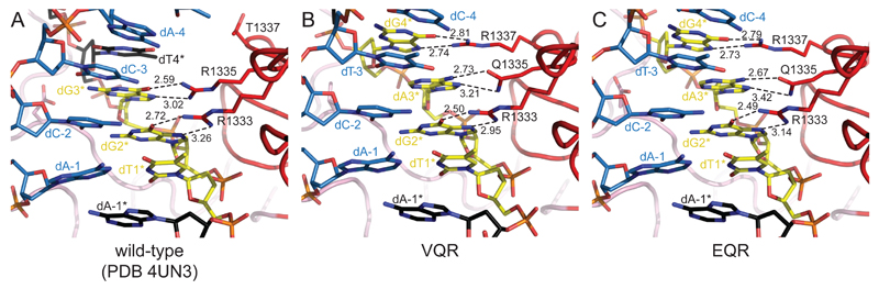
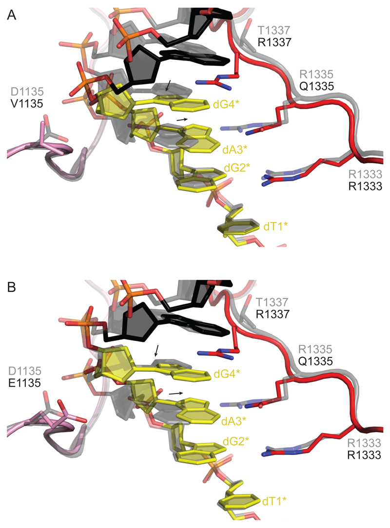
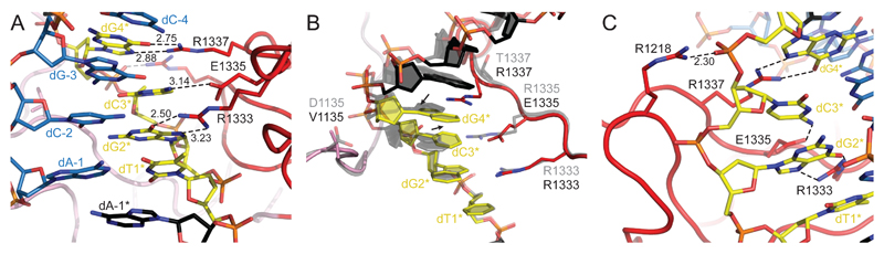
The RNA-guided endonuclease Cas9 from Streptococcus pyogenes (SpCas9) forms the core of a powerful genome editing technology. DNA cleavage by SpCas9 is dependent on the presence of a 5'-NGG-3' protospacer adjacent motif (PAM) in the target DNA, restricting the choice of targetable sequences. To address this limitation, artificial SpCas9 variants with altered PAM specificities have recently been developed. Here we report crystal structures of the VQR, EQR, and VRER SpCas9 variants bound to target DNAs containing their preferred PAM sequences. The structures reveal that the non-canonical PAMs are recognized by an induced fit mechanism. Besides mediating sequence-specific base recognition, the amino acid substitutions introduced in the SpCas9 variants facilitate conformational remodeling of the PAM region of the bound DNA. Guided by the structural data, we engineered a SpCas9 variant that specifically recognizes NAAG PAMs. Taken together, these studies inform further development of Cas9-based genome editing tools.


Figures

Figure 1
Figure 1. TGAG PAM Recognition by VQR and EQR SpCas9 Variants
(A) Zoom-in view of the major groove of the TGG PAM region of the DNA in the structure of the WT SpCas9-sgRNA-target DNA complex. PAM nucleotides are colored yellow. Target DNA strand is colored blue. Non-PAM nucleotides of the non-target DNA strand are colored black. Arginine residues making sequence-specific contacts with the PAM are shown in stick format. Numbers indicate interatomic distances in Å. (B) Zoomed-in view of the TGAG PAM in the VQR variant complex. (C) Zoomed-in view of the TGAG PAM in the EQR variant complex. Both (B) and (C) are shown in the same orientation as the WT SpCas9 complex in (A). Full sequences of the RNA guide and target DNA are provided in Figure S1.
Figure 2
Figure 2. The VQR and EQR Variants Induce a Structural Distortion in the PAM DNA
(A) Close-up view of the superimposed structures of the VQR variant with WT SpCas9. The PAM-interacting regions of the VQR Cas9 variant (colored pink and red) are not perturbed (Figure S2). The non-target DNA strand is colored black with the PAM highlighted in yellow. The superimposed WT SpCas9 structure is shown in gray. Arrows indicate shifts of the base and deoxyribose moieties of dA3* and dG4* in the VQR variant relative to the WT SpCas9 structure. (B) Close-up view of the EQR variant superimposed with WT SpCas9. Color coding is as in (A)
Figure 3
Figure 3. NGCG Recognition and Induced Fit by the VRER SpCas9 Variant
(A) Zoom-in view of the major groove of the TGCG PAM region of the DNA in the structure of the VRER variant complex. PAM nucleotides are colored yellow. The structure is depicted as the WT, VQR, and EQR SpCas9 complexes in Figure 1. (B) Cartoon diagram of the TGCG PAM and the PAM-interacting regions of the VRER SpCas9, overlaid with the structure of the WT SpCas9 complex (gray). The bound DNA is depicted in the same manner as in Figure 2. (C) Close-up view of the salt bridge interaction between Arg1218 and the phosphate group of dG4*.
Figure 4
Figure 4. Structure-Guided Engineering of an NAAG-Specific QQR1 Variant
(A) Close-up view of the PIM of the VQR variant bound to the non-target DNA strand. PAM nucleotides are highlighted in yellow. Hydrogen bonding interactions are indicated with dashed lines. (B) Schematic model of amino acid substitutions introduced in the QQR1 variant in order to induce repositioning of the polypeptide backbone of the PAM-interacting motif and facilitate hydrogen bonding contacts between Gln1335 and an adenine base in the second PAM position. (C) Nuclease activity assays of WT and QQR1 SpCas9 enzymes. WT and QQR1 SpCas9 were programmed using an identical sgRNA (sequence listed in Table S2) and incubated with linearized plasmids containing a target sequence adjacent to either TGGT or TAAG PAMs. Samples were taken at indicated time points and analyzed by agarose gel electrophoresis. (D) PAM specificity of the QQR1 variant. WT and QQR1 SpCas9 proteins were incubated with a sgRNA and linearized plasmid DNAs harboring targets flanked by the indicated PAMs.

Comment in

References

    1. Afonine PV, Grosse-Kunstleve RW, Echols N, Headd JJ, Moriarty NW, Mustyakimov M, Terwilliger TC, Urzhumtsev A, Zwart PH, Adams PD. Towards automated crystallographic structure refinement with phenix.refine. Acta Crystallogr D Biol Crystallogr. 2012;68:352–367. - PMC - PubMed
    1. Anders C, Jinek M. In vitro enzymology of Cas9. Meth Enzymol. 2014;546:1–20. - PMC - PubMed
    1. Anders C, Niewoehner O, Jinek M. In Vitro Reconstitution and Crystallization of Cas9 Endonuclease Bound to a Guide RNA and a DNA Target. Meth Enzymol. 2015;558:515–537. - PMC - PubMed
    1. Anders C, Niewoehner O, Duerst A, Jinek M. Structural basis of PAM-dependent target DNA recognition by the Cas9 endonuclease. Nature. 2014;513:569–573. - PMC - PubMed
    1. Barrangou R. RNA events. Cas9 targeting and the CRISPR revolution. Science. 2014;344:707–708. - PubMed

Publication types