. 2016 Jun;25(6):1115-28.
doi: 10.1002/pro.2924. Epub 2016 Apr 18.

Aggregation of Trp > Glu point mutants of human gamma-D crystallin provides a model for hereditary or UV-induced cataract

Affiliations

Aggregation of Trp > Glu point mutants of human gamma-D crystallin provides a model for hereditary or UV-induced cataract

Eugene Serebryany et al. Protein Sci. 2016 Jun.

Abstract

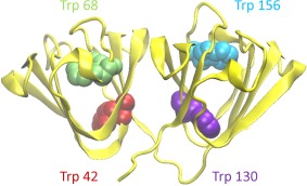
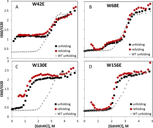
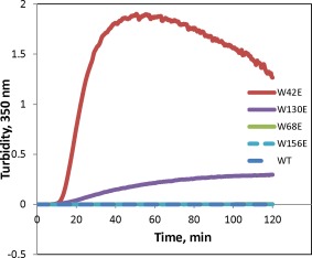
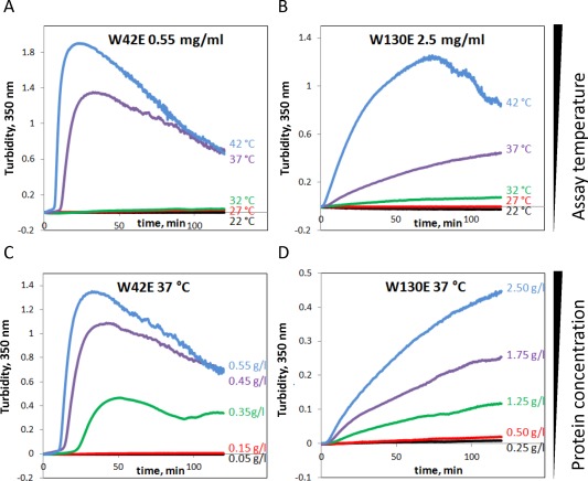
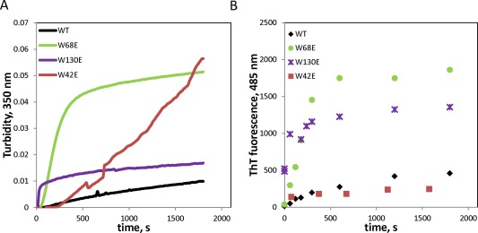
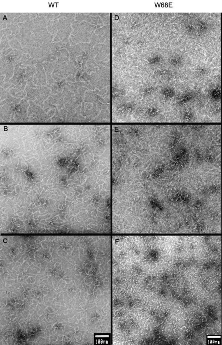
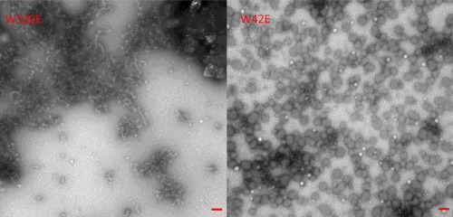
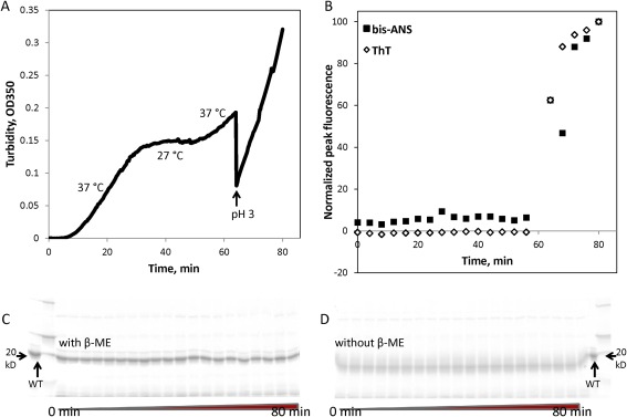
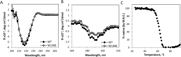
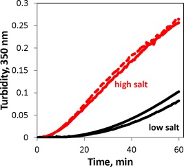
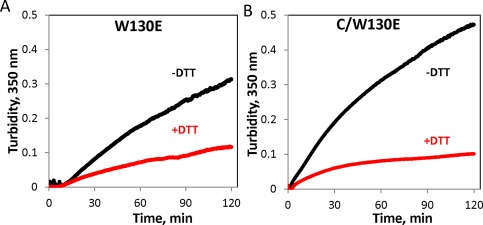
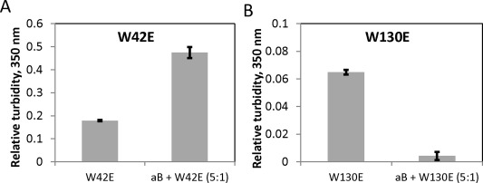
Numerous mutations and covalent modifications of the highly abundant, long-lived crystallins of the eye lens cause their aggregation leading to progressive opacification of the lens, cataract. The nature and biochemical mechanisms of the aggregation process are poorly understood, as neither amyloid nor native-state polymers are commonly found in opaque lenses. The βγ-crystallin fold contains four highly conserved buried tryptophans, which can be oxidized to more hydrophilic products, such as kynurenine, upon UV-B irradiation. We mimicked this class of oxidative damage using Trp→Glu point mutants of human γD-crystallin. Such substitutions may represent a model of UV-induced photodamage-introduction of a charged group into the hydrophobic core generating "denaturation from within." The effects of Trp→Glu substitutions were highly position dependent. While each was destabilizing, only the two located in the bottom of the double Greek key fold-W42E and W130E-yielded robust aggregation of partially unfolded intermediates at 37°C and pH 7. The αB-crystallin chaperone suppressed aggregation of W130E, but not W42E, indicating distinct aggregation pathways from damage in the N-terminal vs C-terminal domain. The W130E aggregates had loosely fibrillar morphology, yet were nonamyloid, noncovalent, showed little surface hydrophobicity, and formed at least 20°C below the melting temperature of the native β-sheets. These features are most consistent with domain-swapped polymerization. Aggregation of partially destabilized crystallins under physiological conditions, as occurs in this class of point mutants, could provide a simple in vitro model system for drug discovery and optimization.

Keywords: amyloid; cataract; crystallin; oxidative damage; protein aggregation; protein misfolding; unfolding intermediate.


Figures

Figure 1
Figure 1
A backbone ribbon representation of the HγD crystal structure (PDB ID 1HK0) (32) showing the four conserved buried Trp residues.
Figure 2
Figure 2
Equilibrium unfolding and refolding curves for the four W > E mutants, with the equilibrium unfolding curve for WT shown on each panel for reference. Deviations in the refolding curves relative to the respective unfolding curves are due to aggregation.
Figure 3
Figure 3
Aggregation of γD at 40.5°C in 10 mM ammonium acetate buffer at pH 7. All proteins are at 1 mg/mL concentration.
Figure 4
Figure 4
(A,B) Temperature‐dependence and (C,D) concentration‐dependence of W130E and W42E aggregation.
Figure 5
Figure 5
Amyloid aggregation of wild‐type and mutant HγD under acidic conditions (pH 3), followed by turbidity (A) and Thioflavin T fluorescence (B).
Figure 6
Figure 6
Electron micrographs of acid‐induced amyloid fibrils formed by (A–C) WT and (D–F) W68E HγD at different times of aggregation at pH 3. The protein was incubated at 1 mg/mL concentration at 37°C, in 50 mM sodium acetate and 100 mM NaCl, pH 3.0. Aliquotes were taken at (A, D) 0 min, (B, E) 2 min, and (C, F) 5 min after the initiation of aggregation. Scale bars are 100 nm.
Figure 7
Figure 7
Representative electron micrographs of aggregates formed by W130E and W42E HγD mutants after 2 h at 37°C. Scale bars are 100 nm.
Figure 8
Figure 8
Characterization of heat‐induced W130E aggregates. In samples taken from the aggregation timecourse as measured by turbidity (A), staining with thioflavin T and bis‐ANS (B) was used to measure amyloid formation and exposure of hydrophobic residues, respectively. SDS‐PAGE analysis in the presence (C) or absence (D) of reducing agent (β‐mercaptoethanol) did not reveal any covalently linked oligomers. Minor bands that may be backbone cleavage products were observed, but their intensity did not vary during the course of aggregation.
Figure 9
Figure 9
Characterization of W130E structure by circular dichroism. (A) Native CD spectrum of the W130E mutant compared to WT in the far‐UV region. (B) Native CD spectra of WT and W130E in the near‐UV region. Error bars represent standard error of the mean of 4 measurements. (C) Thermal melt of W130E as monitored by decrease in the 218 nm peak.
Figure 10
Figure 10
W130E aggregation is sensitive to redox conditions and ionic strength. W130E (1 mg/mL) incubated at 40.5°C in sample buffer alone (10 mM ammonium acetate, black lines), sample buffer with 150 mM NaCl (red solid line), and 150 mM NaCl plus 5 mM MgCl2 (red dashed line).
Figure 11
Figure 11
Redox‐dependence of aggregation of the W130E mutant and its isolated C‐terminal domain. (A) 1.25 mg/mL W130E (59 μM) was incubated at 40.5°C in the presence or absence of 1 mM DTT, (B) Isolated C‐terminal domain carrying the W130E mutation incubated at 0.5 mg/mL (47 μΜ) incubated as in (A).
Figure 12
Figure 12
Aggregation of W130E, but not W42E, can be suppressed by fivefold excess of human αB crystallin. (A) Final relative turbidity of W42E incubated for 2 h at 0.125 mg/mL and 40.5°C with or without fivefold excess (0.625 mg/mL) of the chaperone. (B) 1 mg/mL W130E incubated as in (A) with or without fivefold excess (5 mg/mL) of the chaperone. Error bars represent standard errors of the mean of 3 replicates.

References

    1. Klein R, Klein BEK (2013) The prevalence of age‐related eye diseases and visual impairment in aging: current estimates. Invest Ophthalmol Vis Sci 54:14, ORSF5–ORSF13. - PMC - PubMed
    1. Facts about cataract (2009) National Eye Institute. https://nei.nih.gov/health/cataract/cataract_facts.
    1. Frick KD, Gower EW, Kempen JH, Wolff JL (2007) Economic impact of visual impairment and blindness in the United States. Arch Ophthalmol 125:544–550. - PubMed
    1. Tielsch JM, Kempen JH, Congdon N, Friedman DS (2008) Vision probelms in the U.S.: prevalence of adult visual impairment and age‐related eye disease in America, p. 22. Prevent Blindness America and the National Eye Institute.
    1. Zhao L, Chen XJ, Zhu J, Xi YB, Yang X, Hu LD, Ouyang H, Patel SH, Jin X, Lin DN, Wu F, Flagg K, Cai HM, Li G, Cao GQ, Lin Y, Chen D, Wen C, Chung C, Wang YD, Qiu A, Yeh E, Wang WQ, Hu X, Grob S, Abagyan R, Su ZG, Tjondro HC, Zhao XJ, Luo HR, Hou R, Perry JJP, Gao WW, Kozak I, Granet D, Li YR, Sun XD, Wang J, Zhang LF, Liu YZ, Yan YB, Zhang K (2015) Lanosterol reverses protein aggregation in cataracts. Nature 523:607. - PubMed

Publication types

LinkOut - more resources