. 2016 Aug;64(2):522-34.
doi: 10.1002/hep.28550. Epub 2016 Apr 15.

Soluble Adenylyl Cyclase Regulates Bile Salt-Induced Apoptosis in Human Cholangiocytes

Affiliations

Soluble Adenylyl Cyclase Regulates Bile Salt-Induced Apoptosis in Human Cholangiocytes

Jung-Chin Chang et al. Hepatology. 2016 Aug.

Abstract

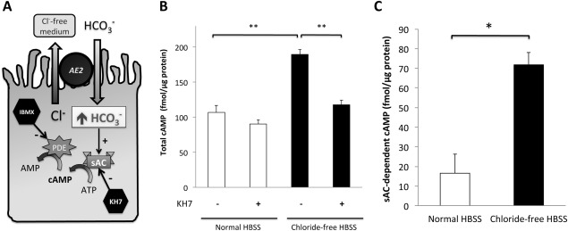
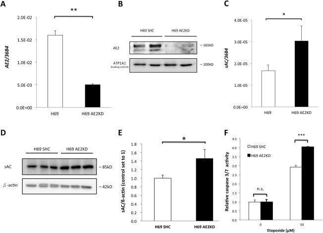
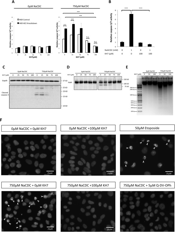
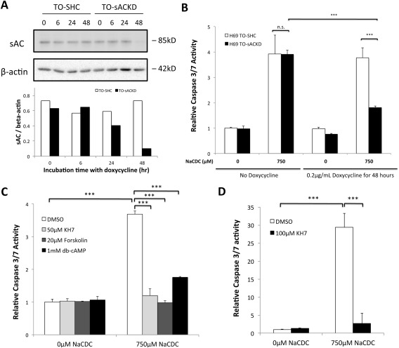
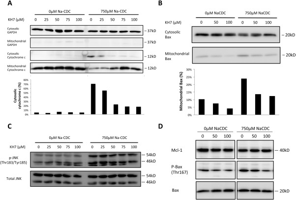
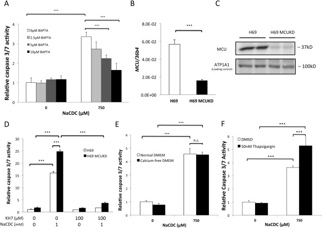
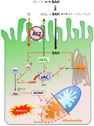
Anion exchanger 2 (AE2), the principal bicarbonate secretor in the human biliary tree, is down-regulated in primary biliary cholangitis. AE2 creates a "bicarbonate umbrella" that protects cholangiocytes from the proapoptotic effects of bile salts by maintaining them deprotonated. We observed that knockdown of AE2 sensitized immortalized H69 human cholangiocytes to not only bile salt-induced apoptosis (BSIA) but also etoposide-induced apoptosis. Because the toxicity of etoposide is pH-independent, there could be a more general mechanism for sensitization of AE2-depleted cholangiocytes to apoptotic stimuli. We found that AE2 deficiency led to intracellular bicarbonate accumulation and increased expression and activity of soluble adenylyl cyclase (sAC), an evolutionarily conserved bicarbonate sensor. Thus, we hypothesized that sAC regulates BSIA. H69 cholangiocytes and primary mouse cholangiocytes were used as models. The sAC-specific inhibitor KH7 not only reversed sensitization to BSIA in AE2-depleted H69 cholangiocytes but even completely prevented BSIA. sAC knockdown by tetracycline-inducible short hairpin RNA also prevented BSIA. In addition, sAC inhibition reversed BSIA membrane blebbing, nuclear condensation, and DNA fragmentation. Furthermore, sAC inhibition also prevented BSIA in primary mouse cholangiocytes. Mechanistically, sAC inhibition prevented Bax phosphorylation at Thr167 and mitochondrial translocation of Bax and cytochrome c release but not c-Jun N-terminal kinase activation during BSIA. Finally, BSIA in H69 cholangiocytes was inhibited by intracellular Ca(2+) chelation, aggravated by thapsigargin, and unaffected by removal of extracellular calcium.

Conclusions: BSIA is regulated by sAC, depends on intracellular Ca(2+) stores, and is mediated by the intrinsic apoptotic pathway; down-regulation of AE2 in primary biliary cholangitis sensitizes cholangiocytes to apoptotic insults by activating sAC, which may play a crucial role in disease pathogenesis. (Hepatology 2016;64:522-534).


Figures

Figure 1
Figure 1
Presence of functional sAC in H69 cholangiocytes was demonstrated by bicarbonate‐stimulated cAMP accumulation assay. (A) Confluent H69 cholangiocyte monolayers were incubated in either normal HBSS or chloride‐free HBSS for 30 minutes. The latter reversed the chloride gradient across the plasma membrane and forced AE2 to take up bicarbonate. (B) Total cAMP was measured by an enzyme‐linked immunosorbent assay in the absence or presence of KH7, a specific inhibitor of sAC. Shown here is the average of two quadruplicate experiments. One‐way ANOVA, P = 0.0007. (C) From each independent quadruplicate experiment in (B), sAC‐derived cAMP was calculated (defined as the difference in total cAMP measured in the absence and presence of KH7). Two‐tailed Student t test, P = 0.03. * P < 0.05, **P ≤ 0.01. Abbreviations: AMP, adenosine monophosphate; ATP, adenosine triphosphate; IBMX, 3‐isobutyl‐1‐methylxanthine; PDE, phosphodiesterase.
Figure 2
Figure 2
Knockdown of AE2 induced sAC expression. AE2 was knocked down by lentivirus‐mediated shRNA interference in H69 cholangiocytes and confirmed by RT‐qPCR (A) and immunoblot (B). Expression of sAC messenger RNA (C) and protein level (D) in both short hairpin control and AE2 knockdown H69 cholangiocytes was examined. Immunoblots were quantified by ImageJ (E). Two‐tailed Student t test, *P < 0.05, **P ≤ 0.01. (F) Control and AE2KD H69 cholangiocytes were incubated with 50 μM etoposide or 0.1% dimethyl sulfoxide (vehicle) for 16 hours. Caspase 3/7 activity was determined. One‐way ANOVA, P < 0.0001. The figure shows a representative experiment from a series with similar results. ***P ≤ .001. Abbreviations: ATP1A1, adenosine triphosphatase, sodium/potassium transporting, alpha 1 polypeptide; KD, knockdown; n.s., not significant; SHC, short hairpin control.
Figure 3
Figure 3
Inhibition of sAC prevented both unconjugated and conjugated BSIA. (A) Control and AE2 knockdown H69 cholangiocytes were incubated with increasing concentrations of sAC‐specific inhibitor KH7 in the absence or presence of 750 μM NaCDC at 37°C and 5% CO2 for 1 hour. Caspase 3/7 activity was determined. One‐way ANOVA, P < 0.0001. (B) Control and AE2 knockdown H69 cholangiocytes were incubated with 1 mM NaGCDC for 4 hours at pH 6.8 with or without KH7 in 37°C, 5% CO2 incubator. One‐way ANOVA, P < 0.0001. (C) H69 cholangiocytes were treated as in (A). Immunoblot for cleaved caspase 3 was performed. The antibody reacts with only cleaved caspase 3 (p17/p19), not procaspase 3. (D) Same treatment condition as (C), immunoblotted for PARP, an endogenous caspase 3/7 substrate. The full‐length PARP (∼113 kDa) was cleaved by caspase 3/7 into the 89‐kDa fragment. (E) DNA fragmentation assay. H69 cholangiocytes were incubated with 0, 50, and 100 μM KH7 in the absence or presence of 750 μM NaCDC for 1 hour. Genomic DNA was extracted and resolved on a 1.5% agarose gel. (F) H69 cholangiocytes were treated with or without 750 μM NaCDC in the presence or absence of 100 μM KH7 and 5 μM Q‐DV‐OPh (pan‐caspase inhibitor). Overnight treatment with 50 μM etoposide served as a positive control. Nuclei were visualized by DAPI staining. Arrowheads indicated apoptotic nuclear condensation. The figure shows a representative experiment from a series with similar results. ***P < 0.001. Abbreviations: Gapdh, glyceraldehyde 3‐phosphate dehydrogenase; KD, knockdown; n.s., not significant; SHC, short hairpin control.
Figure 4
Figure 4
sAC‐derived cAMP promoted BSIA, while tmAC‐derived cAMP inhibited BSIA. (A) Control and inducible sAC knockdown H69 cholangiocytes were treated with 0.2 μg/mL doxycycline for the indicated periods of time. sAC knockdown was examined by immunoblotting. Immunoblot was quantified by ImageJ. Results are normalized to β‐actin. (B) Short hairpin control and sAC knockdown H69 cholangiocytes were induced with or without 0.2 μg/mL doxycycline for 48 hours and subsequently incubated with 750 μM NaCDC or vehicle for 1 hour. Caspase 3/7 activity was determined. One‐way ANOVA, P < 0.0001. The figure shows a representative experiment from a series of three independent experiments with similar results. (C) H69 cholangiocytes were incubated with or without 750 μM NaCDC in the presence of dimethyl sulfoxide (vehicle control), 50 μM KH7 (sAC‐specific inhibitor), 20 μM forskolin (tmAC‐specific activator), or 1 mM dibutyryl‐cAMP (membrane‐permeant cAMP analogue) for 4 hours in a 5% CO2, 37°C incubator. Caspase 3/7 activity was determined. One‐way ANOVA, P < 0.0001. (D) Primary mouse cholangiocytes were incubated with or without 750 μM NaCDC in the presence of dimethyl sulfoxide (vehicle control) or 100 μM KH7 for 3 hours in a 5% CO2, 37°C incubator. One‐way ANOVA, P < 0.0001. ***P < 0.001. Abbreviations: db‐cAMP, dibutyryl‐cAMP; DMSO, dimethyl sulfoxide; KD, knockdown; n.s., not significant; SHC, short hairpin control, TO, Tet‐operator.
Figure 5
Figure 5
sAC inhibition prevented cytochrome c release and mitochondrial translocation of Bax during BSIA. (A) Cytochrome c release assay. H69 cholangiocytes were treated with increasing concentrations of KH7 in the absence or presence of 750 μM NaCDC for 1 hour in a 5% CO2, 37°C incubator. Cell fractionation was performed as described in Materials and Methods. Lysates of equal volume were subjected to sodium dodecyl sulfate‐polyacrylamide gel electrophoresis and immunoblotted for glyceraldehyde 3‐phosphate dehydrogenase, a cytosolic marker, and cytochrome c. Cytochrome c immunoblots were quantified by ImageJ. Ratios of cytosolic signal to total signal (cytosolic plus mitochondrial) were calculated. (B) Bax translocation assay was performed and quantified as in (A). Percentage of mitochondrial Bax was calculated as mitochondrial signals divided by the sum of cytosolic and mitochondrial signals. (C) H69 cholangiocytes were treated as in (A). Whole‐cell lysates were used for immunoblot of total and phosphorylated JNK. (D) H69 cholangiocytes were treated as in (B). Whole‐cell lysates were used for immunoblot for myeloid cell leukemia 1, phospho‐Bax (Thr167), and total Bax. The figure shows a representative experiment from a series with similar results. Abbreviations: GAPDH, glyceraldehyde 3‐phosphate dehydrogenase; Mcl‐1, myeloid cell leukemia 1.
Figure 6
Figure 6
BSIA depends on intracellular calcium stores. (A) H69 cholangiocytes were loaded with increasing concentrations of 1,2‐bis(2‐aminophenoxy)ethane‐N,N,N′,N′‐tetraacetic acid (BAPTA) and then treated with either vehicle or 750 μM NaCDC for 1 hour. Caspase 3/7 activities were determined. One‐way ANOVA, P < 0.0001. (B) MCU was knocked down by lentivirus‐mediated shRNA interference. Knockdown was confirmed by RT‐qPCR. Two‐tailed Student t test, P < 0.0001. (C) MCU knockdown was confirmed by immunoblotting. (D) Short hairpin control and MCU knockdown H69 cholangiocytes were treated with 1 mM NaCDC‐induced apoptosis in the absence or presence of sAC‐specific inhibitor KH7. One‐way ANOVA, P < 0.0001. (E) H69 cholangiocytes were incubated with 750 μM NaCDC in normal or Ca2+‐free medium. One‐way ANOVA, P < 0.0001. (F) H69 cholangiocytes were pretreated with or without 50 nM thapsigargin for 15 minutes and then with 0 μM or 750 μM NaCDC. The figure shows a representative experiment from a series with similar results. ***P < 0.001. Abbreviations: ATP1A1, adenosine triphosphatase, sodium/potassium transporting, alpha 1 polypeptide; BAPTA, 1,2‐bis(2‐aminophenoxy)ethane‐N,N,N′,N′‐tetraacetic acid; DMEM, Dulbecco's modified Eagle's medium; DMSO, dimethyl sulfoxide; KD, knockdown; n.s., not significant.
Figure 7
Figure 7
Working hypothesis of how sAC is involved in the pathogenesis of PBC in the context of AE2 down‐regulation. Protonated bile salts are nonpolar and enter cells at a higher rate than their deprotonated counterparts. AE2 on the apical membrane of cholangiocytes generates a bicarbonate umbrella that deprotonates the otherwise proapoptotic bile salts. Reduced AE2 expression in PBC cholangiocytes causes more bile acids to enter. Once inside the cells, bile salts cause JNK phosphorylation and activating phosphorylation of Bax at Thr167, which promote mitochondrial translocation of Bax, leading to MOMP, cytochrome c release, and apoptosis. Inhibition of sAC prevents bile salt‐induced Bax phosphorylation at Thr167 but not JNK phosphorylation. By acting as a calcium ionophore, bile salts cause Ca2+ release from endoplasmic reticulum, which is necessary but not sufficient for BSIA. On the other hand, the intracellular bicarbonate accumulation secondary to impaired secretion increases sAC activity synergistically with bile salt‐triggered Ca2+ release and enhances MOMP and apoptosis. Abbreviations: ATP, adenosine triphosphate; BA, deprotonated bile salt; BAH, protonated bile salt; ER, endoplasmic reticulum.

Comment in

  • New evidence supporting the biliary bicarbonate umbrella theory.
    Fernandez-Barrena MG, Barcena-Varela M, Banales JM. Fernandez-Barrena MG, et al. Clin Res Hepatol Gastroenterol. 2017 Mar;41(2):126-128. doi: 10.1016/j.clinre.2016.09.004. Epub 2016 Nov 3. Clin Res Hepatol Gastroenterol. 2017. PMID: 27818187 No abstract available.

References

    1. Selmi C, Bowlus CL, Gershwin ME, Coppel RL. Primary biliary cirrhosis. Lancet 2011;377:1600–1609. - PubMed
    1. Selmi C, Mayo MJ, Bach N, Ishibashi H, Invernizzi P, Gish RG, et al. Primary biliary cirrhosis in monozygotic and dizygotic twins: genetics, epigenetics, and environment. Gastroenterology 2004;127:485–492. - PubMed
    1. Hirschfield GM, Liu X, Han Y, Gorlov IP, Lu Y, Xu C, et al. Variants at IRF5‐TNPO3, 17q12‐21 and MMEL1 are associated with primary biliary cirrhosis. Nat Genet 2010;42:655–657. - PMC - PubMed
    1. Mells GF, Floyd JA, Morley KI, Cordell HJ, Franklin CS, Shin SY, et al. Genome‐wide association study identifies 12 new susceptibility loci for primary biliary cirrhosis. Nat Genet 2011;43:329–332. - PMC - PubMed
    1. Medina JF, Martinez A, Vazquez JJ, Prieto J. Decreased anion exchanger 2 immunoreactivity in the liver of patients with primary biliary cirrhosis. Hepatology 1997;25:12–17. - PubMed

Publication types