

Resurfaced cell-penetrating nanobodies: A potentially general scaffold for intracellularly targeted protein discovery
- PMID: 26991318
- PMCID: PMC4941773
- DOI: 10.1002/pro.2926
Resurfaced cell-penetrating nanobodies: A potentially general scaffold for intracellularly targeted protein discovery
Abstract
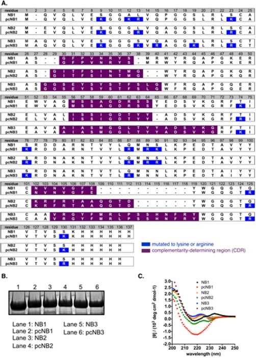
By virtue of their size, functional group diversity, and complex structure, proteins can often recognize and modulate disease-relevant macromolecules that present a challenge to small-molecule reagents. Additionally, high-throughput screening and evolution-based methods often make the discovery of new protein binders simpler than the analogous small-molecule discovery process. However, most proteins do not cross the lipid bilayer membrane of mammalian cells. This largely limits the scope of protein therapeutics and basic research tools to those targeting disease-relevant receptors on the cell surface or extracellular matrix. Previously, researchers have shown that cationic resurfacing of proteins can endow cell penetration. However, in our experience, many proteins are not amenable to such extensive mutagenesis. Here, we report that nanobodies-a small and stable protein that can be evolved to recognize virtually any disease-relevant receptor-are amenable to cationic resurfacing, which results in cell internalization. Once internalized, these nanobodies access the cytosol. Polycationic resurfacing does not appreciably alter the structure, expression, and function (target recognition) of a previously reported GFP-binding nanobody, and multiple nanobody scaffolds are amenable to polycationic resurfacing. Given this, we propose that polycationic resurfaced cell-penetrating nanobodies might represent a general scaffold for intracellularly targeted protein drug discovery.
Keywords: cell-penetrating; nanobody; polycationic resurfacing; supercharging.
© 2016 The Protein Society.
Figures


References
-
- Overington JP, Al‐Lazikani B, Hopkins AL (2006) Opinion ‐ how many drug targets are there? Nat Rev Drug Discov 5:993–996. - PubMed
-
- Smith GP, Petrenko VA (1997) Phage display. Chem Rev 97:391–410. - PubMed
-
- Boder ET, Wittrup KD (2000) Yeast surface display for directed evolution of protein expression, affinity, and stability. Methods Enzymol 328:430–444. - PubMed
-
- DePorter SM, McNaughton BR (2014) Engineered M13 bacteriophage nanocarriers for intracellular delivery of exogenous proteins to human prostate cancer cells. Bioconjug Chem 25:1620–1625. - PubMed
Publication types
MeSH terms
Substances
Associated data
- Actions
LinkOut - more resources
Full Text Sources
Other Literature Sources
Research Materials
