. 2016 Apr 18;55(17):5337-41.
doi: 10.1002/anie.201511657. Epub 2016 Mar 16.

Facile Recoding of Selenocysteine in Nature

Affiliations

Facile Recoding of Selenocysteine in Nature

Takahito Mukai et al. Angew Chem Int Ed Engl. .

Abstract

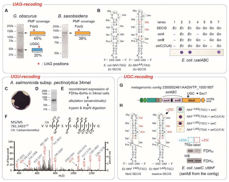
Selenocysteine (Sec or U) is encoded by UGA, a stop codon reassigned by a Sec-specific elongation factor and a distinctive RNA structure. To discover possible code variations in extant organisms we analyzed 6.4 trillion base pairs of metagenomic sequences and 24 903 microbial genomes for tRNA(Sec) species. As expected, UGA is the predominant Sec codon in use. We also found tRNA(Sec) species that recognize the stop codons UAG and UAA, and ten sense codons. Selenoprotein synthesis programmed by UAG in Geodermatophilus and Blastococcus, and by the Cys codon UGU in Aeromonas salmonicida was confirmed by metabolic labeling with (75) Se or mass spectrometry. Other tRNA(Sec) species with different anticodons enabled E. coli to synthesize active formate dehydrogenase H, a selenoenzyme. This illustrates the ease by which the genetic code may evolve new coding schemes, possibly aiding organisms to adapt to changing environments, and show the genetic code is much more flexible than previously thought.

Keywords: genetic code; metagenome; selenocysteine; sense codon recoding; synthetic biology.


Figures

Figure 1
Figure 1. Non-canonical selenocysteine assignments in nature
(A) The tRNASec search pipeline and the manually curated output. The non-canonical tRNASec sequences are grouped by codon recognition; their numbers are given with their (putative) bacterial origins from phylogenetic inference. Green color indicates results from whole genomes, while blue represents results from only metagenomic data. For comparison the number of canonical tRNASecUCA sequences is shown (most of them were not curated). “Y” and “R” denote “C or U” and “A or G”, respectively. (B) Inferred cloverleaf structures of non-canonical tRNASec species. The nucleotide polymorphism among the same tRNASec group is indicated with green letters. The Sec codons and the SECIS elements of formate dehydrogenase mRNAs are shown.
Figure 2
Figure 2. Recoding of UAG and cysteine codons to selenocysteine
(A) Metabolic 75Se labeling of G. obscurus and B. saxobsidens cells. Crude extracts were resolved by SDS-PAGE, and their putative selenoproteins were visualized by PhosphorImager analysis. The results of peptide mass fingerprinting (PMF) analyses of the proteins in the excised gel bands are shown to the right of the bands. (B) FDHH expression in E. coli ΔselABC ΔfdhF cells with the G. obscurus selABC genes and a chimeric fdhF(140TAG) gene variant having a G. obscurus SECIS element with a few nucleotide modifications shown in red. The selC(CUA) genes express tRNASecCUA. The expressed selenoprotein FDHH reduced benzyl viologen, resulting in a purple color. (C) FDHH activity of A. salmonicida subsp. pectinolytica 34mel cells. (D) Metabolic 75Se labeling of the FDHH of 34mel. (E) The procedure of sample preparation for the LC-MS/MS analysis of the FDHH selenoprotein. (F) PMF confirms Sec incorporation at codon 140 in the recombinant FDHH. (G) tRNASecGCA gene (selC) locus in a metagenomic contig. (H) In vivo FDHH assays in E. coli ΔselABC ΔfdhF cells with the selABC genes of the metagenomic contig and a chimeric fdhF(140TGC) gene carrying the contig’s SECIS element with a few nucleotide modifications shown in red. The two transformed strains boxed were metabolically labeled with 75Se, and the radioactive FDHH proteins were analyzed by SDS-PAGE and autoradiography or western blotting (WB).

References

    1. Cone JE, Del Rio RM, Davis JN, Stadtman TC. Proc Natl Acad Sci USA. 1976;73:2659–2663. - PMC - PubMed
    1. Ambrogelly A, Palioura S, Söll D. Nat Chem Biol. 2007;3:29–35. - PubMed
    1. Kryukov GV, Castellano S, Novoselov SV, Lobanov AV, Zehtab O, Guigo R, Gladyshev VN. Science. 2003;300:1439–1443. - PubMed
    1. Metanis N, Hilvert D. Curr Opin Chem Biol. 2014;22:27–34. - PubMed
    1. Böck A, Thanbichler M, Rother M, Resch A. In: Aminoacyl-tRNA Synthetases. Ibba M, Francklyn CS, Cusack S, editors. Landes Bioscience; Georgetown, TX: 2005. pp. 320–327.

Publication types