

Correlations of Medial Joint Space Width on Fixed-Flexed Standing Computed Tomography and Radiographs With Cartilage and Meniscal Morphology on Magnetic Resonance Imaging
- PMID: 26991547
- PMCID: PMC5027176
- DOI: 10.1002/acr.22888
Correlations of Medial Joint Space Width on Fixed-Flexed Standing Computed Tomography and Radiographs With Cartilage and Meniscal Morphology on Magnetic Resonance Imaging
Abstract
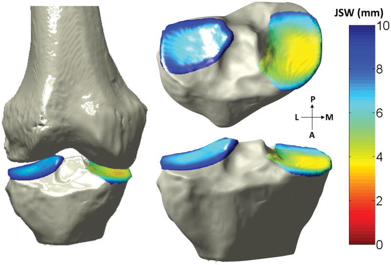
Objective: To assess whether medial tibiofemoral joint space width (JSW) on 3-dimensional (3-D) standing computed tomography (SCT) correlates more closely with magnetic resonance imaging cartilage morphology (CM) and meniscal scores than does radiographic 2-D JSW.
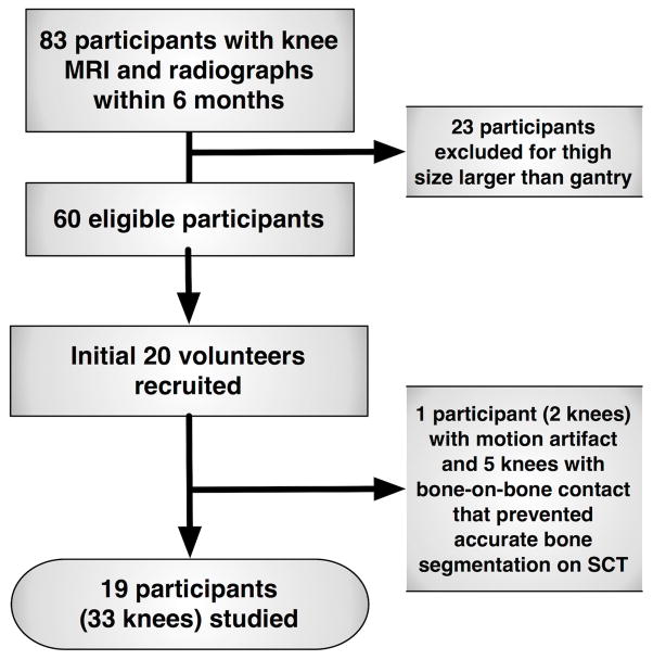
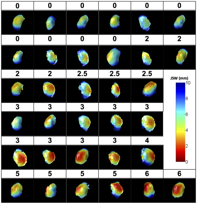
Methods: Participants in the Multicenter Osteoarthritis Study, who had standing fixed-flexion posteroanterior knee radiographs, were recruited. Medial tibiofemoral 3-D JSW on SCT and 2-D JSW on fixed-flexion radiographs were compared with medial tibiofemoral cartilage and meniscal morphology using the Whole-Organ Magnetic Resonance Imaging Score (WORMS). Associations between the area of the articular surface with 3-D JSW <2.5 mm on SCT, radiographic minimal 2-D JSW, and the WORMS-CM and meniscal scores were assessed using Spearman's rho.
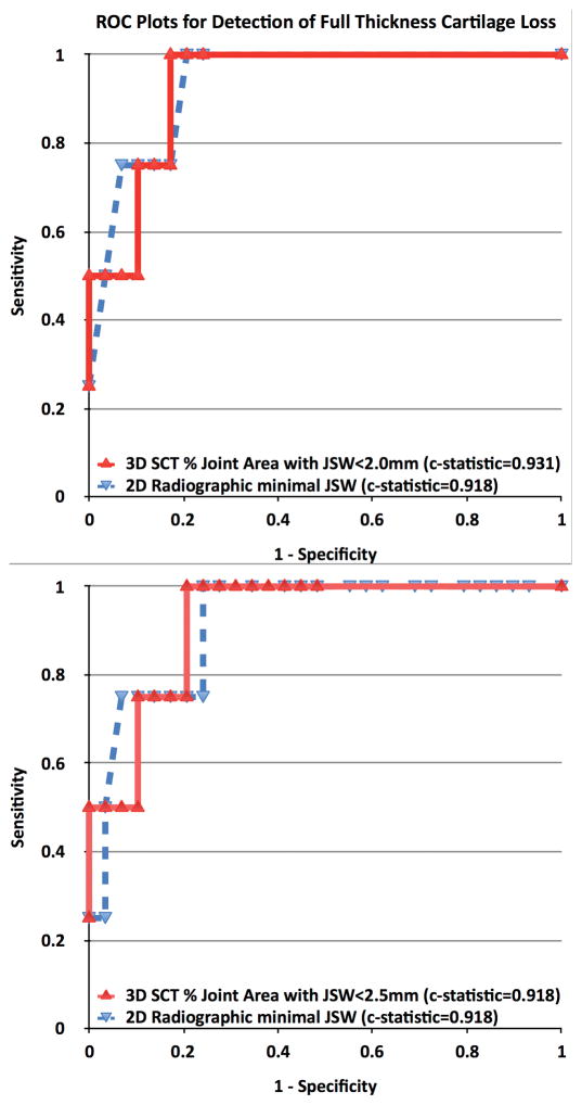
Results: For the 19 participants included (33 knees), mean ± SD age was 66.9 ± 5.4 years, body mass index was 29.5 ± 4.4 kg/m(2) , 42.1% of participants were female, and the Kellgren/Lawrence grades were 0 (21.2%), 1 (36.4%), 2 (18.2%), and 3 (24.2%). The articular surface area with 3-D JSW <2.5 mm on SCT correlated with WORMS-CM scores for the central medial tibia (rs = 0.84, P < 0.001), central medial femur (rs = 0.60, P < 0.007), and posterior medial meniscal tear (rs = 0.39, P < 0.026), as did other cut points for 3-D JSW. Correlations with radiographic minimal 2-D JSW were -0.66, -0.52, and -0.40, respectively, differing from SCT only for tibial cartilage (P = 0.001).
Conclusion: Greater surface area with a low JSW, measured by SCT, correlates more strongly with the severity of tibial cartilage lesions, while correlating with medial femoral cartilage and meniscal damage to a similar extent as radiographic minimal JSW. SCT may enable valid stratification of participants in clinical trials, through quickly and inexpensively characterizing osteoarthritis features.
© 2016, American College of Rheumatology.
Figures
References
-
- FDA, editor. Federal Register. Aug 14, 2007. Clinical Development Programs for Human Drugs, Biological Products, and Medical Devices for the Treatment and Prevention of Osteoarthritis; p. 100. DOCKET NO. 1998D-0077 (FORMERLY 98D-0077)
-
- Guermazi A, Roemer FW, Felson DT, Brandt KD. Motion for debate: osteoarthritis clinical trials have not identified efficacious therapies because traditional imaging outcome measures are inadequate. Arthritis Rheum. 2013;65(11):2748–58. - PubMed
-
- Amin S, LaValley MP, Guermazi A, Grigoryan M, Hunter DJ, Clancy M, et al. The relationship between cartilage loss on magnetic resonance imaging and radiographic progression in men and women with knee osteoarthritis. Arthritis Rheum. 2005;52(10):3152–9. - PubMed
-
- Kijowski R, Blankenbaker DG, Stanton PT, Fine JP, De Smet AA. Radiographic findings of osteoarthritis versus arthroscopic findings of articular cartilage degeneration in the tibiofemoral joint. Radiology. 2006;239(3):818–24. - PubMed
Publication types
MeSH terms
Grants and funding
LinkOut - more resources
Full Text Sources
Other Literature Sources
Medical
