

The specification and wiring of mammalian cutaneous low-threshold mechanoreceptors
- PMID: 26992078
- PMCID: PMC4864430
- DOI: 10.1002/wdev.229
The specification and wiring of mammalian cutaneous low-threshold mechanoreceptors
Abstract
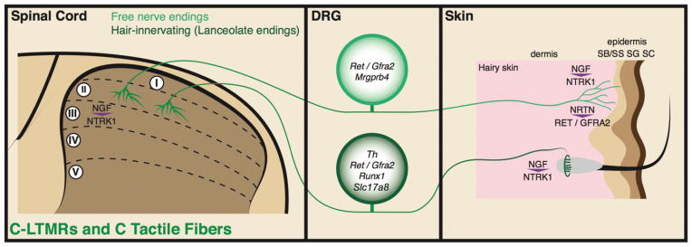
The mammalian cutaneous low-threshold mechanoreceptors (LTMRs) are a diverse set of primary somatosensory neurons that function to sense external mechanical force. Generally, LTMRs are composed of Aβ-LTMRs, Aδ-LTMRs, and C-LTMRs, which have distinct molecular, physiological, anatomical, and functional features. The specification and wiring of each type of mammalian cutaneous LTMRs is established during development by the interplay of transcription factors with trophic factor signalling. In this review, we summarize the cohort of extrinsic and intrinsic factors generating the complex mammalian cutaneous LTMR circuits that mediate our tactile sensations and behaviors. For further resources related to this article, please visit the WIREs website.
© 2016 Wiley Periodicals, Inc.
Conflict of interest statement
Conflict of interest: The authors have declared no conflicts of interest for this article.
Figures
Similar articles
-
The functional organization of cutaneous low-threshold mechanosensory neurons.Cell. 2011 Dec 23;147(7):1615-27. doi: 10.1016/j.cell.2011.11.027. Cell. 2011. PMID: 22196735 Free PMC article.
-
CaMKII Controls Whether Touch Is Painful.J Neurosci. 2015 Oct 21;35(42):14086-102. doi: 10.1523/JNEUROSCI.1969-15.2015. J Neurosci. 2015. PMID: 26490852 Free PMC article.
-
The cellular and molecular basis of direction selectivity of Aδ-LTMRs.Cell. 2014 Dec 18;159(7):1640-51. doi: 10.1016/j.cell.2014.11.038. Cell. 2014. PMID: 25525881 Free PMC article.
-
The gentle touch receptors of mammalian skin.Science. 2014 Nov 21;346(6212):950-4. doi: 10.1126/science.1254229. Science. 2014. PMID: 25414303 Free PMC article. Review.
-
The sensory neurons of touch.Neuron. 2013 Aug 21;79(4):618-39. doi: 10.1016/j.neuron.2013.07.051. Neuron. 2013. PMID: 23972592 Free PMC article. Review.
Cited by
-
Spatial transcriptomics of dorsal root ganglia identifies molecular signatures of human nociceptors.Sci Transl Med. 2022 Feb 16;14(632):eabj8186. doi: 10.1126/scitranslmed.abj8186. Epub 2022 Feb 16. Sci Transl Med. 2022. PMID: 35171654 Free PMC article.
-
The Human Cutaneous Sensory Corpuscles: An Update.J Clin Med. 2021 Jan 10;10(2):227. doi: 10.3390/jcm10020227. J Clin Med. 2021. PMID: 33435193 Free PMC article. Review.
-
Dermal macrophages control tactile perception under physiological conditions via NGF signaling.Sci Rep. 2024 Nov 8;14(1):27192. doi: 10.1038/s41598-024-78683-x. Sci Rep. 2024. PMID: 39516548 Free PMC article.
-
A role for axon-glial interactions and Netrin-G1 signaling in the formation of low-threshold mechanoreceptor end organs.Proc Natl Acad Sci U S A. 2022 Oct 25;119(43):e2210421119. doi: 10.1073/pnas.2210421119. Epub 2022 Oct 17. Proc Natl Acad Sci U S A. 2022. PMID: 36252008 Free PMC article.
-
Skin-type-dependent development of murine mechanosensory neurons.Dev Cell. 2023 Oct 23;58(20):2032-2047.e6. doi: 10.1016/j.devcel.2023.07.020. Epub 2023 Aug 21. Dev Cell. 2023. PMID: 37607547 Free PMC article.
References
-
- Willis WD, Coggeshall RE. Sensory Mechanisms of the Spinal Cord: Volume 1 Primary Afferent Neurons and the Spinal Dorsal Horn. New York: Springer Science & Business Media; 2004.
-
- Serbedzija GN, Fraser SE, Bronner-Fraser M. Pathways of trunk neural crest cell migration in the mouse embryo as revealed by vital dye labelling. Development. 1990;108:605–612. - PubMed
-
- Lawson SN, Biscoe TJ. Development of mouse dorsal root ganglia: an autoradiographic and quantitative study. J Neurocytol. 1979;8:265–274. - PubMed
-
- Kramer I, Sigrist M, de Nooij JC, Taniuchi I, Jessell TM, Arber S. A role for Runx transcription factor signaling in dorsal root ganglion sensory neuron diversification. Neuron. 2006;49:379–393. - PubMed
FURTHER READING
-
- Lallemend F, Ernfors P. Molecular interactions underlying the specification of sensory neurons. Trends Neurosci. 2012;35:373–381. - PubMed
Publication types
MeSH terms
Grants and funding
LinkOut - more resources
Full Text Sources
Other Literature Sources
