Review
. 2016 Apr 10;428(7):1455-64.
doi: 10.1016/j.jmb.2016.03.003. Epub 2016 Mar 16.

Molecular Control of Atypical Protein Kinase C: Tipping the Balance between Self-Renewal and Differentiation

Affiliations
Review

Molecular Control of Atypical Protein Kinase C: Tipping the Balance between Self-Renewal and Differentiation

Michael L Drummond et al. J Mol Biol. .

Abstract

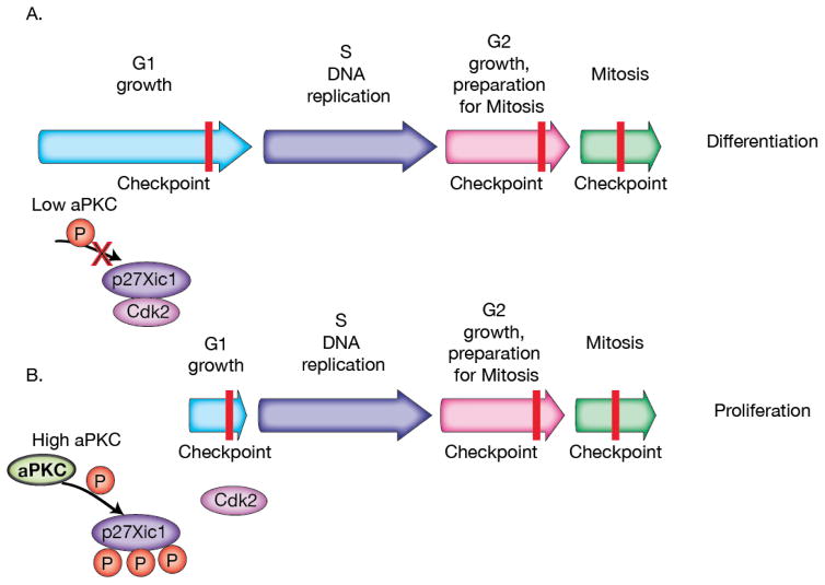
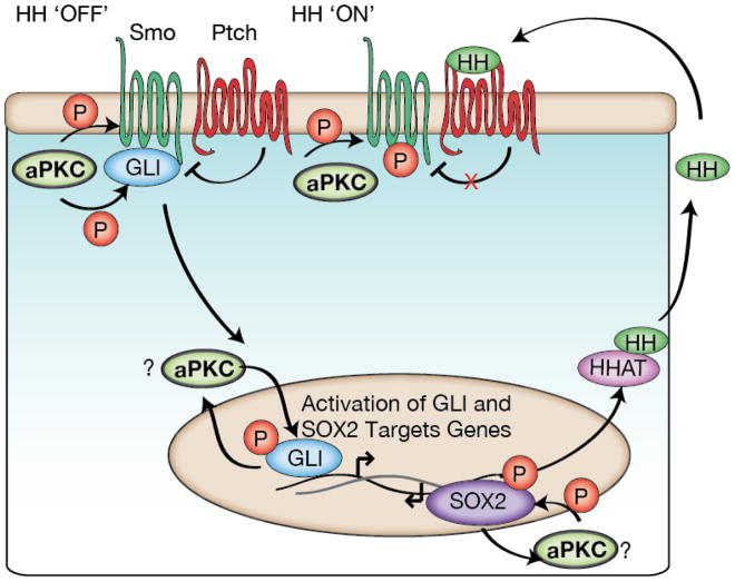
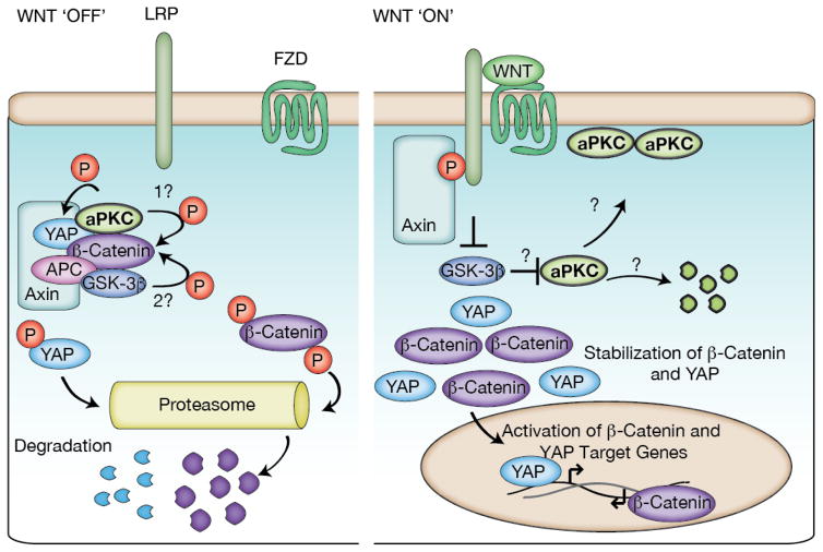
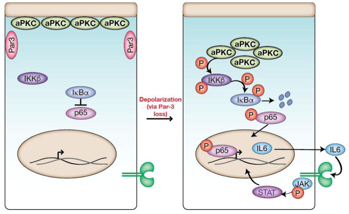
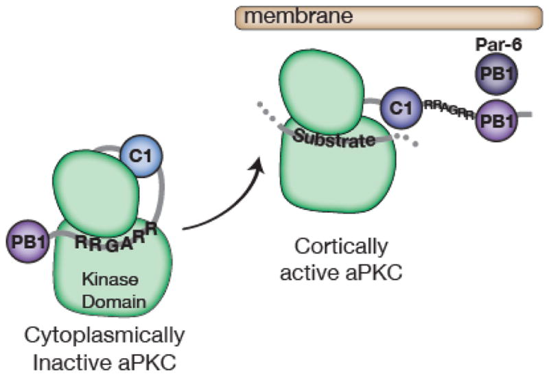
Complex organisms are faced with the challenge of generating and maintaining diverse cell types, ranging from simple epithelia to neurons and motile immune cells [1-3]. To meet this challenge, a complex set of regulatory pathways controls nearly every aspect of cell growth and function, including genetic and epigenetic programming, cytoskeleton dynamics, and protein trafficking. The far reach of cell fate specification pathways makes it particularly catastrophic when they malfunction, both during development and for tissue homeostasis in adult organisms. Furthermore, the therapeutic promise of stem cells derives from their ability to deftly navigate the multitude of pathways that control cell fate [4]. How the molecular components making up these pathways function to specify cell fate is beginning to become clear. Work from diverse systems suggests that the atypical Protein Kinase C (aPKC) is a key regulator of cell fate decisions in metazoans [5-7]. Here, we examine some of the diverse physiological outcomes of aPKC's function in differentiation, along with the molecular pathways that control aPKC and those that are responsive to changes in its catalytic activity.

Keywords: cell proliferation; differentiation; protein kinase.


Figures

Figure 1
Figure 1
PKC family kinases and regulation and function of atypical Protein Kinase C. A. Schematic of the protein kinase C family showing domain architectures, demonstrating both common and unique aspects of each PKC family member (PS = pseudosubstrate; C1 and C2 are cysteine rich domains; PB1 Phox/Bem1 domain). B. Schematic of Par-mediated polarity mechanism. aPKC generates cellular polarity through phosphorylation and exclusion of cortically localized substrates (pink).
Figure 2
Figure 2
aPKC regulation of the cell cycle. A. When aPKC levels are low, p27Xic1 is able to elongate the G1 to S transition by binding to Cdk2, which can lead to differentiation in Xenopus neuroectoderm progenitor cells. B. When aPKC levels are high, p27Xic1 phosphorylation by aPKC blocks p27Xic1 binding of Cdk2, shortening the G1 to S transition to promote proliferation.
Figure 3
Figure 3
aPKC regulation of Hedgehog signaling. In basal cell carcinomas (BCCs) and lung squamous cell carcinomas (LSCCs) aPKC is able to phosphorylate GLI (BCCs) and SOX2 (LSCCs) transcription factors. These phosphorylations can lead to positive feedback, upregulating HH signaling genes including HHAT and aPKC itself. This activation can occur independently of HH ligand receptor binding. In the Drosophlia developing wing, aPKC phosphorylates the Smoothened receptor to regulate its activity and its subsequent proper development.
Figure 4
Figure 4
aPKC regulation of Wnt signaling. aPKC is part of the destruction complex, where it can phosphorylate Catenin to prime it for (2) GSK-3 phosphorylation and subsequent proteasomal degradation. aPKC is also able to phosphorylate YAP, leading to proteasomal degradation. Loss of aPKC or Wnt binding leads to disassembly of the destruction complex and activation of Wnt signaling favoring a proliferative state. The fate of aPKC once the destruction complex is inactivated is unknown.
Figure 5
Figure 5
aPKC regulation of JAK/Stat signaling. Loss of polarity leads to cytoplasmic aPKC which causes activation of IKKβ, degradation of IB, and translocation of p65 to the nucleus to upregulate IL6 production. The increase in IL6 leads to a positive feedback loop with JAK/Stat3 signaling, which, when unregulated, leads to proliferation and tumor progression in a breast cancer model.
Figure 6
Figure 6
Regulation of aPKC localization and activity. Par-6’s interaction with aPKC’s PB1 domain disrupts the pseudosubstrate’s (sequence = RRGARR) inhibition of the kinase domain. The C1 domain may also play a role in regulating aPKC kinase activity.

References

    1. Clevers H, Loh KM, Nusse R. An integral program for tissue renewal and regeneration: Wnt signaling and stem cell control. Science. 2014;346:1248012. - PubMed
    1. Kohwi M, Doe CQ. Temporal Fate Specification and Neural Progenitor Competence During Development. Nat Rev Neurosci. 2013;14:823–838. - PMC - PubMed
    1. Seita J, Weissman IL. Hematopoietic Stem Cell: Self-renewal versus Differentiation. Wiley Interdiscip Rev Syst Biol Med. 2010;2:640–653. - PMC - PubMed
    1. Chen K, Huang Y, Chen J. Understanding and targeting cancer stem cells: therapeutic implications and challenges. Acta Pharmacol Sin. 2013;34:732–740. - PMC - PubMed
    1. Doe CQ. Neural stem cells: balancing self-renewal with differentiation. Development. 2008;135:1575–1587. - PubMed

Publication types