Review
. 2017 Mar:156:22-33.
doi: 10.1016/j.exer.2016.03.010. Epub 2016 Mar 16.

Systems biology of lens development: A paradigm for disease gene discovery in the eye

Affiliations
Review

Systems biology of lens development: A paradigm for disease gene discovery in the eye

Deepti Anand et al. Exp Eye Res. 2017 Mar.

Abstract

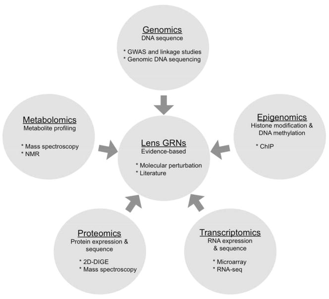
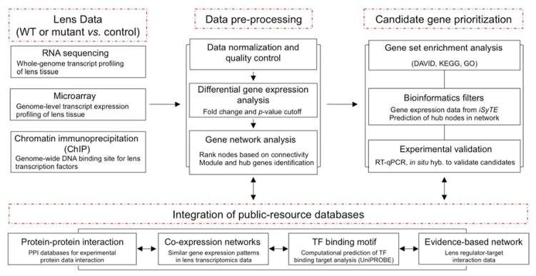
Over the past several decades, the biology of the developing lens has been investigated using molecular genetics-based approaches in various vertebrate model systems. These efforts, involving target gene knockouts or knockdowns, have led to major advances in our understanding of lens morphogenesis and the pathological basis of cataracts, as well as of other lens related eye defects. In particular, we now have a functional understanding of regulators such as Pax6, Six3, Sox2, Oct1 (Pou2f1), Meis1, Pnox1, Zeb2 (Sip1), Mab21l1, Foxe3, Tfap2a (Ap2-alpha), Pitx3, Sox11, Prox1, Sox1, c-Maf, Mafg, Mafk, Hsf4, Fgfrs, Bmp7, and Tdrd7 in this tissue. However, whether these individual regulators interact or their targets overlap, and the significance of such interactions during lens morphogenesis, is not well defined. The arrival of high-throughput approaches for gene expression profiling (microarrays, RNA-sequencing (RNA-seq), etc.), which can be coupled with chromatin immunoprecipitation (ChIP) or RNA immunoprecipitation (RIP) assays, along with improved computational resources and publically available datasets (e.g. those containing comprehensive protein-protein, protein-DNA information), presents new opportunities to advance our understanding of the lens tissue on a global systems level. Such systems-level knowledge will lead to the derivation of the underlying lens gene regulatory network (GRN), defined as a circuit map of the regulator-target interactions functional in lens development, which can be applied to expedite cataract gene discovery. In this review, we cover the various systems-level approaches such as microarrays, RNA-seq, and ChIP that are already being applied to lens studies and discuss strategies for assembling and interpreting these vast amounts of high-throughput information for effective dispersion to the scientific community. In particular, we discuss strategies for effective interpretation of this new information in the context of the rich knowledge obtained through the application of traditional single-gene focused experiments on the lens. Finally, we discuss our vision for integrating these diverse high-throughput datasets in a single web-based user-friendly tool iSyTE (integrated Systems Tool for Eye gene discovery) - a resource that is already proving effective in the identification and characterization of genes linked to lens development and cataract. We anticipate that application of a similar approach to other ocular tissues such as the retina and the cornea, and even other organ systems, will significantly impact disease gene discovery.

Keywords: Bioinformatics; Cataract; Gene regulatory networks; Lens; RNA binding proteins; Transcription factors; iSyTE.


Figures

Figure 1
Figure 1. Systems-level approaches to study lens biology
Information from genomics, epigenomics, transcriptomics, proteomics, metabolomics approaches are already being applied to study lens biology. Integration of these various high-throughput data will require the development of a web-based community resource. Such a resource will enable the derivation, visualization, and analysis of the spatio-temporal gene regulatory networks associated with lens development and homeostasis.
Figure 2
Figure 2. iSyTE predicts SIPA1l3 as a cataract gene
iSyTE predicts SIPA1l3 as high-priority candidate gene in the lens among a 5Mb interval on human chromosome 19. Also see: (Lachke et al. 2012b). The color gradation in the inset offers an estimate of enriched expression of the genes in mouse lens tissue at embryonic day (E)10.5, E11.5 and E12.5. SIPA1l3 is among the top 1% of lens-enriched genes. This prediction was independently proven by two groups that recently reported SIPA1l3 mutations in human congenital cataract (Evers et al. 2015, Greenlees et al. 2015).
Figure 3
Figure 3. An integrated approach flow chart to analyze lens transcriptomics data
High-throughput lens expression and functional data analysis involves distinct steps. The lens transcriptomics data (RNA-seq and microarray) and ChIP data requires data preprocessing steps (normalization and quality control), identification of differentially expressed genes (DEGs) and network analysis. Candidate gene prioritization of DEGs includes functional pathway analysis (functional annotation clustering by DAVID tool and KEGG pathway analysis) and relevance to the lens (iSyTE-analysis). Development of new hypotheses requires integration of knowledge within various publically available informative resources such as protein-protein interactions databases, derivation of co-expression networks (high-throughput expression data of normal lens development), computational prediction of TF-binding motifs (UniPROBE data) and published lens-literature (experimentally validated molecular evidence-based data). This integrative analysis approach of high-throughput gene expression data from wild-type and mutant lens tissue from targeted gene deletion experiments will facilitate prioritization of candidate genes and pathways in the lens.
Figure 4
Figure 4
Integrated network analysis to identify lens-specific Six3 interacting candidates. (A) Six3 lens perturbation data from published literature (evidence-based). (B) Expansion of Six3 evidence-based network using human protein-protein interaction (PPI) data for SIX3 to identify its immediate interacting partners (nodes). (C) Overlay of iSyTE expression data from E10.5 mouse lens tissue and P28 mouse lens isolated epithelium tissue on SIX3 PPI nodes, allowing the identification of high-priority lens relevant candidate nodes. (D) Overlay of iSyTE enriched expression data from E10.5 mouse lens tissue and P28 mouse lens isolated epithelium tissue on SIX3 PPI nodes, further refining the identification of high-priority candidates.

References

    1. Agrawal SA, Anand D, Siddam AD, Kakrana A, Dash S, Scheiblin DA, Dang CA, Terrell AM, Waters SM, Singh A, Motohashi H, Yamamoto M, Lachke SA. Compound mouse mutants of bZIP transcription factors Mafg and Mafk reveal a regulatory network of non-crystallin genes associated with cataract. Hum Genet. 2015;134:717–735. doi: 10.1007/s00439-015-1554-5. - DOI - PMC - PubMed
    1. Aldahmesh MA, Alshammari MJ, Khan AO, Mohamed JY, Alhabib FA, Alkuraya FS. The syndrome of microcornea, myopic chorioretinal atrophy, and telecanthus (MMCAT) is caused by mutations in ADAMTS18. Hum Mutat. 2013;34:1195–1199. doi: 10.1002/humu.22374. - DOI - PubMed
    1. Aldahmesh MA, Khan AO, Mohamed JY, Hijazi H, Al-Owain M, Alswaid A, Alkuraya FS. Genomic analysis of pediatric cataract in Saudi Arabia reveals novel candidate disease genes. Genet Med. 2012;14:955–962. doi: 10.1038/gim.2012.86. - DOI - PubMed
    1. Anand D, Agrawal S, Siddam A, Motohashi H, Yamamoto M, Lachke SA. An integrative approach to analyze microarray datasets for prioritization of genes relevant to lens biology and disease. Genom Data. 2015;5:223–227. doi: 10.1016/j.gdata.2015.06.017. - DOI - PMC - PubMed
    1. Anchan RM, Lachke SA, Gerami-Naini B, Lindsey J, Ng N, Naber C, Nickerson M, Cavallesco R, Rowan S, Eaton JL, Xi Q, Maas RL. Pax6- and Six3-mediated induction of lens cell fate in mouse and human ES cells. PLoS ONE. 2014;9:e115106. doi: 10.1371/journal.pone.0115106. - DOI - PMC - PubMed

Publication types

MeSH terms