. 2016 Jul;65(7):2081-93.
doi: 10.2337/db16-0003. Epub 2016 Mar 18.

The Genetic Program of Pancreatic β-Cell Replication In Vivo

Affiliations

The Genetic Program of Pancreatic β-Cell Replication In Vivo

Agnes Klochendler et al. Diabetes. 2016 Jul.

Abstract

The molecular program underlying infrequent replication of pancreatic β-cells remains largely inaccessible. Using transgenic mice expressing green fluorescent protein in cycling cells, we sorted live, replicating β-cells and determined their transcriptome. Replicating β-cells upregulate hundreds of proliferation-related genes, along with many novel putative cell cycle components. Strikingly, genes involved in β-cell functions, namely, glucose sensing and insulin secretion, were repressed. Further studies using single-molecule RNA in situ hybridization revealed that in fact, replicating β-cells double the amount of RNA for most genes, but this upregulation excludes genes involved in β-cell function. These data suggest that the quiescence-proliferation transition involves global amplification of gene expression, except for a subset of tissue-specific genes, which are "left behind" and whose relative mRNA amount decreases. Our work provides a unique resource for the study of replicating β-cells in vivo.


Figures

Figure 1
Figure 1
Ccnb1-GFP marks replicating cells and allows for FACS sorting of quiescent and replicating β-cells. A: qRT-PCR analysis of sorted unstained, TSQ-stained, and Tomato+ islet cells. Results were normalized to Actb. Pnlip and Ki67 expression levels are presented relative to the expression in unstained islets and GFP sorted cells, respectively. B: GO enrichment analysis of upregulated genes (FDR ≤0.05) in replicating β-cells. Overrepresentation was computed using a statistical test based on hypergeometric distribution. Gene sets with a P value <0.01 and an FDR <0.05 were considered significant.
Figure 2
Figure 2
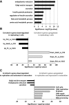
Genes repressed in replicating β-cells are involved in β-cell function and maturation. A: GO analysis of genes downregulated in replicating β-cells. B: Significant overrepresentation of genes upregulated in development in genes repressed in replicating β-cells (FDR ≤0.05) and, conversely, of genes repressed in development in genes induced in replicating β-cells (FDR ≤0.05). C: Genes induced in maturation and repressed in replication are associated with β-cell function. Genes repressed in maturation and activated in replication are associated with cell cycle biological processes. Overrepresentation was computed using a statistical test based on hypergeometric distribution.
Figure 3
Figure 3
Targets of Nkx6.1, Pdx1, and MafA are repressed in replicating β-cells. A: Genes repressed in replicating β-cells are significantly enriched for targets of the β-cell transcription factors. The overrepresentation significance was computed using a statistical test based on hypergeometric distribution. B: FACS histograms of islet cells immunostained for insulin, Ki67, and Nkx6.1 or Pdx1. Cells were gated on insulin+ β-cells. Controls represent cells immunostained with isotype control. wt, wild type.
Figure 4
Figure 4
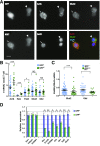
Quantification of RNA molecules by smRNA-FISH in quiescent (GFP) and replicating (GFP+) β-cells. A: Representative image of smRNA-FISH for the detection and measurement of single mRNA molecules of Actb, Glut2, and Ki67, as indicated. Actb level is elevated in GFP+ (arrow) compared with GFP cells (arrowhead) (267 vs. 156 transcripts, respectively), whereas Glut2 levels remain unchanged (60 vs. 61, respectively). Ki67 (marker for replicating cell) is expressed only in GFP+ cells. Scale bar is 10 μm. Image is a maximal projection of 12 optical sections spaced 0.3 μm. B: Quantification based on 15–20 optical sections spaced 0.3 μm in the Z direction. Mean numbers (with SEM) of RNA molecules per optical Z-section per cell (≥20 GFP+ and GFP cells) for the five measured genes. Each dot represents measurements from a single cell. C: The ratios of Glut2/Actb and Glul/Actb transcripts were computed per cell. Statistical significance was assessed using the nonparametric Mann-Whitney test (B and C) (*P < 0.05, ***P < 0.0001). D: qRT-PCR analysis of the indicated mRNAs. Relative mRNA levels (±SE) were calculated after normalization to Actb (n = 3) (*P < 0.05, **P < 0.005, paired t test). NS, not significant.
Figure 5
Figure 5
Metabolic changes in replicating β-cells. A: Glycolytic genes repressed in GFP+ (replicating β-cells). Graph shows the mean expression (±SE) in GFP+ β-cells relative to quiescent GFP β-cells, as derived from the RNA-seq data. All GFP+ values represent significant fold changes compared with GFP values, as computed by DESeq (FDR <0.05). BE: Mitochondrial membrane potential (∆Ψm) measured by TMRE staining in dissociated islet cells after incubation in low (3 mmol/L) and high (16 mmol/L) glucose. TMRE intensity is shown for GFP (B) and GFP+ (C) islet cells incubated at both glucose concentrations; TMRE staining is shown in GFP and GFP+ islet cells after incubation in low (3 mmol/L) and high (16 mmol/L) glucose (D and E, respectively).

Comment in

References

    1. Desgraz R, Bonal C, Herrera PL. β-Cell regeneration: the pancreatic intrinsic faculty. Trends Endocrinol Metab 2011;22:34–43 - PubMed
    1. Prentki M, Nolan CJ. Islet beta cell failure in type 2 diabetes. J Clin Invest 2006;116:1802–1812 - PMC - PubMed
    1. Dor Y, Brown J, Martinez OI, Melton DA. Adult pancreatic beta-cells are formed by self-duplication rather than stem-cell differentiation. Nature 2004;429:41–46 - PubMed
    1. Meier JJ, Butler AE, Saisho Y, et al. . Beta-cell replication is the primary mechanism subserving the postnatal expansion of beta-cell mass in humans. Diabetes 2008;57:1584–1594 - PMC - PubMed
    1. Teta M, Rankin MM, Long SY, Stein GM, Kushner JA. Growth and regeneration of adult beta cells does not involve specialized progenitors. Dev Cell 2007;12:817–826 - PubMed

Publication types