. 2016 Aug;132(2):235-256.
doi: 10.1007/s00401-016-1558-9. Epub 2016 Mar 18.

Presynaptic dystrophic neurites surrounding amyloid plaques are sites of microtubule disruption, BACE1 elevation, and increased Aβ generation in Alzheimer's disease

Affiliations

Presynaptic dystrophic neurites surrounding amyloid plaques are sites of microtubule disruption, BACE1 elevation, and increased Aβ generation in Alzheimer's disease

Katherine R Sadleir et al. Acta Neuropathol. 2016 Aug.

Abstract

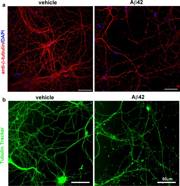
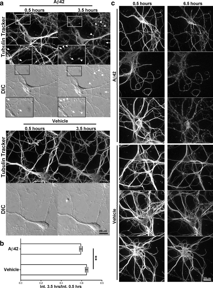
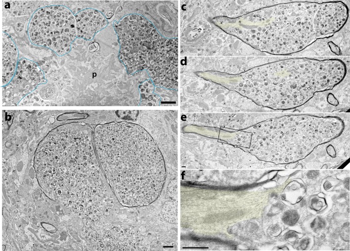
Alzheimer's disease (AD) is characterized by amyloid plaques composed of the β-amyloid (Aβ) peptide surrounded by swollen presynaptic dystrophic neurites consisting of dysfunctional axons and terminals that accumulate the β-site amyloid precursor protein (APP) cleaving enzyme (BACE1) required for Aβ generation. The cellular and molecular mechanisms that govern presynaptic dystrophic neurite formation are unclear, and elucidating these processes may lead to novel AD therapeutic strategies. Previous studies suggest Aβ may disrupt microtubules, which we hypothesize have a critical role in the development of presynaptic dystrophies. To investigate this further, here we have assessed the effects of Aβ, particularly neurotoxic Aβ42, on microtubules during the formation of presynaptic dystrophic neurites in vitro and in vivo. Live-cell imaging of primary neurons revealed that exposure to Aβ42 oligomers caused varicose and beaded neurites with extensive microtubule disruption, and inhibited anterograde and retrograde trafficking. In brain sections from AD patients and the 5XFAD transgenic mouse model of amyloid pathology, dystrophic neurite halos with BACE1 elevation around amyloid plaques exhibited aberrant tubulin accumulations or voids. At the ultrastructural level, peri-plaque dystrophies were strikingly devoid of microtubules and replete with multi-lamellar vesicles resembling autophagic intermediates. Proteins of the microtubule motors, kinesin and dynein, and other neuronal proteins were aberrantly localized in peri-plaque dystrophies. Inactive pro-cathepsin D also accumulated in peri-plaque dystrophies, indicating reduced lysosomal function. Most importantly, BACE1 accumulation in peri-plaque dystrophies caused increased BACE1 cleavage of APP and Aβ generation. Our study supports the hypothesis that Aβ induces microtubule disruption in presynaptic dystrophic neurites that surround plaques, thus impairing axonal transport and leading to accumulation of BACE1 and exacerbation of amyloid pathology in AD.

Keywords: 5XFAD mice; Alzheimer’s disease; Amyloid; Axonal transport; Aβ; BACE1; Cathepsin D; Dystrophic neurite; Microtubule; Tubulin.


Figures

Fig. 1
Fig. 1
Oligomeric Aβ42 causes neurite beading and microtubule disruption in primary neurons. a Primary cortical neurons were cultured from e15.5 mouse embryos and after 9 days in vitro were exposed to 1 μM Aβ42 oligomers or vehicle. After 72 h, coverslips were fixed in 4 % paraformaldehyde, permeabilized, and stained with the antibody TuJ1 against βIII-tubulin (red) and DAPI (blue). Neurite beading is very prominent in Aβ42-treated cultures, while neurites appear smooth and unbeaded in vehicle-treated neurons. b Primary neurons were isolated as described in a, and after 12 days in vitro were exposed to 10 μM Aβ42 oligomers or vehicle. After 22 h of Aβ42 treatment, the fluorescent microtubule probe Tubulin Tracker was added to a final concentration of 250 nM for 30 min. Unfixed, live neurons were then imaged at ×20 using a Nikon Eclipse TS100 microscope with NIS Elements software. Note that Aβ42-induced neurite beading visualized by Tubulin Tracker appears very similar to that observed with βIII-tubulin immunostaining in a, and occurs more rapidly with higher Aβ42 concentration. Taken together, these results strongly suggest that Aβ42 disrupts the organization of microtubules in neurons. Scale bars in all frames 50 μm
Fig. 2
Fig. 2
Microtubule disruption in primary neurons is an early response to oligomeric Aβ42. Primary cortical neurons were cultured as in Fig. 1, and after 12 days in vitro, were labeled for 30 min with 250 nM Tubulin Tracker, rinsed, and then exposed to 10 μM Aβ42 oligomers or vehicle. Live imaging on an Andor spinning disk confocal microscope began 30 min after addition of Aβ42. a Fluorescence and differential interference contrast (DIC) images were acquired every 5 min for 3 h. Neurite beading, microtubule accumulation in varicosities (arrowheads), and microtubule fragmentation (arrows) in neurites were present 3.5 h after Aβ42 addition, in the absence of significant cell death or BACE1 elevation [49]. In contrast, the morphology of vehicle-treated neurons was unaffected, with the exception of a moderate decrease of fluorescence intensity. DIC imaging revealed that neurites of Aβ42-treated neurons were intact and continuous, even though they exhibited beading and microtubule fragmentation, indicating that the observed microtubule disruption was not the result of physical degeneration of neurites. Insets show higher magnification images of the boxed region of the Aβ42-treated culture to accentuate varicosity formation and microtubule fragmentation. b The ratios of Tubulin Tracker fluorescence intensity (Int.) at 3.5 h (hours) to that at 0.5 h after Aβ42 treatment were calculated for 46 image fields from two separate experiments and averaged for Aβ42-treated and vehicle-treated neurons. Overall Tubulin Tracker fluorescence intensity decreased in neurons over time due to photo-bleaching, as indicated by fluorescence intensity ratios below 1. Nevertheless, the Tubulin Tracker fluorescence intensity ratio for Aβ42-treated neurons showed a small but highly significant decrease compared to vehicle treatment (p = 0.004), suggesting microtubule depolymerization and reduced stability of microtubule networks after only 3.5 h of Aβ42 exposure. Error bars SEM; **, p < 0.01. c To investigate longer Aβ42 treatment times and minimize photo-bleaching, primary neuron cultures were prepared as above and images collected once every 60 min for 6 h. Neurite beading, aberrant microtubule localization, and microtubule fragmentation were even more prevalent and pronounced in neurons after 6.5 h compared to 3.5 h of Aβ42 treatment, while vehicle-treated neurites continued to appear normal. Scale bars 20 μm for all images in a, c
Fig. 3
Fig. 3
Tubulin isoforms are aberrantly localized in BACE1-positive peri-plaque dystrophic neurites in vivo. a Post-mortem brain sections of superior frontal gyrus from three AD patients and three cognitively normal controls were stained with antibodies against BACE1 (red) and βIII-tubulin (green), and methoxy XO4 (blue) to label amyloid deposits. Representative plaques from two cases (AD 14–190 and AD 11–193, Columns 1–2) are shown; additional examples in Fig. S2. Cognitively normal controls were negative for plaques, as expected (not shown). As previously reported [86], BACE1 immunostaining was observed in a halo of dystrophic neurites surrounding individual plaques. In contrast, βIII-tubulin immunostaining was almost entirely absent in peri-plaque BACE1-positive dystrophies. Non-dystrophic neuropil in areas adjacent to plaques exhibited normal intensities and patterns of βIII-tubulin immunostaining. Some red fluorescence (white arrows) represents non-specific background from blood vessels. Brain sections from the aggressive amyloid 5XFAD mouse model were stained with antibodies against BACE1 (red) and βIII-tubulin, acetylated α-tubulin or polyglutamylated tubulin (green). Representative 5XFAD amyloid deposits are shown (Columns 3–5), with plaque cores marked by asterisks; additional examples in Fig. S2. Similar to human AD, BACE1 accumulated in peri-plaque dystrophic neurites in 5XFAD brain sections. Additionally, the different tubulin isoforms were generally reduced in dystrophic halos around plaques, although tubulins often occurred in amorphous accumulations that showed little co-localization with BACE1 immunostaining. Scale bars in all frames 10 μm. b Immunofluorescence intensities of BACE1 and tubulin isoforms (as indicated at top of each column) in peri-plaque dystrophic neurites were measured and BACE1/tubulin intensity ratios calculated and compared to those in normal-appearing regions of neuropil within the same frame (Supplementary Methods). For human tissue, 16 dystrophies and corresponding non-dystrophic regions were measured per case. For murine tissue, BACE1/tubulin isoform ratios were determined for 11–20 BACE1-positive dystrophic regions and a corresponding number of nearby neuropil areas. BACE1/tubulin ratios for AD 14–190 and AD 11–193 (Panels 1 and 2, respectively) and 5XFAD mice (Panels 35) are shown. While there was substantial variation for dystrophic neurites, BACE1/tubulin intensity ratios for dystrophies were significantly elevated compared to those for normal neuropil for both AD cases and 5XFAD mice. Error bars SEM; ***p < 0.001. c The quantitative relationships between BACE1 and tubulin isoform immunofluorescence intensities by another method, intensity profiles as a function of distance across the plaque were generated (Supplementary Methods). Representative plaques are shown (upper row) for AD 14–190 and AD 11–193 (Panels 1 and 2, respectively) and 5XFAD mice (Panels 35). White line indicates path through plaque from which the intensity profiles were generated (bottom row). Note the inverse relationships between BACE1 and tubulin intensities as a function of distance across the plaque in each case. Taken together, these results suggest that tubulin, and hence microtubules, is reduced and/or mis-localized in peri-plaque dystrophic neurites. AU arbitrary units. Scale bars in all frames 10 μm
Fig. 4
Fig. 4
Microtubules are strikingly absent in peri-plaque dystrophic axons. 5XFAD brain tissue was prepared for electron microscopy (EM) and assessed ultrastructurally for the presence of microtubules in peri-plaque dystrophic axons. a As previously reported [25], electron microscopy of 5XFAD brain revealed amyloid plaques (p) surrounded by dystrophic neurites (representative examples outlined in blue) filled with electron-dense multi-lamellar vesicles, possibly autophagic intermediates. b EM of a transverse section through a pair of dystrophic myelinated axons filled with multi-lamellar vesicles. Note that the lower portion of the axon on the left has lost its myelin sheath. cf EM of serial ultrathin sections occasionally revealed series of longitudinal sections through dystrophic axons near amyloid plaques. Shown is such a series of a peri-plaque dystrophic myelinated axon with a portion of normal axon appearing on the left side of the dystrophy. Intact, continuous microtubules (yellow shading) are present in the morphologically normal part of the axon, but then microtubules become disorganized and mostly disappear within the dystrophic region. Note the dark outline of the surrounding myelin sheath, definitively confirming axonal identity. These ultrastructural images provide direct evidence that microtubules are disrupted and largely absent in peri-plaque dystrophic regions of axons. Images are spaced ~200 nm apart. f Higher magnification of rectangle in e. Scale bars 1 μm
Fig. 5
Fig. 5
Microtubule motor proteins are aberrantly localized in BACE1-positive peri-plaque dystrophic neurites. Sections from 5XFAD mouse brains were stained with antibodies against BACE1 (red) and various components of the dynein–dynactin retrograde transport complex, dynein intermediate chain (green, a), p150glued (green, b), dynamitin (green, c), or the anterograde motor protein kinesin heavy chain (green, d). Representative amyloid deposits are shown; additional examples in Figs. S3 and S4. Dynein intermediate chain and p150glued were reduced in peri-plaque BACE1-positive dystrophic neurites, while dynamitin and kinesin heavy chain tended to accumulate in dystrophies and showed partial but limited co-localization with BACE1. These results together with evidence of microtubule disruption strongly suggest that axonal transport is impaired in dystrophic regions of peri-plaque axons. DAPI staining (blue) identified nuclei and autofluorescence of amyloid plaque cores (marked by asterisks). Scale bars 10 μm (a, b, d) and 20 μm (c)
Fig. 6
Fig. 6
BACE1-positive peri-plaque dystrophies accumulate the presynaptic protein synaptophysin and the lysosomal protease cathepsin D, but lack the active zone protein bassoon. Sections from 5XFAD mouse brains were stained with antibodies against BACE1 (red) and active zone protein bassoon (green, a), presynaptic protein synaptophysin (green, b), somatodendritic marker MAP2, (green, c), or lysosomal protease cathepsin D (green, d). Representative amyloid deposits are shown; additional examples in Figs. S3 and S4. Both bassoon and synaptophysin co-localized extensively with BACE1 in presynaptic terminals in the stratum lucidum (bottom row), but bassoon and synaptophysin were absent and enriched in peri-plaque dystrophies, respectively (top row). The somatodendritic protein MAP2, which did not co-localize with BACE1 in the stratum lucidum (bottom row), was absent in peri-plaque dystrophies (top row). The lysosomal protease cathepsin D was found in neuronal soma as expected and did not co-localize with BACE1 in stratum lucidum (bottom row). However, cathepsin D tended to accumulate in some dystrophies, indicating aberrant localization of lysosomes in peri-plaque dystrophic neurites. These results suggest that BACE1-positive peri-plaque dystrophies are largely presynaptic axons and terminals, although dystrophic terminals are unlikely to have active synapses as they lack bassoon. DAPI staining (blue) identified nuclei and autofluorescence of amyloid plaque cores (marked by asterisks). Scale bars 10 μm in all frames except for the bottom row, which is 20 μm
Fig. 7
Fig. 7
Levels of immature pro-cathepsin D are increased in 5XFAD mouse brain. a Cortices from 6-month 5XFAD (+) or non-transgenic (−) mice were homogenized and subjected to immunoblot analysis using antibodies against human APP (top panel; antibody 6E10) and the C-terminus of the lysosomal protease cathepsin D (CatD), which recognizes both immature (inactive) pro-CatD and mature (active) CatD heavy chain (middle panel). Ponceau S staining of the immunoblot was used to control for protein loading (bottom panel). Note that 43-kDa immature pro-CatD bands are more intense than 28 kDa mature CatD heavy chain bands, especially in 5XFAD cortices. b Band intensities from the immunoblot in a were quantified and normalized to total protein as determined by ponceau S staining intensity in respective lanes and then displayed as percentage of non-transgenic (non-Tg) control. Note that 5XFAD cortex levels of pro-CatD were nearly twofold higher than those in non-Tg cortex, while levels of CatD heavy chain were not significantly changed, suggesting that lysosomal maturation is impaired in 5XFAD brains. Error bars SEM; **p < 0.01; NS not significant
Fig. 8
Fig. 8
Neoepitope antibodies recognize BACE1-cleaved APP and Aβ but not full-length APP in vivo. Sagittal brain sections of 5XFAD;BACE1+/+ and 5XFAD;BACE1−/− mice were immunostained with neoepitope antibodies to a the free C-terminus of BACE1-cleaved APP ectodomain ending in the Swedish mutation, sAPPβ (antibody ANJJ [45, 46], green), b the free N-terminus of BACE1-cleaved Aβ (antibody 3D6 [23], red), or c the free C-terminus of γ-secretase-cleaved Aβ42 (green). Sections were also co-immunostained with an antibody recognizing full-length APP (antibody Karen [64], red), then imaged by confocal microscopy. All three neoepitope immunoreactivities were present in soma (arrows), and processes in the case of sAPPβ (arrowheads), of pyramidal neurons in the cortex of 5XFAD;BACE1+/+ mice, while none were detected in 5XFAD;BACE1−/− cortex, despite overexpression of transgenic APP in pyramidal neurons of both genotypes. These data confirm that ANJJ, 3D6, and Aβ42 antibodies are selective for their respective neoepitopes and do not cross-react to full-length APP. DAPI staining (blue) identified nuclei and autofluorescence of amyloid deposit cores (marked by asterisks). Note that amyloid deposits are completely lacking in 5XFAD;BACE1−/− cortex, as previously reported [41]. Scale bars 10 μm (b), 20 μm (a)
Fig. 9
Fig. 9
BACE1 accumulation in peri-plaque dystrophic neurites increases BACE1 cleavage of APP and generation of Aβ42. Sagittal brain sections of 5XFAD mice were co-immunostained with antibodies recognizing BACE1 and cleaved APP fragment neoepitopes (sAPPβ, 3D6, Aβ42) validated in Fig. 8. a The BACE1-cleaved free C-terminal neoepitope of sAPPβ (ANJJ, green) showed robust co-localization with elevated BACE1 (red) in peri-plaque dystrophic neurites. Note the intense sAPPβ immunoreactivity in dystrophies, while little if any sAPPβ signal is present in surrounding neuropil, demonstrating dramatically elevated BACE1 cleavage of APP in BACE1-positive dystrophic neurites near amyloid deposits. b Antibody 3D6 (green), which recognizes the free N-terminal neoepitope of BACE1-cleaved Aβ, also co-localized with elevated BACE1 (red) in dystrophic neurites, confirming increased BACE1 activity and Aβ generation in dystrophies. Antibody 3D6 also detects amyloid plaques, as expected (example marked with asterisk). c Like 3D6, an antibody recognizing the free C-terminal neoepitope generated after γ-secretase cleavage of Aβ42 (green) co-localized with elevated BACE1 (red) in peri-plaque dystrophies, and detected amyloid deposits, as expected (asterisk). d Co-immunostaining also demonstrated that both sAPPβ (green) and 3D6 (Aβ, red) neoepitopes co-localized in peri-plaque dystrophies. e Co-immunostaining with antibodies against BACE1 (red) and APP (Karen, green) also showed a high level of co-localization. Note that all cleaved APP fragment neoepitopes and BACE1 show a punctate immunostaining pattern in dystrophies, suggesting that they are localized to vesicles such as endosomes, while APP immunostaining appears more uniform, implying greater cell surface localization. Panels in the right column of each set are high-magnification images of the boxed regions in the frames of the left column. DAPI staining (blue) identified nuclei and autofluorescence of amyloid deposit cores (marked by asterisks). Scale bar sizes are indicated in bottom panels for images in each column

References

    1. Arriagada PV, Growdon JH, Hedley-Whyte ET, Hyman BT. Neurofibrillary tangles but not senile plaques parallel duration and severity of Alzheimer’s disease. Neurology. 1992;42:631–639. doi: 10.1212/WNL.42.3.631. - DOI - PubMed
    1. Blasko I, Beer R, Bigl M, Apelt J, Franz G, Rudzki D, Ransmayr G, Kampfl A, Schliebs R. Experimental traumatic brain injury in rats stimulates the expression, production and activity of Alzheimer’s disease beta-secretase (BACE-1) J Neural Transm. 2004;111:523–536. doi: 10.1007/s00702-003-0095-6. - DOI - PubMed
    1. Boissonneault V, Plante I, Rivest S, Provost P. MicroRNA-298 and microRNA-328 regulate expression of mouse beta-amyloid precursor protein-converting enzyme 1. J Biol Chem. 2009;284:1971–1981. doi: 10.1074/jbc.M807530200. - DOI - PMC - PubMed
    1. Cai H, Wang Y, McCarthy D, Wen H, Borchelt DR, Price DL, Wong PC. BACE1 is the major beta-secretase for generation of Abeta peptides by neurons. Nat Neurosci. 2001;4:233–234. doi: 10.1038/85064. - DOI - PubMed
    1. Cheong FK, Feng L, Sarkeshik A, Yates JR, 3rd, Schroer TA. Dynactin integrity depends upon direct binding of dynamitin to Arp1. Mol Biol Cell. 2014;25:2171–2180. doi: 10.1091/mbc.E14-03-0842. - DOI - PMC - PubMed

Publication types

MeSH terms

Substances

LinkOut - more resources