Review
. 2016 Mar;36(2):181-94.
doi: 10.1007/s10571-015-0273-8. Epub 2016 Mar 18.

Lymphatic Clearance of the Brain: Perivascular, Paravascular and Significance for Neurodegenerative Diseases

Affiliations
Review

Lymphatic Clearance of the Brain: Perivascular, Paravascular and Significance for Neurodegenerative Diseases

Erik N T P Bakker et al. Cell Mol Neurobiol. 2016 Mar.

Abstract

The lymphatic clearance pathways of the brain are different compared to the other organs of the body and have been the subject of heated debates. Drainage of brain extracellular fluids, particularly interstitial fluid (ISF) and cerebrospinal fluid (CSF), is not only important for volume regulation, but also for removal of waste products such as amyloid beta (Aβ). CSF plays a special role in clinical medicine, as it is available for analysis of biomarkers for Alzheimer's disease. Despite the lack of a complete anatomical and physiological picture of the communications between the subarachnoid space (SAS) and the brain parenchyma, it is often assumed that Aβ is cleared from the cerebral ISF into the CSF. Recent work suggests that clearance of the brain mainly occurs during sleep, with a specific role for peri- and para-vascular spaces as drainage pathways from the brain parenchyma. However, the direction of flow, the anatomical structures involved and the driving forces remain elusive, with partially conflicting data in literature. The presence of Aβ in the glia limitans in Alzheimer's disease suggests a direct communication of ISF with CSF. Nonetheless, there is also the well-described pathology of cerebral amyloid angiopathy associated with the failure of perivascular drainage of Aβ. Herein, we review the role of the vasculature and the impact of vascular pathology on the peri- and para-vascular clearance pathways of the brain. The different views on the possible routes for ISF drainage of the brain are discussed in the context of pathological significance.

Keywords: Cerebrospinal fluid; Cerebrovascular basement membranes; Interstitial fluid; Paravascular; Perivascular.


Conflict of interest statement

The authors, Erik N.T.P. Bakker, Brian J. Bacskai, Michal Arbel-Ornath, Roxana Aldea, Beatrice Bedussi, Alan WJ Morris, Roy O Weller, Roxana O Carare, declare no conflict of interest for this manuscript.

Figures

Fig. 1
Fig. 1
Electron micrograph showing the continuity between the extracellular spaces and the basement membrane of a cerebral capillary. The extracellular space between the astrocyte end-feet (Ast) is emphasized by a star and communicates directly with the cerebrovascular basement membranes (CVBM). 24-week Wiskar rat cortical capillary. Magnification: ×50,000, scale bar 500 nm
Fig. 2
Fig. 2
Morphology of the spaces around arteries as observed by fluorescence microscopy. Left panel: the dimensions of the space around a cortical artery in vivo in the brain of a mouse. Right panel: the same vessel immediately after sacrifice of the mouse, showing collapse of the paravascular space. Scale bar represents 50 μm (Bakker, Levitski and VanBavel, unpublished data)
Fig. 3
Fig. 3
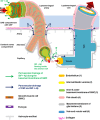
Schematic representation of the peri- and para-vascular spaces in the human cerebral cortex (not to scale). The subarachnoid space (SAS), filled with cerebrospinal fluid (CSF), is separated from the subpial space by the pia matter (Alcolado et al. 1988). A leptomeningeal sheath (the pial sheath shown in light pink) derived from the pia matter is also reflected on the walls of both arteries and veins that are crossing the SAS, thus separating the SAS from the cerebral cortex. Bundles of collagen are interposed in the subpial space and form the adventitia of leptomeningeal blood vessels, which is an expandable perivascular space. The pial sheath surrounding the leptomeningeal arteries in SAS continues to coat the arteries as they penetrate the cortex and it is closely applied to the outer basement membrane of smooth muscle cells (SMCs). Nonetheless, the veins from the cerebral cortex do not have a coating pial sheath. The middle layers of basement membrane (dark green), situated between layers of SMCs, represent the pathway for the perivascular drainage of solutes out of the brain. As emphasized, solutes do not drain along the inner or outer basement membrane layers of SMCs (light green). For the sake of simplicity, we illustrated only one layer of SMCs spiralling within the arterial wall. The perivascular drainage pathway (green arrows) follows a similar spiral trajectory which is spanned by a matrix of proteins. The basement membrane of the glia limitans (dark grey) is closely applied to the pia matter as the artery enters the brain. Thus another narrow perivascular space is created between the irregular surface of the pial sheath and the outer basement membrane of the SMCs and that of the glia limitans, respectively (Zhang et al. 1990). The narrow perivascular space has also been presented under the nomenclature of “paravascular space” and it has been proposed to be part of the glymphatic pathway (dark blue arrows). The cortical para-arterial pathway was referred to as the Virchow–Robin space (VRS). However, in the era of electron microscopy the exact boundaries of the VRS are not clearly defined (Color figure online)

References

    1. Abbott NJ (2004) Evidence for bulk flow of brain interstitial fluid: significance for physiology and pathology. Neurochem Int 45(4):545–552 - PubMed
    1. Abbott NJ (2013) Blood–brain barrier structure and function and the challenges for CNS drug delivery. J Inherit Metab Dis 36(3):437–449 - PubMed
    1. Alcolado R, Weller R, Parrish E, Garrod D (1988) The cranial arachnoid and pia mater in man: anatomical and ultrastructural observations. Neuropathol Appl Neurobiol 14(1):1–17 - PubMed
    1. Alperin N, Oliu CJ, Bagci AM, Lee SH, Kovanlikaya I, Adams D, Katzen H, Ivkovic M, Heier L, Relkin N (2014) Low-dose acetazolamide reverses periventricular white matter hyperintensities in iNPH. Neurology 82(15):1347–1351 - PMC - PubMed
    1. Andres K, Von Düring M, Muszynski K, Schmidt R (1987) Nerve fibres and their terminals of the dura mater encephali of the rat. Anat Embryol 175(3):289–301 - PubMed