. 2016 May;139(Pt 5):1394-416.
doi: 10.1093/brain/aww039. Epub 2016 Mar 17.

Neuregulin-1 controls an endogenous repair mechanism after spinal cord injury

Affiliations

Neuregulin-1 controls an endogenous repair mechanism after spinal cord injury

Katalin Bartus et al. Brain. 2016 May.

Abstract

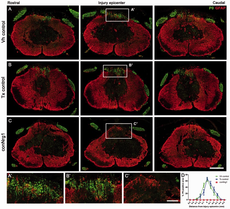
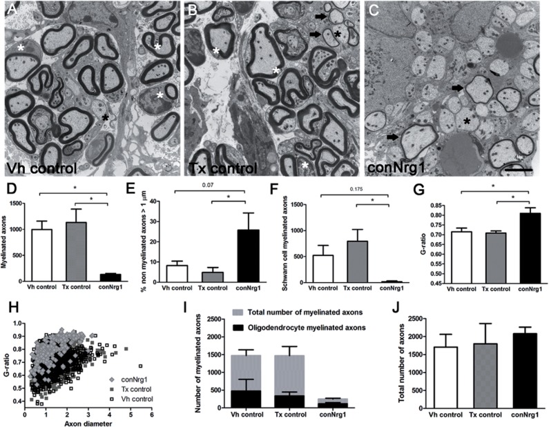
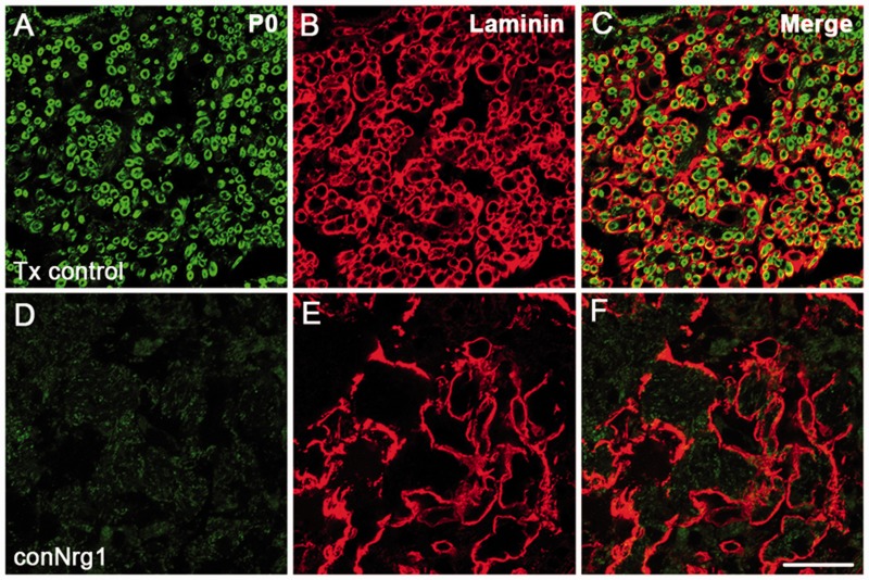
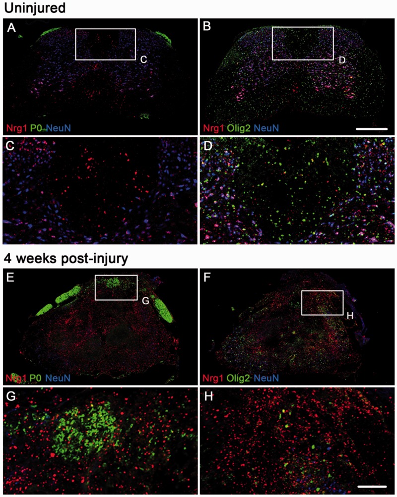
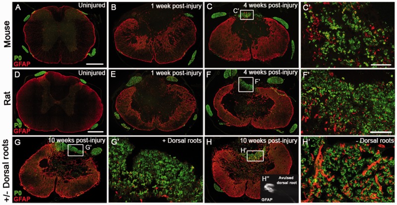
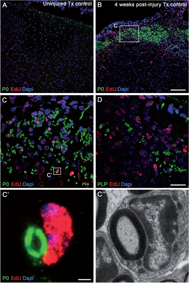
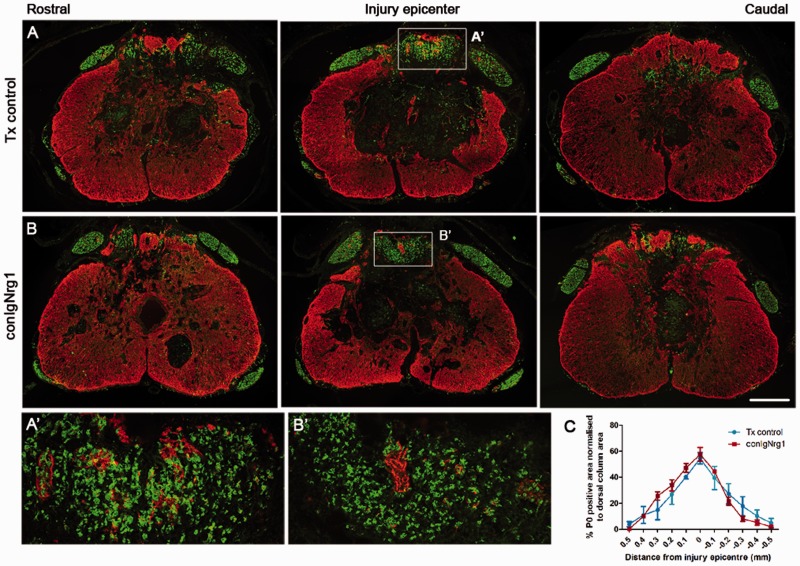
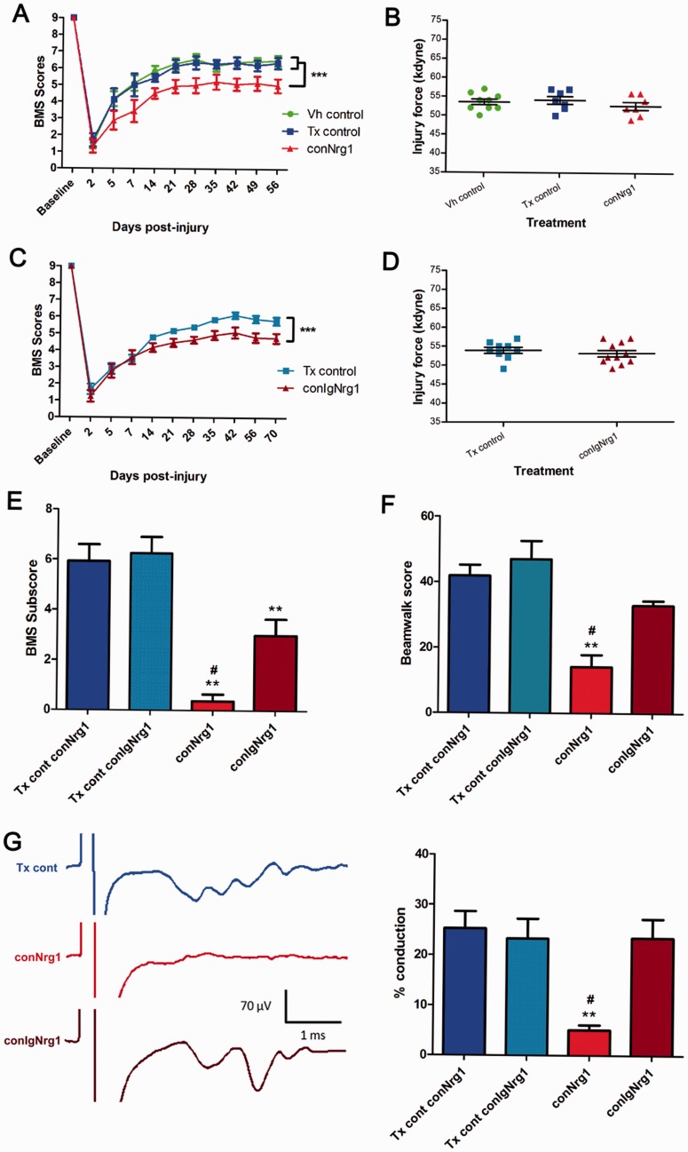
Following traumatic spinal cord injury, acute demyelination of spinal axons is followed by a period of spontaneous remyelination. However, this endogenous repair response is suboptimal and may account for the persistently compromised function of surviving axons. Spontaneous remyelination is largely mediated by Schwann cells, where demyelinated central axons, particularly in the dorsal columns, become associated with peripheral myelin. The molecular control, functional role and origin of these central remyelinating Schwann cells is currently unknown. The growth factor neuregulin-1 (Nrg1, encoded by NRG1) is a key signalling factor controlling myelination in the peripheral nervous system, via signalling through ErbB tyrosine kinase receptors. Here we examined whether Nrg1 is required for Schwann cell-mediated remyelination of central dorsal column axons and whether Nrg1 ablation influences the degree of spontaneous remyelination and functional recovery following spinal cord injury. In contused adult mice with conditional ablation of Nrg1, we found an absence of Schwann cells within the spinal cord and profound demyelination of dorsal column axons. There was no compensatory increase in oligodendrocyte remyelination. Removal of peripheral input to the spinal cord and proliferation studies demonstrated that the majority of remyelinating Schwann cells originated within the injured spinal cord. We also examined the role of specific Nrg1 isoforms, using mutant mice in which only the immunoglobulin-containing isoforms of Nrg1 (types I and II) were conditionally ablated, leaving the type III Nrg1 intact. We found that the immunoglobulin Nrg1 isoforms were dispensable for Schwann cell-mediated remyelination of central axons after spinal cord injury. When functional effects were examined, both global Nrg1 and immunoglobulin-specific Nrg1 mutants demonstrated reduced spontaneous locomotor recovery compared to injured controls, although global Nrg1 mutants were more impaired in tests requiring co-ordination, balance and proprioception. Furthermore, electrophysiological assessments revealed severely impaired axonal conduction in the dorsal columns of global Nrg1 mutants (where Schwann cell-mediated remyelination is prevented), but not immunoglobulin-specific mutants (where Schwann cell-mediated remyelination remains intact), providing robust evidence that the profound demyelinating phenotype observed in the dorsal columns of Nrg1 mutant mice is related to conduction failure. Our data provide novel mechanistic insight into endogenous regenerative processes after spinal cord injury, demonstrating that Nrg1 signalling regulates central axon remyelination and functional repair and drives the trans-differentiation of central precursor cells into peripheral nervous system-like Schwann cells that remyelinate spinal axons after injury. Manipulation of the Nrg1 system could therefore be exploited to enhance spontaneous repair after spinal cord injury and other central nervous system disorders with a demyelinating pathology.media-1vid110.1093/brain/aww039_video_abstractaww039_video_abstract.

Keywords: axon degeneration; demyelination; remyelination; spinal cord injury; transgenic model.


Figures

None
Spontaneous remyelination after spinal cord injury is mediated largely by Schwann cells of unknown origin. Bartus et al. show that neuregulin-1 promotes differentiation of spinal cord-resident precursor cells into PNS-like Schwann cells, which remyelinate central axons and promote functional recovery. Targeting the neuregulin-1 system could enhance endogenous regenerative processes.
Figure 1
Figure 1
Ablation of Nrg1 prevents remyelination of spinal axons by Schwann cells after spinal cord injury. (A–C) Co-staining of astrocytes (GFAP, red) and Schwann cell-associated myelin (P0, green) in serial sections of the spinal cord that span the rostrocaudal axis of the injury in vehicle control (Vh control, A), tamoxifen control (Tx control, B) and Nrg1-ablated (conNrg1, C) contused mouse spinal cords at 10 weeks post-injury. In all animals, the peripheral myelin protein P0 is apparent outside the spinal cord, in the peripheral dorsal and ventral roots, as expected. However, 10 weeks after contusion injury, Schwann cell-associated myelin (P0) is also observed in the spinal dorsal columns of control animals, being particularly abundant in the epicentre of the lesion (A and B). Strikingly, P0 is absent in the spinal dorsal columns of injured mice lacking Nrg1 (conNrg1; C). (A’C’) High magnification of boxed areas indicated in AC. (D) Quantification of P0-positive area in the dorsal columns assessed in sections that span the rostrocaudal axis of the injury site reveals undetectable levels of P0 in conNrg1 mice, compared to control groups. Data are presented as mean ± SEM. (**P < 0.007, two-way ANOVA, post hoc Bonferroni n = 4–5 animals/group). Scale bars = 250 µm (C); 50 µm (C’). Images not using the red/green colour scheme are available in the Supplementary material.
Figure 2
Figure 2
Ultrastructural analysis of Schwann cell-mediated remyelination of spinal axons after injury and its dependence on Nrg1 signalling. (AC) Electron micrographs of transverse sections of the dorsal column 10 weeks after spinal contusion injury in vehicle control (Vh control; A), tamoxifen control (Tx control; B) and conditional Nrg1 mutant (conNrg1; C) mice. In control animals, axons are undergoing remyelination and Schwann cells (white asterisk) can be seen to mediate remyelination; Schwann cells and their myelin are identified by the signet ring-like appearance of Schwann cell myelin, thicker and more compact myelin, and basal laminae around the Schwann cells. In conNrg1 mutant animals, many large diameter unmyelinated axons are visible (black asterisk), Schwann cells were rarely detected and the small degree of remyelination is mediated by oligodendrocytes that produce a thin myelin sheath (arrows). Remyelinating oligodendrocytes do not have nuclei directly apposed to the myelin or surrounding basal lamina and oligodendrocyte-associated myelin is less dense than the myelin associated with Schwann cells. (D) The number of myelinated axons in the dorsal column was significantly decreased in conNrg1 mice compared with control (Vh control = 998 ± 160, Tx control = 1132 ± 256 and conNrg1 = 134 ± 20). (E) The percentage of unmyelinated axons with a diameter >1 µm was increased in conNrg1 animals versus control (Vh control = 8% ± 2, Tx control = 5% ± 2 and conNrg1 = 26% ± 8). (F) A dramatic reduction in the number of Scwann cell-myelinated axons in conNrg1 mutant animals was observed (Vh control = 522 ± 192, Tx control = 794 ± 227 and conNrg1 = 17 ± 16). (G) A significant increase in the G-ratio in the conNrg1 animals (Vh control = 0.72 ± 0.01, Tx control = 0.71 ± 0.02 and conNrg1 = 0.81 ± 0.02) indicate very thin myelin sheaths. Data shown in DG are presented as mean ± SEM. (*P < 0.05, one-way ANOVA, post hoc Tukey’s n = 3–4 animals/group). No significant differences were observed between the two control groups in any of the measures analysed. Scale bar = 2 µm. (H) Scatter plot relating G-ratio and axon diameter of all the axons analysed show a shift to higher G-ratios in conNrg1 animals (P < 0.001 Kolmogorov-Smirnov test) without significant changes in axon calibre. (I) Comparison of counts of total myelinated axons and axons myelinated by oligodendrocytes in vehicle control, tamoxifen control and conNrg1 animals. After ablation of Nrg1 the total number of myelinated axons is decreased (data are presented as mean ± SEM, P < 0.05, one-way ANOVA, post hoc Tukey’s, n = 3–4 animals/group) but the number of axons myelinated by oligodendrocytes is not altered (Vh control = 475 ± 326, Tx control = 338 ± 184 and conNrg1 = 117 ± 60). (J) The total number of axons present in the dorsal column after spinal cord contusion remains similar in all groups (data are presented as mean ± SEM, one-way ANOVA, post hoc Tukey’s, n = 3–4 animals/group).
Figure 3
Figure 3
Nrg1-dependent central axon remyelination after spinal cord injury is mediated by typical Schwann cells. (AF) Co-staining of basal lamina (laminin, red) and Schwann cell-associated myelin (P0, green) in tamoxifen control (Tx control, AC) and Nrg1-ablated (conNrg1, DF) mouse spinal cords 10 weeks after contusion injury reveals defined ring-like structures immunoreactive for laminin. These ring-like structures represent the typical basal lamina associated with Schwann cells, and are apparent in close proximity to P0-positive myelin rings in injured control spinal cord. No Schwann cell-associated myelin and only sparse and diffuse laminin staining is observed in spinal cords from conNrg1 mice. Scale bar = 25 µm. Images not using the red/green colour scheme are available in the Supplementary material.
Figure 4
Figure 4
Expression of Nrg1 before and after spinal cord injury. Fluorescent in situ hybridization using a pan-Nrg1 probe (red) co-stained with markers for neurons (NeuN, blue), Schwann cell associated myelin (P0, green, A, C, E, G) and oligodendrocytes (Olig2, green, B, D, F and H). In the uninjured spinal cord (AD) Nrg1 labelling can be seen within neurons. The highest level of expression is seen in motor neurons within the ventral horn; however Nrg1 is also expressed by neurons of the dorsal horn. In the uninjured state there is no co-localization of Nrg with P0 (A and C) and very few oligodendrocytes express Nrg1 (B and D). Four weeks after spinal contusion injury, compact myelin, which is P0 immunoreactive, can be seen within the dorsal column and there is little co-localization with Nrg1 (E and G). Similarly, few Olig2 immunoreactive profiles express Nrg1 (F and H). Scale bars = 250 µm (B); 100 µm (H). Images not using the red/green colour scheme are available in the Supplementary material.
Figure 5
Figure 5
Schwann cell-mediated remyelination in mouse and rat spinal cords follows the same time course and is unhindered by avulsion of multiple dorsal roots at and adjacent to the injury site. (AC) Co-staining of astrocytes (GFAP, red) and Schwann cell-associated myelin (P0, green), shows the time course of the appearance of Schwann cell myelin the injured mouse spinal course. Peripheral myelin (P0) is not apparent in uninjured mouse spinal cord (A) or at 1 week post-injury (B), but is present in the dorsal columns by 4 weeks after injury (C). C’ shows a high magnification of the boxed area in panel C. (DF) Co-staining of astrocytes (GFAP, red) and Schwann cell-associated myelin (P0, green), shows the time course of the appearance of Schwann cell myelin the injured rat spinal course. Peripheral myelin (P0) is not apparent in uninjured rat spinal cord (D) or at 1 week post-injury (E), but is present by 4 weeks after injury (F). F’ shows high magnification of the boxed area in panel F. (G and H) At 10 weeks post-injury Schwann cell-mediated remyelination is apparent after spinal contusion injury irrespective of presence (G) or absence (H) of dorsal roots, one of the main possible peripheral sources of the Schwann cells. G’ and H’ show high magnifications of boxed areas in panels G and H. (H’’) Example of an avulsed dorsal root, which encompasses the entire rootlet including the dorsal root entry zone that is visualized by staining of the astroglial marker GFAP. Scale bars = 250 µm (A and D); 50 µm (C’ and F’). Images not using the red/green colour scheme are available in the Supplementary material.
Figure 6
Figure 6
Centrally remyelinating Schwann cells are produced de novo in the injured spinal cord. (A and B) Immunohistochemical staining for Schwann cell myelin (P0, green), nuclear EdU (red) and DAPI (blue) shows abundant P0-positive myelin rings in close apposition with EdU-positive cell nuclei 4 weeks after contusion injury (B) but not in control uninjured spinal cords (A). (C) High magnification image showing the boxed area in B, demonstrating remyelinating EdU-positive Schwann cells in association with P0-positive Schwann cell-derived myelin. (D) High magnification image of dorsal column axons associated with central PLP-positive myelin. (C’ and C’’) High magnification image showing the boxed area in C. Co-labelling clearly reveals direct apposition of a EdU-positive Schwann cell with a P0-positive myelin ring (C’) alongside an electron microscopic comparison of a Schwann cell in the dorsal columns that has remyelinated a CNS axon (C’’). Scale bars = 100 µm (B); 20 µm (D); 2 µm (C’). Images not using the red/green colour scheme are available in the Supplementary material.
Figure 7
Figure 7
Ablation of Ig-containing isoforms of Nrg1 (Nrg1 types 1 and 2) does not interfere with Schwann cell-mediated remyelination after spinal contusion injury. (A and B) Co-staining of astrocytes (GFAP, red) and Schwann cell-associated myelin (P0, green) shows appearance of Schwann cell myelin the injured mouse spinal cord of tamoxifen control (Tx control, A) and IgNrg1-ablated mice (conIgNrg1, B). Ten weeks after contusion injury, Schwann cell-associated myelin (P0) is abundant in the dorsal column of spinal cords from both control animals and mice lacking the IgNrg1 isoforms, being particularly abundant in the lesion epicentre. (A’ and B’) High magnification of boxed areas in A and B. (C) Quantification of P0-positive area in sections spanning the rostrocaudal axis of the injury site reveals similar levels in conIgNrg1 and control mice (nsP > 0.05, two-way ANOVA, post hoc Bonferroni, n = 4 animals/group). Scale bars = 250 µm (B); 50 µm (B’). Images not using the red/green colour scheme are available in the Supplementary material.
Figure 8
Figure 8
Ablation of Nrg1 leads to impaired spontaneous functional recovery after spinal contusion injury. Functional recovery assessed by BMS open field locomotion scores in conNrg1 (A) and conIgNrg1 (C) mice show a similar initial deficit in all groups acutely after spinal contusion injury. BMS scores gradually improve over the first few weeks and begin to plateau ∼3 weeks post-injury. Spontaneous functional recovery is significantly impaired in both conNrg1 (A) and conIgNrg1 (C) mutant mice, compared to vehicle and tamoxifen controls. Baseline BMS scores were not different between groups. Data are presented as mean ± SEM. (***P < 0.001, two-way ANOVA, post hoc Bonferroni, n = 9–11 animals/group). (B and D) Contusion impact data showing the actual force applied to individual mice was within 10% of the intended force of 50 kdyne and mean values for each group were not significantly different (P > 0.05; one-way ANOVA), confirming that any group differences were not due to differences in the impact force during surgery. (E) BMS subscores reveal significantly reduced functional recovery both in conNrg1 and conIgNrg1 animals compared to controls at 8 weeks post-injury. However, conNrg1 mice were significantly more impaired than conIgNrg1 mice in areas including stepping frequency, coordination, paw position, trunk stability, and tail position. (F) Beam-walking scores reveal reduced beam-walking performance both in conNrg1 and conIgNrg1 animals compared to controls at 8 weeks post-injury. However, conNrg1 mice were significantly more impaired than conIgNrg1 mice. Data are presented as mean ± SEM (**P < 0.01, #P < 0.05, one-way ANOVA, post hoc Tukey’s, n = 6–9 animals/group). *Significantly different to tamoxifen controls; #significantly different to conIgNrg1. (G) In vivo electrophysiological recordings assessing dorsal column function. Example traces (each averaged from 16 raw traces) show conduction through the lesion site in control, conNrg1 and conIgNrg1 animals (the representative control trace is taken from a conNrg1 tamoxifen control mouse). In control and conIgNrg1 injured animals stimulation artefacts, which have been cropped on the x-axis to allow appropriate scaling, were followed by evoked afferent activity at a latency of ∼1.5 ms, whereas little or no activity was evoked in conNrg1 animals. All stimulation was supramaximal. Quantification of the percentage of axons capable of conducting through the contusion site confirmed significant levels of dorsal column function in injured controls and conIgNrg1 mice at 10 weeks post-injury (when significant Schwann cell-mediated remyelination of the dorsal columns is apparent), which was dramatically reduced in conNrg1 animals (where Schwann cell-mediated remyelination is absent). Data are presented as mean ± SEM (**P < 0.01, #P < 0.05, one-way ANOVA, post hoc Tukey’s, n = 5–6 animals/group); **significantly different to tamoxifen controls; #significantly different to conIgNrg1.

Similar articles

Cited by

References

    1. Assinck PL, Duncan G, Plemel J, Lee M, Liu J, Bergles D, et al. PDGFRα-positive progenitor cells form myelinating oligodendrocytes and Schwann cells following contusion spinal cord injury. Society for Neuroscience Abstract 2015, Program No. 338.03. Chicago; 2015.
    1. Basso DM, Fisher LC, Anderson AJ, Jakeman LB, McTigue DM, Popovich PG. Basso Mouse Scale for locomotion detects differences in recovery after spinal cord injury in five common mouse strains. J Neurotrauma 2006; 23: 635–59. May; - PubMed
    1. Beattie MS, Bresnahan JC, Komon J, Tovar CA, Van Meter M, Anderson DK, et al. Endogenous repair after spinal cord contusion injuries in the rat. Exp Neurol 1997; 148: 453–63.; - PubMed
    1. Beattie MS, Harrington AW, Lee R, Kim JY, Boyce SL, Longo FM, et al. ProNGF induces p75-mediated death of oligodendrocytes following spinal cord injury. Neuron 2002; 36: 375–86. - PMC - PubMed
    1. Bermingham-McDonogh O, Xu YT, Marchionni MA, Scherer SS. Neuregulin expression in PNS neurons: isoforms and regulation by target interactions. Mol Cell Neurosci 1997; 10: 184–95. - PubMed

Publication types

MeSH terms