

Experimental Evaluation of Proposed Small-Molecule Inhibitors of Water Channel Aquaporin-1
- PMID: 26993802
- PMCID: PMC4885500
- DOI: 10.1124/mol.116.103929
Experimental Evaluation of Proposed Small-Molecule Inhibitors of Water Channel Aquaporin-1
Abstract
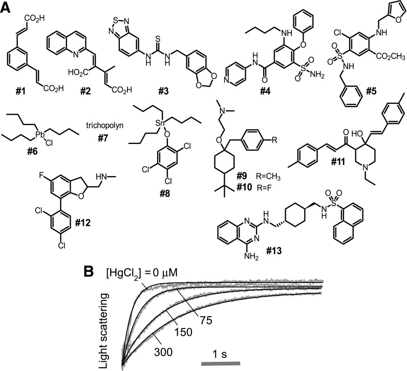
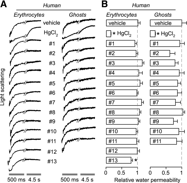
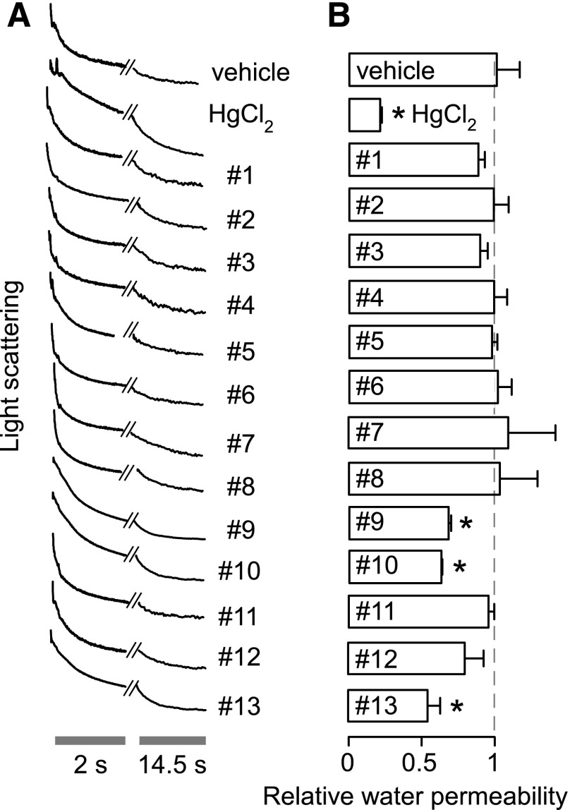
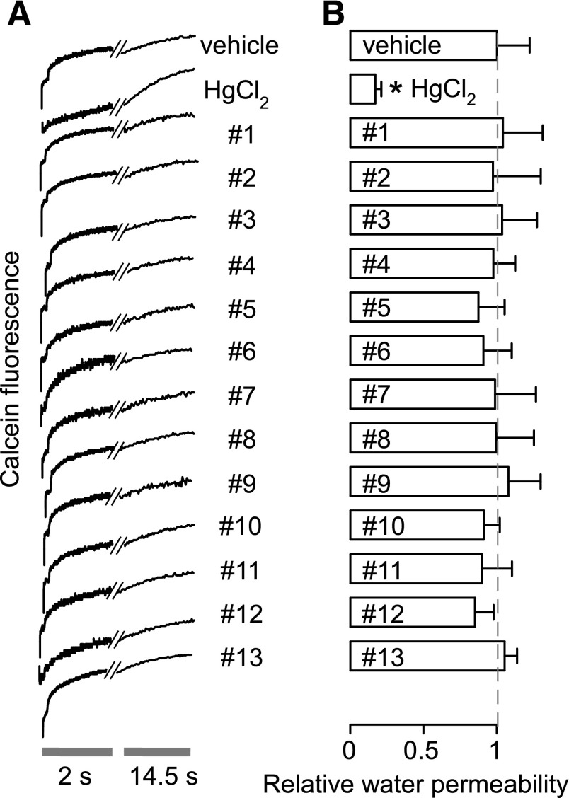
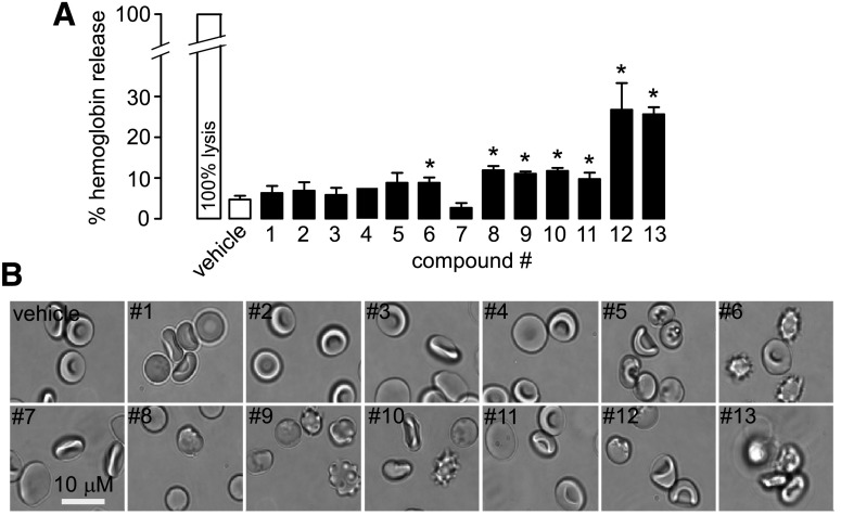
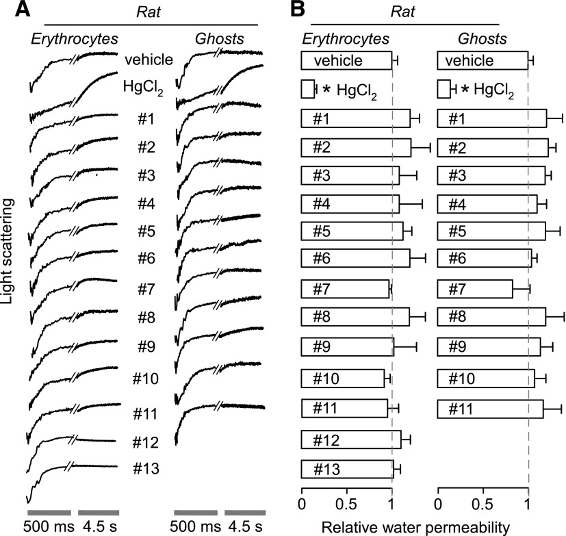
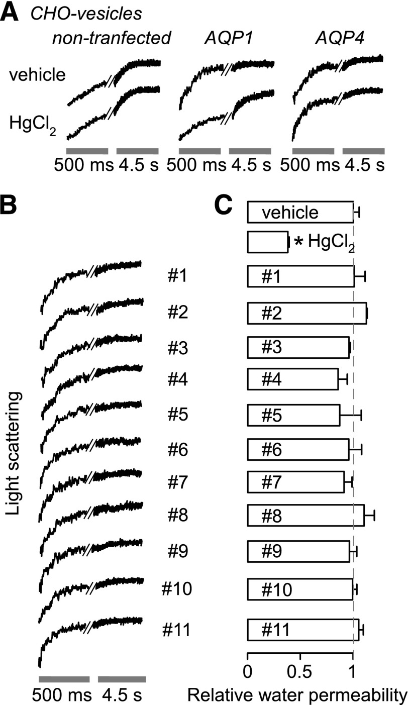
The aquaporin-1 (AQP1) water channel is a potentially important drug target, as AQP1 inhibition is predicted to have therapeutic action in edema, tumor growth, glaucoma, and other conditions. Here, we measured the AQP1 inhibition efficacy of 12 putative small-molecule AQP1 inhibitors reported in six recent studies, and one AQP1 activator. Osmotic water permeability was measured by stopped-flow light scattering in human and rat erythrocytes that natively express AQP1, in hemoglobin-free membrane vesicles from rat and human erythrocytes, and in plasma membrane vesicles isolated from AQP1-transfected Chinese hamster ovary cell cultures. As a positive control, 0.3 mM HgCl2 inhibited AQP1 water permeability by >95%. We found that none of the tested compounds at 50 µM significantly inhibited or increased AQP1 water permeability in these assays. Identification of AQP1 inhibitors remains an important priority.
Copyright © 2016 by The American Society for Pharmacology and Experimental Therapeutics.
Figures
Similar articles
-
Comparative efficacy of HgCl2 with candidate aquaporin-1 inhibitors DMSO, gold, TEA+ and acetazolamide.FEBS Lett. 2006 Dec 11;580(28-29):6679-84. doi: 10.1016/j.febslet.2006.11.025. Epub 2006 Nov 20. FEBS Lett. 2006. PMID: 17126329 Free PMC article.
-
Acetazolamide inhibits osmotic water permeability by interaction with aquaporin-1.Anal Biochem. 2006 Mar 15;350(2):165-70. doi: 10.1016/j.ab.2006.01.003. Epub 2006 Jan 24. Anal Biochem. 2006. PMID: 16480680
-
Automated cell-based assay for screening of aquaporin inhibitors.Anal Chem. 2009 Oct 1;81(19):8219-29. doi: 10.1021/ac901526k. Anal Chem. 2009. PMID: 19705854 Free PMC article.
-
The first discovered water channel protein, later called aquaporin 1: molecular characteristics, functions and medical implications.Mol Aspects Med. 2012 Oct-Dec;33(5-6):518-34. doi: 10.1016/j.mam.2012.06.001. Epub 2012 Jun 15. Mol Aspects Med. 2012. PMID: 22705445 Review.
-
Aquaporin-1 in the peritoneal membrane: Implications for water transport across capillaries and peritoneal dialysis.Biochim Biophys Acta. 2006 Aug;1758(8):1078-84. doi: 10.1016/j.bbamem.2006.02.025. Epub 2006 Mar 20. Biochim Biophys Acta. 2006. PMID: 16581016 Review.
Cited by
-
HAuCl4, Putative General Aquaporins Blocker, Reduces Platelet Spreading, Filopodia Formation, Procoagulant Response, and Thrombus Formation Under Flow.Front Physiol. 2020 Aug 21;11:1025. doi: 10.3389/fphys.2020.01025. eCollection 2020. Front Physiol. 2020. PMID: 32973556 Free PMC article.
-
Signaling Mechanisms and Pharmacological Modulators Governing Diverse Aquaporin Functions in Human Health and Disease.Int J Mol Sci. 2022 Jan 26;23(3):1388. doi: 10.3390/ijms23031388. Int J Mol Sci. 2022. PMID: 35163313 Free PMC article. Review.
-
Human Aquaporins: Functional Diversity and Potential Roles in Infectious and Non-infectious Diseases.Front Genet. 2021 Mar 16;12:654865. doi: 10.3389/fgene.2021.654865. eCollection 2021. Front Genet. 2021. PMID: 33796134 Free PMC article. Review.
-
Characterization of the Aquaporin-9 Inhibitor RG100204 In Vitro and in db/db Mice.Cells. 2022 Oct 4;11(19):3118. doi: 10.3390/cells11193118. Cells. 2022. PMID: 36231080 Free PMC article.
-
Identification and characterization of potent and selective aquaporin-3 and aquaporin-7 inhibitors.J Biol Chem. 2019 May 3;294(18):7377-7387. doi: 10.1074/jbc.RA118.006083. Epub 2019 Mar 11. J Biol Chem. 2019. PMID: 30862673 Free PMC article.
References
-
- Beitz E, Golldack A, Rothert M, von Bülow J. (2015) Challenges and achievements in the therapeutic modulation of aquaporin functionality. Pharmacol Ther 155:22–35. - PubMed
-
- Brooks HL, Regan JW, Yool AJ. (2000) Inhibition of aquaporin-1 water permeability by tetraethylammonium: involvement of the loop E pore region. Mol Pharmacol 57:1021–1026. - PubMed
-
- Carbrey JM, Agre P. (2009) Discovery of the aquaporins and development of the field. Handbook Exp Pharmacol 190:3–28. - PubMed
-
- Chang LW. (1990) The neurotoxicology and pathology of organomercury, organolead, and organotin. J Toxicol Sci 15 (Suppl 4):125–151. - PubMed
-
- de Groot BL, Engel A, Grubmüller H. (2001) A refined structure of human aquaporin-1. FEBS Lett 504:206–211. - PubMed
Publication types
MeSH terms
Substances
Grants and funding
LinkOut - more resources
Full Text Sources
Other Literature Sources
Molecular Biology Databases
Research Materials
