Review
. 2016 Jul;73(13):2467-90.
doi: 10.1007/s00018-016-2172-7. Epub 2016 Mar 18.

Cell migration in the developing rodent olfactory system

Affiliations
Review

Cell migration in the developing rodent olfactory system

Dhananjay Huilgol et al. Cell Mol Life Sci. 2016 Jul.

Abstract

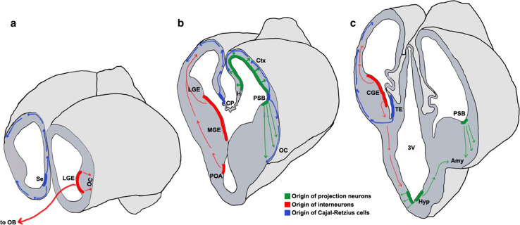
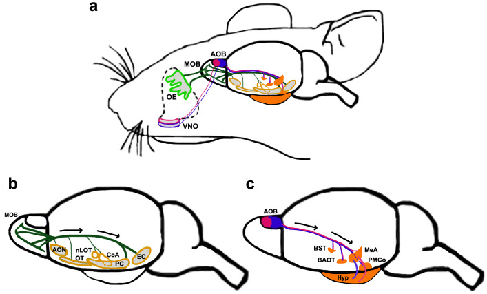
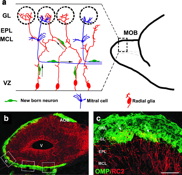
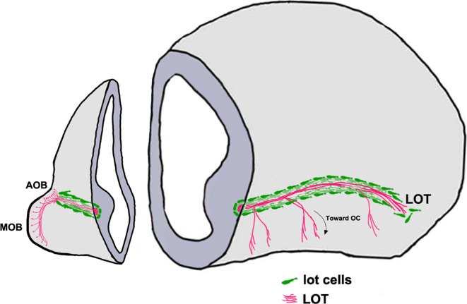
The components of the nervous system are assembled in development by the process of cell migration. Although the principles of cell migration are conserved throughout the brain, different subsystems may predominantly utilize specific migratory mechanisms, or may display unusual features during migration. Examining these subsystems offers not only the potential for insights into the development of the system, but may also help in understanding disorders arising from aberrant cell migration. The olfactory system is an ancient sensory circuit that is essential for the survival and reproduction of a species. The organization of this circuit displays many evolutionarily conserved features in vertebrates, including molecular mechanisms and complex migratory pathways. In this review, we describe the elaborate migrations that populate each component of the olfactory system in rodents and compare them with those described in the well-studied neocortex. Understanding how the components of the olfactory system are assembled will not only shed light on the etiology of olfactory and sexual disorders, but will also offer insights into how conserved migratory mechanisms may have shaped the evolution of the brain.

Keywords: Domains; Evolution; Migration; Neocortex; Olfactory; Vomeronasal.


Figures

Fig. 1
Fig. 1
Neuronal migrations in the embryonic forebrain [, , , –, , , , –305]. Schematics representing the mouse brain at embryonic day (E)12.5 sectioned in the coronal plane at the rostral (a), mid (b), and caudal (c) levels. Domains of origin and migration routes for projection neurons (green), interneurons (red), and Cajal–Retzius cells (blue) are illustrated. Colored bands represent the ventricular zone and progenitors residing therein; arrows represent the route and direction of migration. 3V third ventricle, Amy amygdala, CGE caudal ganglionic eminence, CP choroid plexus, Ctx cortex, H hippocampus, Hyp hypothalamus, LGE lateral ganglionic eminence, MGE medial ganglionic eminence, OB olfactory bulb, OC olfactory cortex, POA pre-optic area, PSB pallial–subpallial boundary, Se septum, TE thalamic eminence
Fig. 2
Fig. 2
The main and accessory olfactory system [, –, , –192, 194, 205]. a Schematic depicting both systems of the mouse olfactory circuit. OSNs located in the OE (light green) project to the MOB, whereas VSNs in the VNO project to the AOB. Apical (pink outline) and basal (blue outline) VNO neurons project to the aAOB (solid pink) and pAOB (solid blue), respectively. b The main olfactory system. MOB M/T cells send their axons along the LOT in the direction of the black arrows to multiple targets (yellow outlined structures). These include different components of the olfactory cortex: the AON, OT, PC, EC, and the olfactory amygdaloid nuclei, CoA and nLOT. c The accessory olfactory system. Both the aAOB (solid pink) and the pAOB (solid blue) projection neurons send axons along the LOT (in the direction of the black arrows) to different parts of the vomeronasal amygdala, including the MeA, PMCo, and components of the extended amygdala, BST, BAOT (solid orange structures). AOB accessory olfactory bulb, aAOB anterior AOB, pAOB posterior AOB, AON anterior olfactory nucleus, BAOT bed nucleus of accessory olfactory tract, BST bed nucleus of stria terminalis, CoA cortical amygdaloid nucleus, EC entorhinal cortex, Hyp hypothalamus, LOT lateral olfactory tract, MeA medial amygdaloid nucleus, MOB main olfactory bulb, nLOT nucleus of the lateral olfactory tract, OE olfactory epithelium, OT olfactory tubercle, PC piriform cortex, PMCo posteromedial cortical nucleus, VNO vomeronasal organ
Fig. 3
Fig. 3
Migration of new neurons within the MOB [51, 52, 58]. a Schematic showing a sagittal section of an embryonic day (E)18.5 MOB. The radial glia (red) are convoluted and intertwined, with their endfeet merging in the glomeruli or in the EPL. The cell bodies of M/T cells (blue) are in the MCL, and their axons extend parallel to the ventricular zone. Newborn neurons (green) migrate radially to their destined laminar positions using the radial glia, or tangentially using M/T cells axons as scaffolds. b Confocal image of an E18.5 mouse MOB sagittal section immunostained for a radial glial marker, RC2 (red), and an OSN marker, the olfactory marker protein (OMP, green). c Magnified view of boxed area in b showing radial glial endfeet (arrows) penetrating glomeruli formed by OSN axons. Scale bar in c is 400 μm. Additional boxes in b are from the original artwork in [58]. AOB accessory olfactory bulb, EPL external plexiform layer, GL glomerular layer, MCL mitral cell layer, MOB main olfactory bulb, ONL olfactory nerve layer, OSN olfactory sensory neuron, V ventricle, VZ ventricular zone. The images in b, c are from Fig. 1 of [58], copyright 2001 Wiley-Liss, Inc. Reprinted with permission
Fig. 4
Fig. 4
“Lot cell” array and formation of the LOT [152, 153]. Schematic representing one hemisphere of an embryonic day (E)14.5 mouse brain. The projection neurons of the MOB and the AOB extend their axons along the LOT and innervate different olfactory cortical and vomeronasal structures. The “lot cells” (green) form a “permissive corridor” along the lateral face of the telencephalon through which the LOT axons (pink) grow. AOB accessory olfactory bulb, LOT lateral olfactory tract, MOB main olfactory bulb
Fig. 5
Fig. 5
Cell migrations to the olfactory cortex [26, 29, 30, 34, 35, 37, 172]. Schematics representing an embryonic day (E)12.5 brain sectioned at rostral (a) and mid (b) levels in the coronal plane to reveal neuroepithelial domains and cell migrations (arrows) that populate different olfactory cortical structures. CoA cortical amygdaloid nucleus, LP lateral pallium, dLGE dorsal lateral ganglionic eminence, vLGE ventral lateral ganglionic eminence, MGE medial ganglionic eminence, OT olfactory tubercle, PC piriform cortex, RMTW rostromedial telencephalic wall, VP ventral pallium
Fig. 6
Fig. 6
The caudal amygdaloid stream and migration to the nLOT2/3 [180]. a A sagittal section of an E17.5 mouse brain shows the caudal amygdaloid stream (CAS; yellow arrow) arising in the caudal telencephalic VZ and terminating in the globular nLOT2/3 (yellow circle, black arrow). Dashed arrow depicts the migration route from the VP to other amygdaloid nuclei. b Both the CAS (arrowhead) and the nLOT2/3 (arrow) are identified by NeuroD expression, c NeuroD expression in a lateral-to-medial series of sagittal sections at E15.5, d in utero electroporation of an EGFP-expressing construct in the caudal telencephalic neuroepithelium at E11.5, and examination of the brain at E15.5, reveals GFP-positive cells migrating along the CAS (white arrow). Note the residual GFP-positive neuroepithelium at the site of electroporation indicating the origin of the nLOT2/3 cells (white arrowheads). Scale bars are 200 μm. nLOT nucleus of lateral olfactory tract, Hi hippocampus, Ncx neocortex. All images in this figure are from [180], copyright 2007 Nature Publishing Group. Reprinted with permission
Fig. 7
Fig. 7
Neuroepithelial domains contributing to components of the vomeronasal system [, , , , –214, 225, 226]. ac Schematics of sections of the forebrain at three rostrocaudal levels in the coronal plane showing the different neuroepithelial domains that contribute to the hypothalamic nuclei of the VNS. Newborn neurons migrate radially from the designated neuroepithelial ventricular zones (vMPN, vAH, vVMH, vPM) to populate the MPN, AH, VMH, and PM, respectively. a The AEP and POA provide neurons to the BST, MeA, and PMCo. The interganglionic sulcus, between the LGE and MGE, generates interneurons destined for the AH/POA. b The vPVH produces neurons for the vomeronasal amygdala (MeA, PMCo and BST). c the TE (asterisks, b, c) generates M/T neurons destined for the pAOB, which migrate to the telencephalic surface at caudal levels (pink arrows). These neurons undertake a tangential migration along the telencephalic surface (pink arrows, a, b) to the rostrally located pAOB which is not seen in the schematic. The pAOB, MeA, PMCo, BST, and the hypothalamic nuclei are all generated from the VZ of the third ventricle and are all part of the VNS. 3V third ventricle, AEP anterior entopeduncular area, BST bed nucleus of stria terminalis, MeA medial amygdaloid nucleus, pAOB accessory olfactory bulb, posterior division, PMCo posteromedial cortical nucleus, POA preoptic area, TE thalamic eminence, vAH ventricular zone for anterior hypothalamic nucleus, vMPN ventricular zone for medial preoptic nucleus, vPM ventricular zone for pre-mammillary nucleus, vPVH ventricular zone for paraventricular hypothalamic nucleus, vVMH ventricular zone for ventromedial hypothalamic nucleus

References

    1. Haas CA, et al. Role for reelin in the development of granule cell dispersion in temporal lobe epilepsy. J Neurosci. 2002;22(14):5797–5802. - PMC - PubMed
    1. Bozzi Y, Casarosa S, Caleo M. Epilepsy as a neurodevelopmental disorder. Front Psychiatry. 2012;3:19. doi: 10.3389/fpsyt.2012.00019. - DOI - PMC - PubMed
    1. Jamadar S, et al. Genetic influences of cortical gray matter in language-related regions in healthy controls and schizophrenia. Schizophr Res. 2011;129(2–3):141–148. doi: 10.1016/j.schres.2011.03.027. - DOI - PMC - PubMed
    1. Lee FH, et al. Abnormal interneuron development in disrupted-in-schizophrenia-1 L100P mutant mice. Mol Brain. 2013;6:20. doi: 10.1186/1756-6606-6-20. - DOI - PMC - PubMed
    1. Penagarikano O, et al. Absence of CNTNAP2 leads to epilepsy, neuronal migration abnormalities, and core autism-related deficits. Cell. 2011;147(1):235–246. doi: 10.1016/j.cell.2011.08.040. - DOI - PMC - PubMed

Publication types

LinkOut - more resources