. 2016 May 1;196(9):3653-64.
doi: 10.4049/jimmunol.1501176. Epub 2016 Mar 18.

Transient Surface CCR5 Expression by Naive CD8+ T Cells within Inflamed Lymph Nodes Is Dependent on High Endothelial Venule Interaction and Augments Th Cell-Dependent Memory Response

Affiliations

Transient Surface CCR5 Expression by Naive CD8+ T Cells within Inflamed Lymph Nodes Is Dependent on High Endothelial Venule Interaction and Augments Th Cell-Dependent Memory Response

David Askew et al. J Immunol. .

Abstract

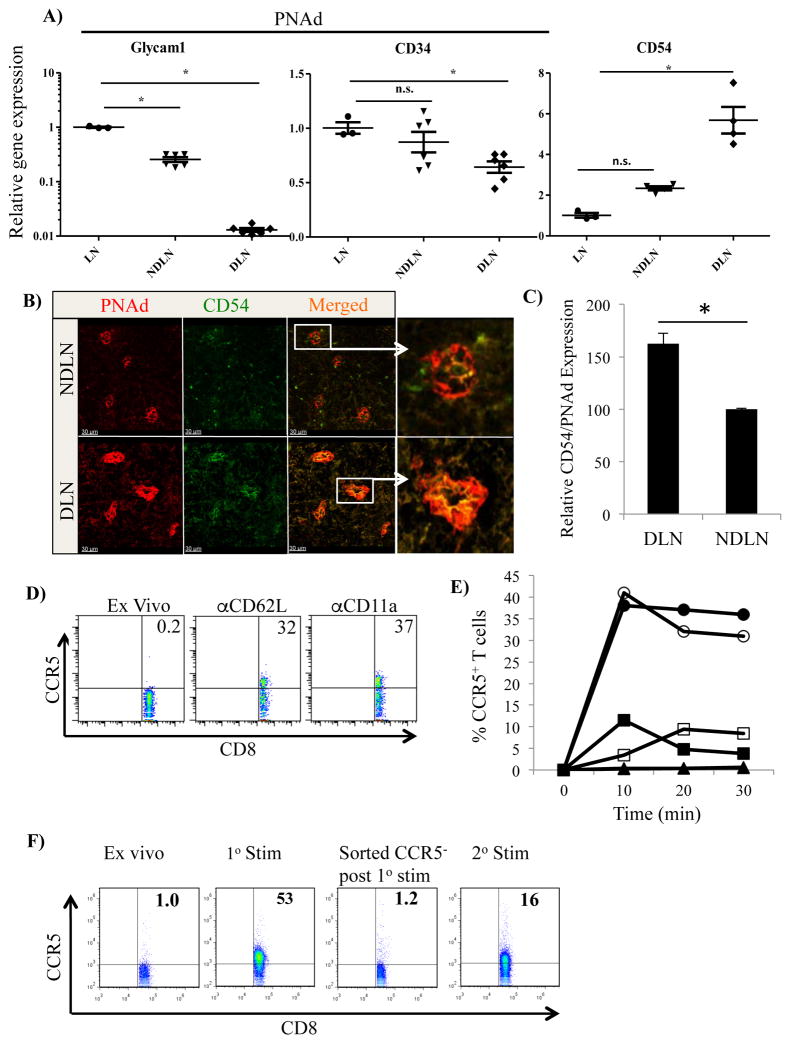
In inflamed lymph nodes, Ag-specific CD4(+) and CD8(+) T cells encounter Ag-bearing dendritic cells and, together, this complex enhances the release of CCL3 and CCL4, which facilitate additional interaction with naive CD8(+) T cells. Although blocking CCL3 and CCL4 has no effect on primary CD8(+) T cell responses, it dramatically impairs the development of memory CD8(+) T cells upon Ag rechallenge. Despite the absence of detectable surface CCR5 expression on circulating native CD8(+) T cells, these data imply that naive CD8(+) T cells are capable of expressing surface CCR5 prior to cognate Ag-induced TCR signaling in inflamed lymph nodes; however, the molecular mechanisms have not been characterized to date. In this study, we show that CCR5, the receptor for CCL3 and CCL4, can be transiently upregulated on a subset of naive CD8(+) T cells and that this upregulation is dependent on direct contact with the high endothelial venule in inflamed lymph node. Binding of CD62L and CD11a on T cells to their ligands CD34 and CD54 on the high endothelial venule can be enhanced during inflammation. This enhanced binding and subsequent signaling promote the translocation of CCR5 molecules from intracellular vesicles to the surface of the CD8(+) T cell. The upregulation of CCR5 on the surface of the CD8(+) T cells increases the number of contacts with Ag-bearing dendritic cells, which ultimately results in increased CD8(+) T cell response to Ag rechallenge.


Figures

Figure 1
Figure 1. Naïve CD8+ T cells transiently express CCR5 early upon entry into an inflamed LN and requires direct contact
Congenic (CD45.1+) mice were immunized with Alum/CpG (20 μg) on the right footpad and Alum/vehicle on the left footpad 48 hours before adoptive i.v. transfer of 107 naïve CD8+ T cells (CD45.2+). Four hours later, draining LNs were harvested and stained with antibodies to CD45.2, CD8, and CCR5. A) Shown are flow cytometry analyses of CCR5 expression on CD45.2+CD8+ T cells. B) Compilation of 10 transfer experiments described in A. C) As before, CD45.1 mice were immunized with CpG/Alum or PBS/Alum 48 prior to transfer of 107 naïve CD45.2+ CD8+ T cells. At 2, 4, and 6 h post-injection LN were removed and CD45.2+ cells were stained for CD8 and CCR5 expression. D) Congenic (CD45.1+) mice were immunized with LPS/Alum (10 μg) in the footpad 36 hours before harvesting. The inflamed draining LNs (DLN) as well as non-inflamed LNs distant from immunization sites (NDLN) were placed in separate wells of a 96-well plate. Freshly isolated, CD45.2+ naïve T cells were placed either directly on top of the LNs (DC = “Direct contact”) or bathed in LN-containing medium via a transwell with 0.8um-pore membrane (TW = “Transwell”). Only T cells in direct contact with inflamed LN express enhanced surface CCR5. E) C57BL/6, MHC-II KO, or β2M KO mice were immunized with Alum/CpG (20 μg) on the right footpad and Alum/vehicle on the left footpad 48 hours before adoptive i.v. transfer of 107 naïve CD8+ T cells (CD45.1+). Four hours later, draining LNs were harvested and stained with antibodies to CD45.1, CD8, and CCR5. F and G) CD45.1+ mice were injected in right dorsal footpad with CpG/Alum 48 h prior to injection of 0.5, 5.0, or 20 x 106 CD45.2 T cells. Four hours later DLN and NDLN were collected and stained for expression of CD45.2, CD8, and CCR5 and examined by flow cytometry, and both percentage and number of CCR5+ cells were determined for each LN. Data presented is representative of three separate experiments.
Figure 2
Figure 2. Expression of HEV-associated molecules important for T cell attachment are influenced during inflammation and can promote CCR5 expression in OT-I T cells
Both draining and non-draining popliteal LN were removed from mice 48 h after injection of CpG/Alum into right footpad. RNA was isolated from tissue and converted into cDNA. The relative expressions of selected genes were compared to GAPDH levels in normal LN as control. Gene expression in LN was further normalized using relative expression of CD31 as a marker of endothelial cells to account for infiltration of immune cells. Results are from 4–6 separate samples run in triplicate. B) Mice received injection of CpG into right footpad 48 h prior to LNs being removed, fixed, frozen, and sectioned onto microscope slide. Sections were then stained for expression of CD54 and PNAd by immunofluorescence. LN were collected from 6 mice with 3–5 areas scanned per LN. Average intensities for both PNAd and CD54 were used to determine relative expression of CD54 compared to PNAd as described in materials and methods. C) Relative CD54/PNAd expression was compared for 5 ROIs for DLN and NDLN. D) Naïve OT-I CD8+ T cells were incubated with anti-CD11a, or anti-CD62L coated beads for 30 min at 4°C, transferred to 37°C for 10 minutes, placed on ice prior to staining for CD8 and CCR5 expression, and then examined by flow cytometry. Experiment was repeated 5 times with representative experiment being shown. E) Percentage of CCR5+ expressed in OT-I T cells that were incubated with different doses (△▲=10 μl, □■ = 30 μl, ○● = 50 μl) of either anti-CD11a (open symbols) or anti-CD62L-coated (filled symbols) beads during 10 minute increments. Experiment was repeated three times with representative experiment shown. F) OT-I T cells were cultured with 50 μl anti-CD11a beads for 20 min (1° stim). Bead-bound cells were isolated and then flushed off beads. CCR5+ cells were removed and remaining CCR5 cells were cultured with 50 μl anti-CD11a beads for 20 min (2° stim). Samples were then stained for CD8 and CCR5 expression.
Figure 3
Figure 3. Expression of CCR5 is due to transport from pre-formed vesicles
A) CD45.1+ mice were injected with CpG/Alum in the right dorsal footpad and 48 h later, CD45.2+ OT-I T cells were adoptively transferred. Three h later, T cells were recovered from treated CD45.1 mice DLN and NDLN. Recovered T cells and naïve OT-I T cells (ex vivo) were allowed to attach to poly-lysine coated cover slips, fixed, permeabilized and stained for expression of CD8 (green), CCR5 (red). After being selected for CD45.2+ expression, cells were examined by confocal microscopy. Cells from 6 mice were placed on poly-lysine coated plate and 3–5 regions/slide were examined. B) Percentage of CCR5 that was co-localized with CD8 was determined using Imaris software. C) To determine expression of intracellular CCR5 by flow cytometry, OT-I T cells were fixed, permeabilized, and stained with antibodies against CD8 and CCR5. D) To examine effects of inhibitors of protein synthesis or transport on CCR5 expression, OT-I T cells were incubated in the presence of specific inhibitors for 2 h at 37°C. Cells were then transferred to 4°C and anti-CD11a coated beads were added for 30 min with mixing. Cells were transferred to 37°C for 15 min and then placed on ice. Cells were then stained for CD8 and CCR5 expression and then examined by flow cytometry. Experiment was repeated three times with representative experiment being shown. E) Relative expression of CCR5 was determined by comparing the number of CCR5+ cells with OT-I stimulated with anti-CD11a beads in the presence of inhibitors. Results are the average of three separate experiments.
Figure 4
Figure 4. CCR5 expression enhances DC-T cell interaction and promotes development of long-term memory
A) To determine the effect of CCR5 expression on development of memory CD8+ T cells, naïve OT-1 (CD45.2+) T cells were exposed to CpG-inflamed CD45.1+ LNs in vitro for 4 hours. 5x10⁴ each of CCR5+ (“CCR5+ sort”) and CCR5 (“CCR5 sort”) fractions of OT-I T cells were separately introduced into independent CD45.1+ naïve mice via tail-vein injection, along with 1x10⁶ OT-II CD4+ T cells. As controls, similar number of OT-I T cells on a CCR5−/− background (“CCR5ko”), as well as unfractionated naïve OT-I T cells (“Bulk”) were introduced into separate recipient animals. One day after transfer, the recipient animals were immunized with CpG + SIINFEKL + OVA323–339 in alum. At 7 days post immunization, LN and spleen were examined for B) the number of OT-I T cells and C) the number of IFNγ-producing OT-I T cells. Twenty-eight days after immunization, LN/Spleen cells were enumerated for D) the number of CD45.2+ CD8+ OT-I T cells in the recipient mice, and E) the number of intracellular IFNγ–producing OTI T cells. F) OVA-pulsed, Cell-Tracker Blue-labeled DCs were injected via footpad 24 h prior to the injection of 1 x 10⁷ CFSE-labeled CCR5+OT-I T cells and 1x 10⁷ SNARF-labeled CCR5OT-I T cells. LNs were isolated 5 h later, fixed, sliced into 5 μm sections and examined using 2-photon microscopy. G) The numbers of contacts between CCR5+ T cells or CCR5 OT-I T cells with Ag-pulsed DCs were then determined for each slice.
Figure 5
Figure 5. Induction of surface CCR5 expression in T cells that enter inflamed LN
Model showing naïve OT-I T cells crossing HEV under homeostatic and inflamed conditions. A) Under homeostatic conditions T cells encounter HEV that are releasing Glycam-1 which bind CD62L on the surface of T cell and reduce signaling mediated by surface bound CD34. T cells can then bind CCL21, which in combination with CD62L signaling promotes conformational changes in CD11a/CD18. The T cells can now bind CD54 and migrate through the HEV into the LN. B) Under inflammatory conditions there is limited soluble Glycam-1, which will enhance CD62L binding to HEV-associated CD34, as increased expression of HEV associated CCL21 will enhance binding through CCR7 and provide enhanced signaling to activate CD11a/CD18. Increased expression of CD54 on HEV provides both increased attachment to HEV and increased signaling through CD11a/CD18. This increased signaling through both CD62L and CD11a promote the expression of CCR5 in a subset of CD8+ T cells that cross HEV and enter the inflamed LN.

References

    1. Blattman JN, Antia R, Sourdive DJ, Wang X, Kaech SM, Murali-Krishna K, Altman JD, Ahmed R. Estimating the precursor frequency of naive antigen-specific CD8 T cells. The Journal of experimental medicine. 2002;195:657–664. - PMC - PubMed
    1. Castellino F, Germain RN. Cooperation between CD4+ and CD8+ T cells: when, where, and how. Annual review of immunology. 2006;24:519–540. - PubMed
    1. Castellino F, Huang AY, Altan-Bonnet G, Stoll S, Scheinecker C, Germain RN. Chemokines enhance immunity by guiding naive CD8+ T cells to sites of CD4+ T cell-dendritic cell interaction. Nature. 2006;440:890–895. - PubMed
    1. Hugues S, Scholer A, Boissonnas A, Nussbaum A, Combadiere C, Amigorena S, Fetler L. Dynamic imaging of chemokine-dependent CD8+ T cell help for CD8+ T cell responses. Nature immunology. 2007;8:921–930. - PubMed
    1. von Andrian UH, Mempel TR. Homing and cellular traffic in lymph nodes. Nat Rev Immunol. 2003;3:867–878. - PubMed

MeSH terms