

Impact of placental insufficiency on fetal skeletal muscle growth
- PMID: 26994511
- PMCID: PMC5014698
- DOI: 10.1016/j.mce.2016.03.017
Impact of placental insufficiency on fetal skeletal muscle growth
Abstract
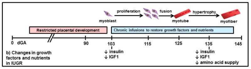
Intrauterine growth restriction (IUGR) caused by placental insufficiency is one of the most common and complex problems in perinatology, with no known cure. In pregnancies affected by placental insufficiency, a poorly functioning placenta restricts nutrient supply to the fetus and prevents normal fetal growth. Among other significant deficits in organ development, the IUGR fetus characteristically has less lean body and skeletal muscle mass than their appropriately-grown counterparts. Reduced skeletal muscle growth is not fully compensated after birth, as individuals who were born small for gestational age (SGA) from IUGR have persistent reductions in muscle mass and strength into adulthood. The consequences of restricted muscle growth and accelerated postnatal "catch-up" growth in the form of adiposity may contribute to the increased later life risk for visceral adiposity, peripheral insulin resistance, diabetes, and cardiovascular disease in individuals who were formerly IUGR. This review will discuss how an insufficient placenta results in impaired fetal skeletal muscle growth and how lifelong reductions in muscle mass might contribute to increased metabolic disease risk in this vulnerable population.
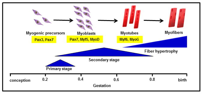
Keywords: Amino acids; Developmental programming; Muscle protein synthesis; Myoblast; Myofiber; Myogenesis.
Copyright © 2016 Elsevier Ireland Ltd. All rights reserved.
Conflict of interest statement
Declaration of interest: The authors have no conflicts of interest to disclose.
Figures

Similar articles
-
Skeletal muscle protein accretion rates and hindlimb growth are reduced in late gestation intrauterine growth-restricted fetal sheep.J Physiol. 2018 Jan 1;596(1):67-82. doi: 10.1113/JP275230. Epub 2017 Oct 26. J Physiol. 2018. PMID: 28940557 Free PMC article.
-
Rates of myogenesis and myofiber numbers are reduced in late gestation IUGR fetal sheep.J Endocrinol. 2019 Dec 19;244(2):339-352. doi: 10.1530/JOE-19-0273. J Endocrinol. 2019. PMID: 31751294 Free PMC article.
-
Postnatal development of skeletal muscle in pigs with intrauterine growth restriction: morphofunctional phenotype and molecular mechanisms.J Anat. 2020 May;236(5):840-853. doi: 10.1111/joa.13152. Epub 2020 Jan 29. J Anat. 2020. PMID: 31997379 Free PMC article.
-
Developmental programming in response to intrauterine growth restriction impairs myoblast function and skeletal muscle metabolism.J Pregnancy. 2012;2012:631038. doi: 10.1155/2012/631038. Epub 2012 Jul 31. J Pregnancy. 2012. PMID: 22900186 Free PMC article. Review.
-
Skeletal Muscle Damage in Intrauterine Growth Restriction.Adv Exp Med Biol. 2018;1088:93-106. doi: 10.1007/978-981-13-1435-3_5. Adv Exp Med Biol. 2018. PMID: 30390249 Review.
Cited by
-
Reduced Na+ K+ -ATPase activity may reduce amino acid uptake in intrauterine growth restricted fetal sheep muscle despite unchanged ex vivo amino acid transporter activity.J Physiol. 2020 Apr;598(8):1625-1639. doi: 10.1113/JP278933. Epub 2020 Feb 3. J Physiol. 2020. PMID: 31909825 Free PMC article.
-
Human Placenta Buffers the Fetus from Adverse Effects of Perceived Maternal Stress.Cells. 2021 Feb 12;10(2):379. doi: 10.3390/cells10020379. Cells. 2021. PMID: 33673157 Free PMC article.
-
KLB dysregulation mediates disrupted muscle development in intrauterine growth restriction.J Physiol. 2022 Apr;600(7):1771-1790. doi: 10.1113/JP281647. Epub 2022 Feb 17. J Physiol. 2022. PMID: 35081669 Free PMC article.
-
Why is human uterine artery blood flow during pregnancy so high?Am J Physiol Regul Integr Comp Physiol. 2022 Nov 1;323(5):R694-R699. doi: 10.1152/ajpregu.00167.2022. Epub 2022 Sep 12. Am J Physiol Regul Integr Comp Physiol. 2022. PMID: 36094446 Free PMC article.
-
Changes in microvascular perfusion of heart and skeletal muscle in sheep around the time of birth.Exp Physiol. 2023 Jan;108(1):135-145. doi: 10.1113/EP090809. Epub 2022 Nov 24. Exp Physiol. 2023. PMID: 36420621 Free PMC article.
References
-
- ACOG. ACOG Practice bulletin no. 134: fetal growth restriction. Obstet Gynecol. 2013;121:1122–33. - PubMed
-
- Abuzzahab MJ, Schneider A, Goddard A, Grigorescu F, Lautier C, Keller E, Kiess W, Klammt J, Kratzsch J, Osgood D, Pfaffle R, Raile K, Seidel B, Smith RJ, Chernausek SD, Intrauterine Growth Retardation Study, G. IGF-I receptor mutations resulting in intrauterine and postnatal growth retardation. N Engl J Med. 2003;349:2211–22. - PubMed
-
- Amato MC, Guarnotta V, Giordano C. Body composition assessment for the definition of cardiometabolic risk. J Endocrinol Invest. 2013;36:537–43. - PubMed
-
- Anderson MS, Thamotharan M, Kao D, Devaskar SU, Qiao L, Friedman JE, Hay WW., Jr Effects of acute hyperinsulinemia on insulin signal transduction and glucose transporters in ovine fetal skeletal muscle. Am J Physiol Regul Integr Comp Physiol. 2005;288:R473–81. - PubMed
-
- Anthony RV, Scheaffer AN, Wright CD, Regnault TR. Ruminant models of prenatal growth restriction, Reprod Suppl. 2003;61:183–94. - PubMed
Publication types
MeSH terms
Grants and funding
LinkOut - more resources
Full Text Sources
Other Literature Sources
