. 2016 Mar 16;8(1):1-28.
doi: 10.1002/9780470559277.ch150174.

RNA Imaging with Dimeric Broccoli in Live Bacterial and Mammalian Cells

Affiliations

RNA Imaging with Dimeric Broccoli in Live Bacterial and Mammalian Cells

Grigory S Filonov et al. Curr Protoc Chem Biol. .

Abstract

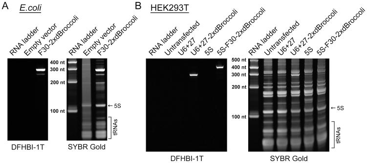
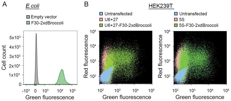
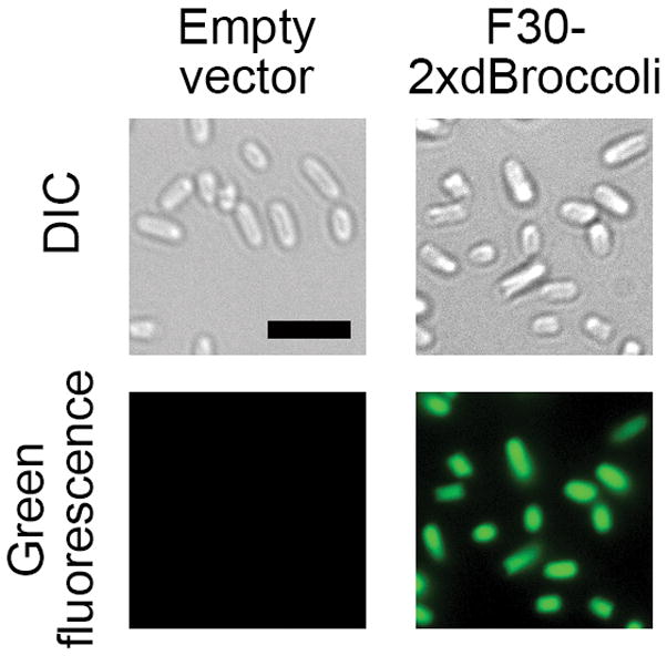
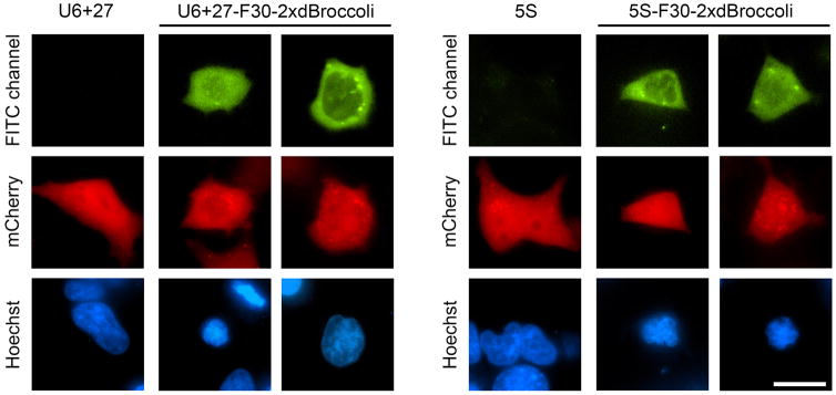
RNA spatial dynamics play a crucial role in cell physiology, and thus the ability to monitor RNA localization in live cells can provide insight into important biological problems. This unit focuses on imaging RNAs using an RNA mimic of GFP. This approach relies on an RNA aptamer called dimeric Broccoli, which binds to and switches on the fluorescence of DFHBI, a small molecule mimicking the fluorophore in GFP. Dimeric Broccoli is tagged to heterologously expressed RNAs and, upon DFHBI binding, the fluorescent signal of dimeric Broccoli reports the transcript's localization in cells. This protocol describes the process of validating the fluorescence of dimeric Broccoli--labeled transcripts in vitro and in cells, flow cytometry analysis to determine overall fluorescence levels in cells, and fluorescence imaging in bacterial and mammalian cells. Overall, the protocol should be useful for researchers seeking to image high-abundance RNAs, such as those transcribed off the T7 promoter in bacteria or off Pol III--dependent promoters in mammalian cells.

Keywords: RNA aptamer; RNA imaging; aptamer expression; fluorescence microscopy.


Figures

Figure 1
Figure 1
Sequence and schematic representation of the F30-2xdBroccoli tag. (A) Sequence of F30-2xdBroccoli is presented. Green indicates the dBroccoli units, orange indicates the F30 sequence. An RNA of interest can be appended on either 5′ or 3′ end, or F30-2xdBroccoli can be inserted inside of an RNA of interest. (B) Schematic drawing of F30-2xdBroccoli. Two Broccoli units (green) are inserted into each of two stem-loops of F30 (orange).
Figure 2
Figure 2
Broccoli expression is readily detected using the in-gel staining protocol. (A) Bacterial cells were transformed with either pET28c plasmid (negative control) or with pET28c-F30-2xdBroccoli and the RNA was expressed for four hours. Then total RNA was isolated, separated using denaturing PAGE and stained with DFHBI-1T and then with SYBR Gold, as described the Basic Protocol 1. DFHBI-1T specifically reveals Broccoli-containing bands while SYBR Gold detects total cellular RNA. (B) Mammalian (HEK293T) cells were either untransfected or transfected with pAV5S or pAVU6+27 (negative controls) or with pAV5S-F30-2xdBroccoli or pAVU6+27-F30-2xdBroccoli. After 72 h expression total RNA was isolated, separated using denaturing PAGE and stained with DFHBI-1T and then with SYBR Gold, as described in the Alternate Protocol 1. Broccoli-containing bands are clearly detectable even in such a complex mixture as total mammalian cell RNA.
Figure 3
Figure 3
Flow cytometry analysis reveals bright fluorescent cell population. (A) Flow cytometry analysis of the pET28c- or pET28c-F30-2xdBroccoli-transformed bacteria. The RNA was expressed for 4 h and then the cells were treated with 40 μM DFHBI-1T and analyzed using a FACSAria II instrument as described in the Basic Protocol 2. Fluorescent events were detected in the modified FITC channel (488 nm excitation and 530±25 emission filter). The data is presented as an overlay of the histograms for the pET28c- and pET28c-F30-2xdBroccoli-transformed bacteria. Broccoli-expressing cells are more than 100 fold brighter than the negative cell population. (B) Flow cytometry analysis of the untransfected mammalian cells or the cells transfected with pAV5S or pAVU6+27 (negative controls) or with pAV5S-F30-2xdBroccoli or pAVU6+27-F30-2xdBroccoli. mCherry-expressing plasmid was co-transfected as a transfection efficiency control. The cells were harvested 72 h after transfection and treated with 40 μM DFHBI-1T as described in the Alternate Protocol 2. The data is presented as a two-parameter dot plot with both FITC (488 nm excitation and 530±25 emission filter) and PE-Texas Red (561 nm excitation and 610±10 nm emission) channels presented. Broccoli-expressing cells show high transfection efficiency and robust fluorescent signal in both channels, compared to the controls.
Figure 4
Figure 4
F30-2xdBroccoli imaging in E. coli. The pET28c or pET28c-F30-2xdBroccoli plasmids were transformed into bacteria, then RNA was expressed for 4 h and the cells were processed as described in the Basic Protocol 3 and imaged. Broccoli fluorescence was detected in the FITC channel (excitation filter 470±20 nm and emission filter 525±25 nm). Exposure time 200 ms. Scale bar, 5 μm.
Figure 5
Figure 5
5S-F30-2xdBroccoli and U6+27-F30-2xdBroccoli imaging in HEK293T cells. Cells were transfected and processed as described in the Alternate Protocol 3, treated with 40 μM DFHBI-1T and 5 μg/ml Hoechst 33342 and imaged using the microscope. Broccoli fluorescence was detected in the FITC channel (excitation filter 470±20 nm and emission filter 525±25 nm), mCherry-filled cells were observed in the Texas Red channel (excitation filter 560±20 nm and emission filter 630±37.5 nm) and Hoechst 33342-stained nuclei were imaged in the DAPI channel (excitation filter 350±25 nm and emission filter 460±25 nm). Exposure times are 0.5–1 s for Broccoli and 100–400 ms for Hoechst 33342 and mCherry. Scale bar, 20 μm.

References

    1. Bao G, Tsourkas A, Santangelo PJ. Engineering nanostructured probes for sensitive intracellular gene detection. Mech Chem Biosyst. 2004;1:23–36. - PubMed
    1. Belly A, Bodon G, Blot B, Bouron A, Sadoul R, Goldberg Y. CHMP2B mutants linked to frontotemporal dementia impair maturation of dendritic spines. J Cell Sci. 2010;123:2943–2954. - PMC - PubMed
    1. Bertrand E, Chartrand P, Schaefer M, Shenoy SM, Singer RH, Long RM. Localization of ASH1 mRNA particles in living yeast. Molecular Cell. 1998;2:437–445. - PubMed
    1. Bratu DP, Cha BJ, Mhlanga MM, Kramer FR, Tyagi S. Visualizing the distribution and transport of mRNAs in living cells. Proceedings of the National Academy of Sciences of the United States of America. 2003;100:13308–13313. - PMC - PubMed
    1. Filonov GS, Kam CW, Song W, Jaffrey SR. In-gel imaging of RNA processing using broccoli reveals optimal aptamer expression strategies. Chemistry & biology. 2015;22:649–660. - PMC - PubMed

Publication types