Review
. 2016 Jul;26(7):535-546.
doi: 10.1016/j.tcb.2016.02.007. Epub 2016 Mar 16.

Targeting Fat: Mechanisms of Protein Localization to Lipid Droplets

Affiliations
Review

Targeting Fat: Mechanisms of Protein Localization to Lipid Droplets

Nora Kory et al. Trends Cell Biol. 2016 Jul.

Abstract

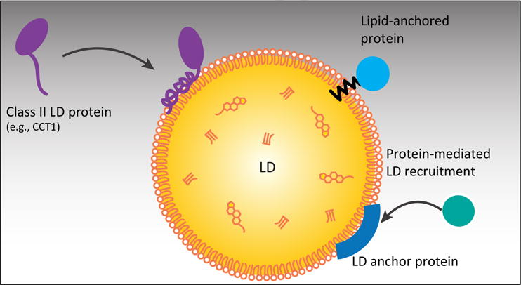
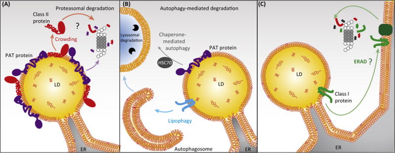
How proteins specifically localize to the phospholipid monolayer surface of lipid droplets (LDs) is being unraveled. We review here the major known pathways of protein targeting to LDs and suggest a classification framework based on the localization origin for the protein. Class I proteins often have a membrane-embedded, hydrophobic 'hairpin' motif, and access LDs from the endoplasmic reticulum (ER) either during LD formation or after formation via ER-LD membrane bridges. Class II proteins access the LD surface from the cytosol and bind through amphipathic helices or other hydrophobic domains. Other proteins require lipid modifications or protein-protein interactions to bind to LDs. We summarize knowledge for targeting and removal of the different classes, and highlight areas needing investigation.

Keywords: amphipathic α-helix; hydrophobic hairpin; lipolysis; organelle protein composition; triglyceride storage.


Figures

Figure 1
Figure 1. Mechanisms of Class I Protein Targeting to Lipid Droplets (LDs) from the Endoplasmic Reticulum (ER)
Class I LD proteins containing a hydrophobic hairpin motif are present in the ER in the absence of LDs. (A) Some class I proteins accumulate on nascent LDs in the ER and translocate to the LD surface during LD formation [15]. (B) After formation, other class I LD proteins target expanding LDs through membrane bridges from the ER [12]. (C) Electron micrograph showing ER-LD bridges in Drosophila S2 cells (image by Florian Wilfling, Morven Graham, and Xinran Liu). Abbreviations: ACSL3, acyl-CoA synthetase long-chain family member 3; eLD, expanding LD; GPAT4, glycerol-3-phosphate acyltransferase 4; LD, initial LD.
Figure 2
Figure 2. Mechanisms of Class II Protein Targeting Lipid Droplets (LDs) from the Cytosol
Class II proteins containing an amphipathic helix LD-binding motif or multiple amphipathic and hydrophobic domains target LDs probably from the cytosol. Some proteins bind to LDs with lipid anchors or by binding to other LD proteins; however, the mechanisms that regulate their targeting are not known.
Figure 3
Figure 3. Mechanisms of Protein Removal from Lipid Droplets (LDs)
LD proteins are removed from LDs by relocalization or degradation. (A) Class II LD proteins are displaced from the LD surface by protein crowding during lipolysis [11] and could be degraded by the ubiquitin/proteasome system, which for instance degrades the class II protein perilipin 2 [116,117]. (B) Whole LDs or parts can undergo autophagy leading to protein as well as lipid degradation [120,121]. Perilipins 2 and 3 undergo chaperone-mediated autophagy during lipolysis [106]. (C) Mechanisms removing class I proteins from LDs are not known but might include relocalization back to the ER and ER-associated degradation (ERAD), or extraction and degradation directly from the LD surface by unknown factors. Abbreviations: ER, endoplasmic reticulum; PAT protein, member of the perilipin/adipose differentiation-related protein (ADRP)/tail-interacting protein of 47 kDa (TIP47) family; HSC70, heat shock cognate 70 kDa.

References

    1. Thiam AR, et al. The biophysics and cell biology of lipid droplets. Nat Rev Mol Cell Biol. 2013;14:775–786. - PMC - PubMed
    1. Walther TC, Farese RV., Jr Lipid droplets and cellular lipid metabolism. Annu Rev Biochem. 2012;81:687–714. - PMC - PubMed
    1. Wilfling F, et al. Lipid droplet biogenesis. Curr Opin Cell Biol. 2014;29:39–45. - PMC - PubMed
    1. Welte MA. Expanding roles for lipid droplets. Curr Biol. 2015;25:R470–R481. - PMC - PubMed
    1. Gross DA, Silver DL. Cytosolic lipid droplets: from mechanisms of fat storage to disease. Crit Rev Biochem Mol Biol. 2014;49:304–326. - PubMed

Publication types