A two-component system regulates gene expression of the type IX secretion component proteins via an ECF sigma factor
- PMID: 26996145
- PMCID: PMC4800418
- DOI: 10.1038/srep23288
A two-component system regulates gene expression of the type IX secretion component proteins via an ECF sigma factor
Abstract
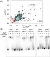
The periodontopathogen Porphyromonas gingivalis secretes potent pathogenic proteases, gingipains, via the type IX secretion system (T9SS). This system comprises at least 11 components; however, the regulatory mechanism of their expression has not yet been elucidated. Here, we found that the PorY (PGN_2001)-PorX (PGN_1019)-SigP (PGN_0274) cascade is involved in the regulation of T9SS. Surface plasmon resonance (SPR) analysis revealed a direct interaction between a recombinant PorY (rPorY) and a recombinant PorX (rPorX). rPorY autophosphorylated and transferred a phosphoryl group to rPorX in the presence of Mn(2+). These results demonstrate that PorX and PorY act as a response regulator and a histidine kinase, respectively, of a two component system (TCS), although they are separately encoded on the chromosome. T9SS component-encoding genes were down-regulated in a mutant deficient in a putative extracytoplasmic function (ECF) sigma factor, PGN_0274 (SigP), similar to the porX mutant. Electrophoretic gel shift assays showed that rSigP bound to the putative promoter regions of T9SS component-encoding genes. The SigP protein was lacking in the porX mutant. Co-immunoprecipitation and SPR analysis revealed the direct interaction between SigP and PorX. Together, these results indicate that the PorXY TCS regulates T9SS-mediated protein secretion via the SigP ECF sigma factor.
Figures


Similar articles
-
The PorX Response Regulator of the Porphyromonas gingivalis PorXY Two-Component System Does Not Directly Regulate the Type IX Secretion Genes but Binds the PorL Subunit.Front Cell Infect Microbiol. 2016 Aug 31;6:96. doi: 10.3389/fcimb.2016.00096. eCollection 2016. Front Cell Infect Microbiol. 2016. PMID: 27630829 Free PMC article.
-
A PorX/PorY and σP Feedforward Regulatory Loop Controls Gene Expression Essential for Porphyromonas gingivalis Virulence.mSphere. 2021 Jun 30;6(3):e0042821. doi: 10.1128/mSphere.00428-21. Epub 2021 May 28. mSphere. 2021. PMID: 34047648 Free PMC article.
-
PorA of the Type IX Secretion Is a Ligand of the PorXY Two-Component Regulatory System in Porphyromonas gingivalis.Mol Microbiol. 2025 Jun;123(6):569-585. doi: 10.1111/mmi.15363. Epub 2025 Apr 8. Mol Microbiol. 2025. PMID: 40195800 Free PMC article.
-
Type IX secretion: the generation of bacterial cell surface coatings involved in virulence, gliding motility and the degradation of complex biopolymers.Mol Microbiol. 2017 Oct;106(1):35-53. doi: 10.1111/mmi.13752. Epub 2017 Aug 9. Mol Microbiol. 2017. PMID: 28714554 Review.
-
The Type IX Secretion System: Advances in Structure, Function and Organisation.Microorganisms. 2020 Aug 1;8(8):1173. doi: 10.3390/microorganisms8081173. Microorganisms. 2020. PMID: 32752268 Free PMC article. Review.
Cited by
-
Post-translational Modifications in Oral Bacteria and Their Functional Impact.Front Microbiol. 2021 Dec 2;12:784923. doi: 10.3389/fmicb.2021.784923. eCollection 2021. Front Microbiol. 2021. PMID: 34925293 Free PMC article. Review.
-
Structure-function analysis of PorXFj, the PorX homolog from Flavobacterium johnsioniae, suggests a role of the CheY-like domain in type IX secretion motor activity.Sci Rep. 2024 Mar 19;14(1):6577. doi: 10.1038/s41598-024-57089-9. Sci Rep. 2024. PMID: 38503809 Free PMC article.
-
Amino acids as wetting agents: surface translocation by Porphyromonas gingivalis.ISME J. 2019 Jun;13(6):1560-1574. doi: 10.1038/s41396-019-0360-9. Epub 2019 Feb 19. ISME J. 2019. PMID: 30783212 Free PMC article.
-
Phylogenetic comparison between Type IX Secretion System (T9SS) protein components suggests evidence of horizontal gene transfer.PeerJ. 2020 Jun 26;8:e9019. doi: 10.7717/peerj.9019. eCollection 2020. PeerJ. 2020. PMID: 32617187 Free PMC article.
-
Response regulator PorX coordinates oligonucleotide signalling and gene expression to control the secretion of virulence factors.Nucleic Acids Res. 2022 Nov 28;50(21):12558-12577. doi: 10.1093/nar/gkac1103. Nucleic Acids Res. 2022. PMID: 36464236 Free PMC article.
References
-
- Papapanou P. N. Epidemiology of periodontal diseases: an update. J. Int. Acad. Periodontol. 1, 110–116 (1999). - PubMed
-
- Irfan U. M., Dawson D. V. & Bissada N. F. Epidemiology of periodontal disease: a review and clinical perspectives. J. Int. Acad. Periodontol. 3, 14–21 (2001). - PubMed
-
- Armitage G. C. Periodontal diseases: diagnosis. Ann. Periodontol. 1, 37–215 (1996). - PubMed
-
- Page R. C., Offenbacher S., Schroeder H. E., Seymour G. J. & Kornman K. S. Advances in the pathogenesis of periodontitis: summary of developments, clinical implications and future directions. Periodontol. 2000 14, 216–248 (1997). - PubMed
Publication types
MeSH terms
Substances
LinkOut - more resources
Full Text Sources
Other Literature Sources
Molecular Biology Databases
Research Materials

