Review
. 2016 Aug;105(8):449-62.
doi: 10.1002/bip.22836.

Invited review: Activation of G proteins by GTP and the mechanism of Gα-catalyzed GTP hydrolysis

Affiliations
Review

Invited review: Activation of G proteins by GTP and the mechanism of Gα-catalyzed GTP hydrolysis

Stephen R Sprang. Biopolymers. 2016 Aug.

Abstract

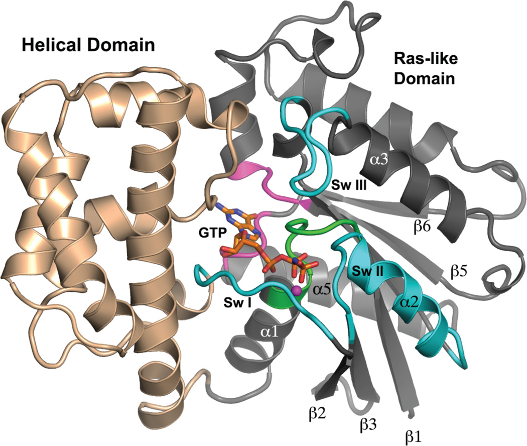
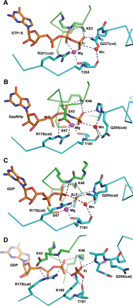
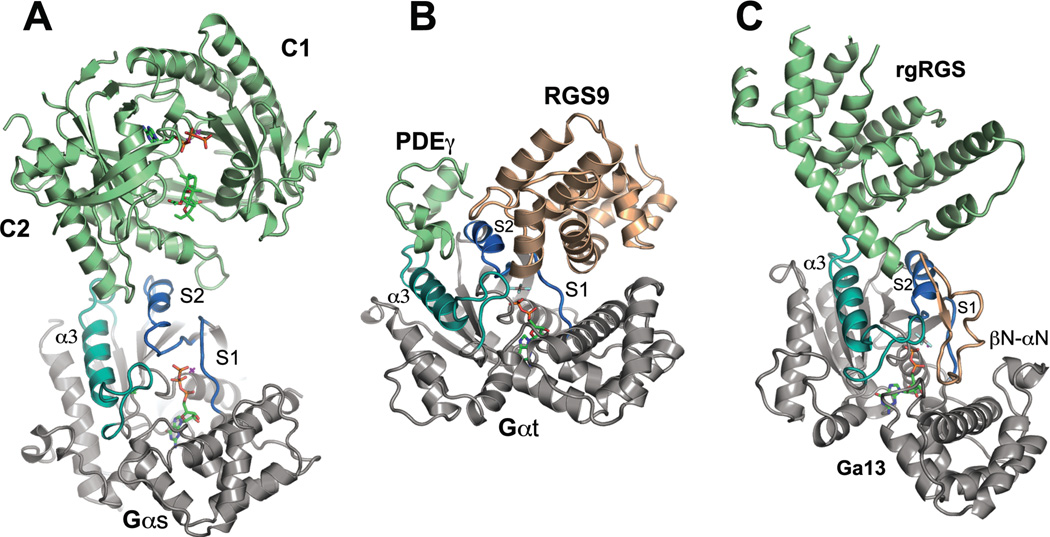
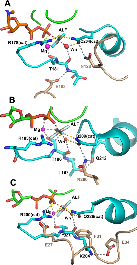
This review addresses the regulatory consequences of the binding of GTP to the alpha subunits (Gα) of heterotrimeric G proteins, the reaction mechanism of GTP hydrolysis catalyzed by Gα and the means by which GTPase activating proteins (GAPs) stimulate the GTPase activity of Gα. The high energy of GTP binding is used to restrain and stabilize the conformation of the Gα switch segments, particularly switch II, to afford stable complementary to the surfaces of Gα effectors, while excluding interaction with Gβγ, the regulatory binding partner of GDP-bound Gα. Upon GTP hydrolysis, the energy of these conformational restraints is dissipated and the two switch segments, particularly switch II, become flexible and are able to adopt a conformation suitable for tight binding to Gβγ. Catalytic site pre-organization presents a significant activation energy barrier to Gα GTPase activity. The glutamine residue near the N-terminus of switch II (Glncat ) must adopt a conformation in which it orients and stabilizes the γ phosphate and the water nucleophile for an in-line attack. The transition state is probably loose with dissociative character; phosphoryl transfer may be concerted. The catalytic arginine in switch I (Argcat ), together with amide hydrogen bonds from the phosphate binding loop, stabilize charge at the β-γ bridge oxygen of the leaving group. GAPs that harbor "regulator of protein signaling" (RGS) domains, or structurally unrelated domains within G protein effectors that function as GAPs, accelerate catalysis by stabilizing the pre-transition state for Gα-catalyzed GTP hydrolysis, primarily by restraining Argcat and Glncat to their catalytic conformations. © 2016 Wiley Periodicals, Inc. Biopolymers 105: 449-462, 2016.

Keywords: GTPase activating proteins; Heterotrimeric G proteins; Reaction mechanism; Signal Transduction; X-ray crystallography.


Figures

Figure 1
Figure 1
Tertiary structure of Gα. A model of a Gα subunit bound to GTP and Mg2+ is depicted as a ribbon drawing and is based on the crystal structure of Gαi1•GppNHp (PDB 1CIP). The N-terminal 31, and C-terminal 7 amino acid residues are disordered in this structure, and adopt a variety of conformations in several crystal structures, depending on crystal contacts and binding partners. The Helical domain is colored light brown and the Ras-like domain is rendered in gray. Switch segments involved in effector recognition and GTPase activity are labeled and colored cyan. The P-loop is colored green, and loop regions involved in recognition and binding of the guanine moiety of GDP and GDP are colored pink. GppNHp is shown as a stick figure, and the Mg2+ is represented by magenta sphere. Selected secondary structure elements in the Ras domain are labeled.
Figure 2
Figure 2
Snapshots of the Gα catalytic site along the trajectory of GTP hydrolysis, derived from crystal structures. The coloring scheme is the same as that used in Figure 1. Nitrogen, oxygen and phosphorus atoms are colored blue, red and yellow respectively. Sulfer atoms are colored yellow. The magenta and red spheres represent magnesium ion and the water nucleophile, respectively. Residues of interest are labeled. The catalytic Gln and Arg residues are indicated with appropriate residue numbers and “cat” in parentheses. Hydrogen bonds (2.7–3.1Å) and metal-ligand coordination bonds (1.9–2.2Å) are shown as gray dashed lines. A, the structure of Gαs bound to GTPγS and Mg2+ (PDB 1AZT, 2.3Å resolution). Note that neither of the catalytic residues Gln 227 nor Arg 201 form direct contacts with the nucleotide; B, the complex of Gαi1 with GppNHp and Mg2+ (PDB 1CIP, 1.5Å resolution). Here Arg 178 (Argcat) is restrained in a hydrogen—bonded ionic interaction with the P-loop Glu 43. Gln 204 (Glncat) is a hydrogen bond donor to the water nucleophile, thereby orienting its lone pair electrons away from the γ phosphorus. This apparently stable ground-state conformation is expected to be anti-catalytic; C, Gαi1 bound to GDP, Mg2+ and AlF4 (labeled ALF), a model of the pre-organized or pre-transition state (PDB 1GFI, 2.2Å resolution; the AlF4 moiety was not rigidly restrained to planarity during refinement). Argcat is within hydrogen bonding distance of the leaving group β-γ bridge oxygen and Glncat is a hydrogen bond donor to a fluorine (or O) Al substituent and accepts a hydrogen bond from the presumptive water nucleophile. The hydrogen bond network (yellow dashed lines) involving Argcat, Glncat, Wnuc and the the γ phosphate (modeled by AlF) orient Wnuc for nucleophilic attack and stabilize developing charge at the β-γ bridge leaving group oxygen (note also hydrogen bond to the latter from a P-loop amide, present also in GTP analog-bound structures); D, a model of the GDP, Pi ternary complex of Gα from the crystal structure of the G203A mutant of Gαi1 (PDB 1GIT, 2.6Å resolution). Note that switch II has reoriented and is refolded into an α helix at its N-terminus, forming an electropositive binding site for Pi. Both the β phosphate of GDP and Pi are retained in the catalytic site with multiple hydrogen bonds. The Mg2+ binding site is dismantled due to conformational changes in switches I and II.
Figure 3
Figure 3
Structures of Gα bound to effectors and effector-GAPs. In all panels, Gα is rendered in gray except switch I and II, which are colored slate blue, and the α3 helix and switch II, which are rendered in turquoise. Effector domains are colored sage green and GAP domains are rendered in light brown. Ligands and nucleotides are rendered as stick models. A, structure of the catalytic domains (C1 and C2) of adenylyl cyclase bound to the GTPγS complex of Gαs (PDB 1AZS). Two helical segments of adenylyl cyclase and connecting loops engage the middle of switch II and trough between switch II and α3; B, the complex between cyclic GMP phosphodiesterase γ subunit (PDEγ), the RGS domain of RGS9 and Gαt/i1(PDB 1FQK). PDEγ binds at the switch II - α3 interface, while RGS9 occupies a distinct interface between the N-terminal half of switch II and switch I. PDEγ potentiates the GAP activity of RGS9 by stabilizing its interaction with Gα; C, complex of the rgRGS domain of p115RhoGEF with Gα13/i1 (PDB 1SHZ). The RGS-like domain of p115RhoGEF occupies the effector binding region of Gα at the switch II - α3 interface. The βN-αN hairpin domain that conveys GAP activity docks at the interface between the N-terminal half of switch II and switch I.
Figure 4
Figure 4
Interactions between Gα•GDP•MgAlF active site and critical residues of RGS and effector-GAP domains. The coloring scheme used in Figures 1–3 is used. A, contacts between switch I and switch II of Gαi1 and RGS4 (PDB 1AGR, 2.8Å resolution) are shown. RGS residues Asn 128 and Glu163, respectively, constrain the conformation of Gαi1 Q204 (Glncat) to the pre-transition state conformation and stabilize switch I though a hydrogen bond to the backbone amide of Thr 181; B, Asn 260 from the loop between the EF 3 and EF 4 domains of PLC−β3 form a network of hydrogen bonds with residues of switch II at the catalytic site of Gαq (PDB 3OHM, 2.7Å resolution). Interactions between Asn 260 and Gαq mimic that of Asn 128 of RGS4 with Gαi1. The latter are strengthened by hydrogen bond network with residue Gln 212 and Thr 187 of switch I in Gαq. Not shown is the extensive interaction surface of PLC-β3 and the effector-binding surface of Gαq; C, The αN segment of the βN-αN hairpin of p115RhoGEF forms hydrogen bonds with switch I and switch II in Gα13/i1. Acidic residues Glu 27 and Glu 34 form ion pair contacts with Argcat (Arg 200) and Lys 204, respectively, in switch I, stabilizing the interaction between Argcat and the fluoroaluminate, and potentially, the β-γ bridge oxygen of GTP. Phe 31 sterically restrains the position of Glncat as do Asn 128 and Asn 260 in RGS4 and PLC-β3.

Similar articles

Cited by

References

    1. Cassel D, Selinger Z. Biochim Biophys Acta. 1976;452:538–551. - PubMed
    1. Pfeuffer T, Helmreich EJ. J Biol Chem. 1975;250:867–876. - PubMed
    1. Ross EM, Howlett AC, Ferguson KM, Gilman AG. J Biol Chem. 1978;253:6401–6412. - PubMed
    1. Pfeuffer T. J Biol Chem. 1977;252:7224–7234. - PubMed
    1. Gilman AG. Anual Reveiws in Biochemistry. 1987;56:615–649. - PubMed

MeSH terms

Substances

LinkOut - more resources