. 2016 Apr 7;98(4):615-26.
doi: 10.1016/j.ajhg.2016.02.007. Epub 2016 Mar 17.

Pathogenic Variants in PIGG Cause Intellectual Disability with Seizures and Hypotonia

Affiliations

Pathogenic Variants in PIGG Cause Intellectual Disability with Seizures and Hypotonia

Periklis Makrythanasis et al. Am J Hum Genet. .

Abstract

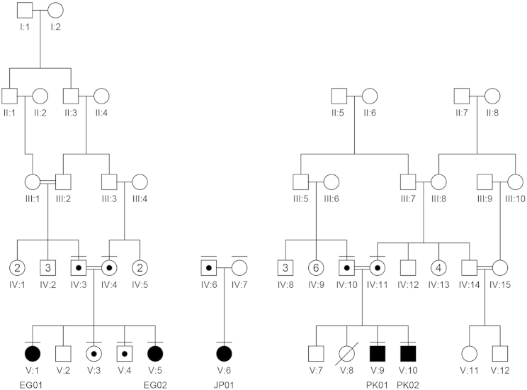
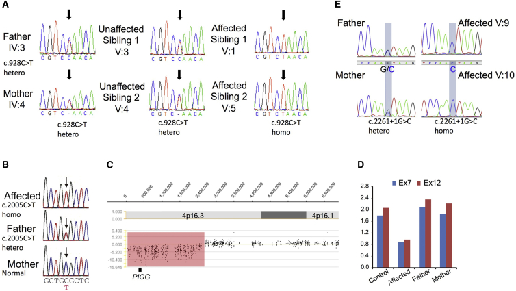
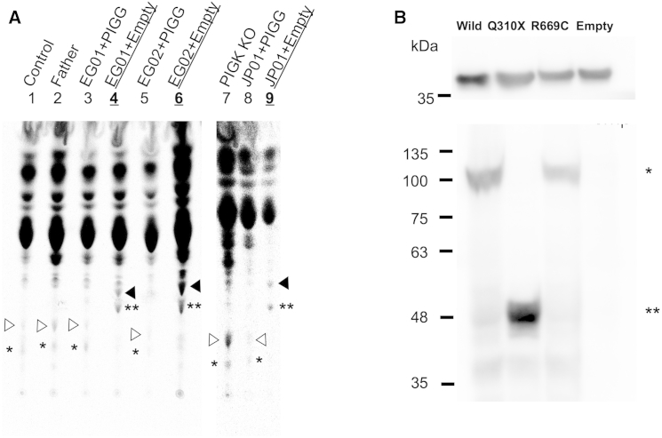
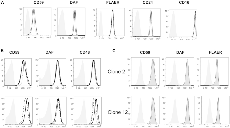
Glycosylphosphatidylinositol (GPI) is a glycolipid that anchors >150 various proteins to the cell surface. At least 27 genes are involved in biosynthesis and transport of GPI-anchored proteins (GPI-APs). To date, mutations in 13 of these genes are known to cause inherited GPI deficiencies (IGDs), and all are inherited as recessive traits. IGDs mainly manifest as intellectual disability, epilepsy, coarse facial features, and multiple organ anomalies. These symptoms are caused by the decreased surface expression of GPI-APs or by structural abnormalities of GPI. Here, we present five affected individuals (from two consanguineous families from Egypt and Pakistan and one non-consanguineous family from Japan) who show intellectual disability, hypotonia, and early-onset seizures. We identified pathogenic variants in PIGG, a gene in the GPI pathway. In the consanguineous families, homozygous variants c.928C>T (p.Gln310(∗)) and c.2261+1G>C were found, whereas the Japanese individual was compound heterozygous for c.2005C>T (p.Arg669Cys) and a 2.4 Mb deletion involving PIGG. PIGG is the enzyme that modifies the second mannose with ethanolamine phosphate, which is removed soon after GPI is attached to the protein. Physiological significance of this transient modification has been unclear. Using B lymphoblasts from affected individuals of the Egyptian and Japanese families, we revealed that PIGG activity was almost completely abolished; however, the GPI-APs had normal surface levels and normal structure, indicating that the pathogenesis of PIGG deficiency is not yet fully understood. The discovery of pathogenic variants in PIGG expands the spectrum of IGDs and further enhances our understanding of this etiopathogenic class of intellectual disability.


Figures

Figure 1
Figure 1
Family Pedigrees The affected individuals are noted as EG01, EG02, JP01, PK01, and PK02. The family members for whom DNA was available are noted with a horizontal bar above their respective symbols. Affected individuals are homozygous or compound heterozygous for the causative variants, and family members are verified carriers. In family JP, the second variant is a de novo CNV that encompasses PIGG.
Figure 2
Figure 2
MRI and EEG of the Affected Individuals (A) MRI of individual EG02 when she was 2.5 years old. (Left panel) Mid-sagittal T1-weighted MRI shows the thin corpus callosum. (Right panel) Axial plane in T1- and T2-weighted MRI shows asymmetry of the lateral ventricles. (B) MRI and EEG of individual JP01. (Upper left panel) Interictal EEG during sleep at the age of 3 years shows focal spikes on the right posterior area (C4-P4-O2-T6, arrows). (Lower panel) Ictal EEG at 14 months shows that low-amplitude fast waves arose from right centrotemporal lesions (C4-T4, arrowheads) and then propagated to the entire right hemisphere. According to the ictal discharge, the individual developed eye deviation and head turning to the right after clonic seizures of the left upper extremity. (Upper right panel) T2-weighted brain MRI at the level of basal ganglia shows normal appearance at 11 months. (C) MRI of individual PK01. (Left panel) Mid-sagittal T1-weighted MRI shows cerebellar hypoplasia. (Right panel) Axial plane in T2-weighted MRI shows mild cerebral atrophy.
Figure 3
Figure 3
Identified Variants (A) Sequencing chromatograms of the identified variant in family EG. The parents and unaffected siblings are heterozygous, whereas the affected individuals are homozygous for the c.928C>T mutation (noted by the black arrow). (B) Sequencing chromatograms of the identified variant in family JP. The father has the heterozygous c.2005C>T variant, but the mother does not (noted by the black arrow). (C) XHMM analysis using WES data of individual JP01. A microdeletion involving PIGG (chr4: 60,226–2,452,836) is indicated. (D) qPCR analysis of exons 7 and 12 of the PIGG genome from individual JP01 and her parents shows a heterozygous PIGG deletion in the affected individual, but not in her mother, suggesting that the deletion occurred de novo on the maternal chromosome. (E) Sequencing chromatograms of the identified variant in family PK are noted by the blue transparent boxes. The parents are heterozygous, and the affected individuals are homozygous for the c.2261+1G>C mutation.
Figure 4
Figure 4
Functional Assays (A) Analysis of mannolipids accumulated in LCLs from the affected individuals. Lanes are as follows: 1, LCLs from a normal individual; 2, LCLs from the father of individuals EG01 and EG02 (IV:3); 3, LCLs (transfected with PIGG cDNA) from individual EG01; 4, LCLs (transfected with an empty vector) from individual EG01; 5, LCLs (transfected with PIGG cDNA) from individual EG02; 6, LCLs (transfected with an empty vector) from individual EG02; 7, K562 PIGK-deficient cells; 8, LCLs (transfected with PIGG cDNA) from individual JP01; and 9, LCLs (transfected with an empty vector) from individual JP01. White arrowheads represent H8, a complete GPI intermediate, and black arrowheads represent H7, a GPI intermediate without ethanolaminephosphate at the second mannose. Asterisks indicate H7′ () and H8′ (∗∗), H7 and H8, respectively, with the fourth mannose. (B) Amounts of each mutant PIGG. (Upper panel) Western blot of GAPDH for loading controls. (Lower panel) Western blot of wild-type () or mutant (∗∗) HA-tagged PIGG.
Figure 5
Figure 5
Flow Cytometric Analysis of Affected Individuals’ Blood and Cell Lines (A) Surface level of GPI-anchored proteins on the granulocytes from individual JP01. Thick lines represent individual JP01, dotted lines represent a healthy control individual, and gray shadows represent the isotype control. (B) Surface level of GPI-anchored proteins on the LCLs from individuals EG01 and EG02 (upper panel) and individual JP01 (lower panel). In the upper panels, thick and thin lines represent individuals EG01 and EG02, respectively, and dotted lines represent their father. In the lower panels, thick lines represent affected individual JP01, and dotted and dashed lines represent healthy individuals. Gray shadows represent the isotype control. (C) Surface level of GPI-anchored proteins on HEK293 cells. PIGG-knockout HEK293 cells were permanently transfected with PIGG cDNA (dotted lines) or an empty vector (thick lines). Dark-gray shadows represent wild-type HEK293 cells, and light-gray shadows represent the isotype control.

References

    1. Maulik P.K., Mascarenhas M.N., Mathers C.D., Dua T., Saxena S. Prevalence of intellectual disability: a meta-analysis of population-based studies. Res. Dev. Disabil. 2011;32:419–436. - PubMed
    1. American Psychiatric Association (2013). DSM-5 Intellectual Disability Fact Sheet, http://www.dsm5.org/documents/intellectual%20disability%20fact%20sheet.pdf.
    1. World Health Organization Division of Mental Health and Prevention of Substance Abuse (1996). ICD-10 Guide for Mental Retardation, http://www.who.int/mental_health/media/en/69.pdf.
    1. Bamshad M.J., Ng S.B., Bigham A.W., Tabor H.K., Emond M.J., Nickerson D.A., Shendure J. Exome sequencing as a tool for Mendelian disease gene discovery. Nat. Rev. Genet. 2011;12:745–755. - PubMed
    1. Gilissen C., Hoischen A., Brunner H.G., Veltman J.A. Disease gene identification strategies for exome sequencing. Eur. J. Hum. Genet. 2012;20:490–497. - PMC - PubMed

Publication types

MeSH terms

Substances