. 2016 Apr 7;98(4):627-42.
doi: 10.1016/j.ajhg.2016.02.008. Epub 2016 Mar 17.

GTF2E2 Mutations Destabilize the General Transcription Factor Complex TFIIE in Individuals with DNA Repair-Proficient Trichothiodystrophy

Affiliations

GTF2E2 Mutations Destabilize the General Transcription Factor Complex TFIIE in Individuals with DNA Repair-Proficient Trichothiodystrophy

Christiane Kuschal et al. Am J Hum Genet. .

Abstract

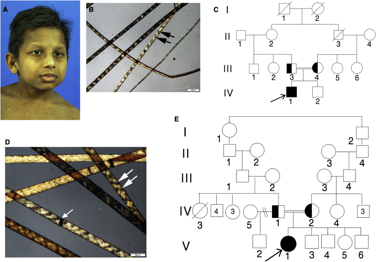
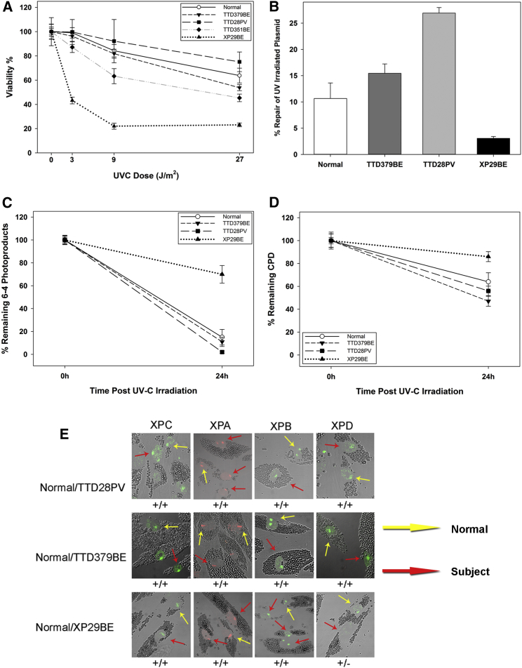
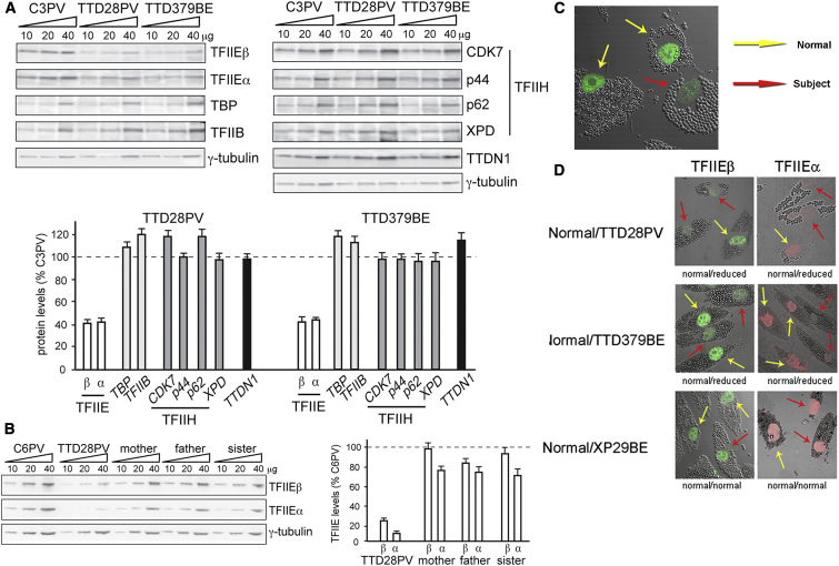
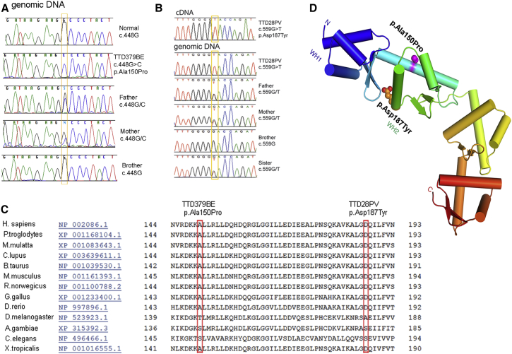
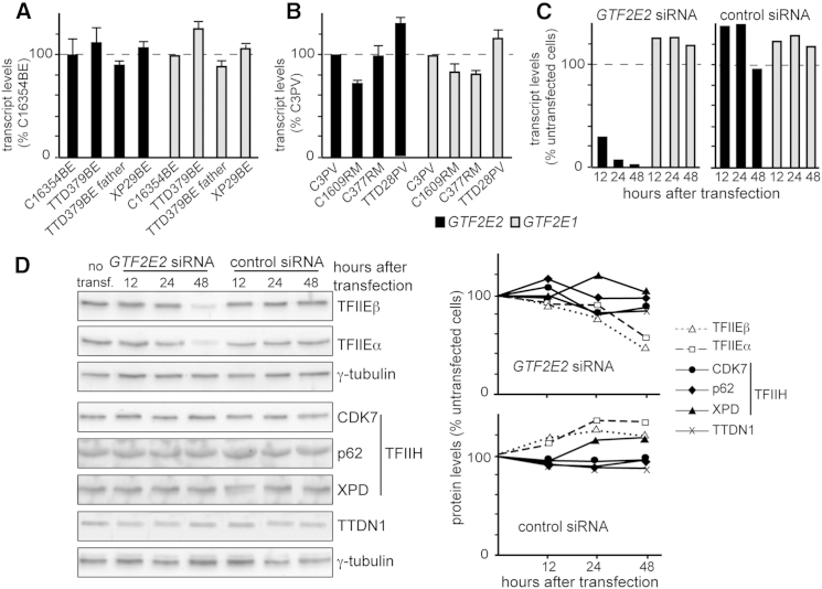
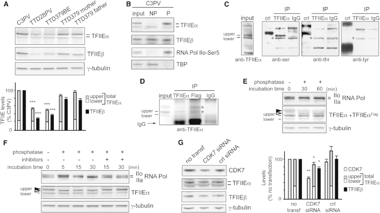
The general transcription factor IIE (TFIIE) is essential for transcription initiation by RNA polymerase II (RNA pol II) via direct interaction with the basal transcription/DNA repair factor IIH (TFIIH). TFIIH harbors mutations in two rare genetic disorders, the cancer-prone xeroderma pigmentosum (XP) and the cancer-free, multisystem developmental disorder trichothiodystrophy (TTD). The phenotypic complexity resulting from mutations affecting TFIIH has been attributed to the nucleotide excision repair (NER) defect as well as to impaired transcription. Here, we report two unrelated children showing clinical features typical of TTD who harbor different homozygous missense mutations in GTF2E2 (c.448G>C [p.Ala150Pro] and c.559G>T [p.Asp187Tyr]) encoding the beta subunit of transcription factor IIE (TFIIEβ). Repair of ultraviolet-induced DNA damage was normal in the GTF2E2 mutated cells, indicating that TFIIE was not involved in NER. We found decreased protein levels of the two TFIIE subunits (TFIIEα and TFIIEβ) as well as decreased phosphorylation of TFIIEα in cells from both children. Interestingly, decreased phosphorylation of TFIIEα was also seen in TTD cells with mutations in ERCC2, which encodes the XPD subunit of TFIIH, but not in XP cells with ERCC2 mutations. Our findings support the theory that TTD is caused by transcriptional impairments that are distinct from the NER disorder XP.


Figures

Figure 1
Figure 1
TTD-Affected Children TTD379BE and TTD28PV (A) TTD379BE, a 10-year-old boy from India with mild micrognathia, low-set ears, and short hair. (B) Hair of affected child TTD379BE showing alternating dark and light “tiger tail” banding (arrows) with polarized microscopy typical of TTD. Hair shafts vary in diameter (). (C) Pedigree of TTD379BE (IV-1). His clinically normal parents (III-3 and III-4) are first cousins. (D) Hair of 16-year-old affected girl, TTD28PV, showing tiger tail banding with polarized microscopy (arrows). The hair shafts also have dark transverse bands that indicate sites of hair fractures (trichoschisis) (arrowhead). (E) Pedigree of TTD28PV (V-1). Her parents (IV-1 and IV-2) are consanguineous.
Figure 2
Figure 2
Normal DNA Repair in Primary Fibroblasts from TTD379BE and TTD28PV (A) Post-UV cell survival in normal, TTD379BE, TTD28PV, XP29BE (XP/XP-D), and TTD351BE (TTD/XP-D) cells. Bars indicate SEM. (B) Post-UV host cell reactivation in normal, TTD379BE, TTD28PV, and XP29BE (XP/XP-D) fibroblasts. Cells were transfected with an UV-C-irradiated (1,000 J/m2) luciferase reporter vector and incubated for 48 hr. Repair of the plasmid is expressed as induced light units of active luciferase compared to a non-irradiated luciferase plasmid. Two experiments each in triplicate were performed. Bars indicate SEM. (C and D) Repair of 6-4 photoproducts (6-4PP) (C) and cyclobutane pyrimidine dimers (CPD) (D) measured by immunofluorescence in normal, TTD379BE, TTD28PV, and XP29BE (XP/XP-D) fibroblasts. Cells were irradiated with 100 J/m2 UVC through a filter with 5 μm pores to generate localized DNA damage. 100 nuclei were scored. Bars indicate SEM. (E) Immunofluorescence analysis of NER proteins in normal (incubated with 1 μm beads, yellow arrows) and TTD379BE or TTD28PV (incubated with 2 μm beads, red arrows) cells loaded on the same coverslip and irradiated with 100 J/m2 UVC through a filter with 5 μm pores to generate localized DNA damage. Post-UV localization of XPD protein to the damaged sites is not detected (−) in XP29BE (XP/XP-D) cells but is present (+) at normal levels in TTD28PV and TTD379BE cells.
Figure 3
Figure 3
Reduced Steady-State Levels of TFIIE and Normal Levels of Other General Transcription Factors in Primary Fibroblasts from Affected Individuals TTD379BE and TTD28PV (A) Immunoblot analysis of whole-cell lysates with antibodies against the α and β subunits of TFIIE, TBP, TFIIB, the CDK7, p44, p62, and XPD subunits of TFIIH, and TTDN1. γ-tubulin was used as loading control. The amount of each protein was first expressed as the mean value of the levels observed in the three increasing concentrations of the cell lysate and normalized to the γ-tubulin content. The protein levels in both TTD cell strains were then expressed as percentages of the corresponding values in the normal (C3PV) cells. The reported values are the means of at least two independent experiments. Bars indicate the SE. (B) Anti-TFIIEα and anti-TFIIEβ immunoblot analysis of whole-cell lysates from lymphoblastoid cells of TTD28PV and her unaffected mother, father, and sister. The amount of the analyzed proteins was determined as described in (A); the levels of TFIIEα and TFIIEβ are expressed as percentages of the corresponding values in the normal C6PV lymphoblasts. The mean levels of two independent experiments are reported. Bars indicate the SE. (C) Reduced immunofluorescence of TFIIEβ protein in TTD379BE cells (incubated with 2 μm beads, red arrow) compared to normal cells (incubated with 1 μm beads, yellow arrows). (D) Immunofluorescence detection of reduced levels of TFIIEβ and TFIIEα in TTD28PV and TTD379BE cells. TTD and normal cells were labeled as in Figure 2C and irradiated with 100 J/m2 UVC through a filter with 5 μm pores to generate localized DNA damage. The TFIIE proteins were reduced in the TTD cells. There was no localization of TFIIE proteins at the site of localized DNA damage in the TTD or normal cells.
Figure 4
Figure 4
Identification of Missense Mutations in GTF2E2 in Affected Children TTD379BE and TTD28PV (A) Electropherograms showing the change identified in GTF2E2 in TTD379BE and his unaffected father and mother but not in the unaffected brother. (B) Electropherograms showing the change identified in GTF2E2 (GenBank: NM_002095.4, NC_000008.10) in TTD28PV and unaffected father, mother, and sister but not in the unaffected brother. For cDNA numbering, +1 corresponds to the A of the ATG translation initiation codon in the reference sequence. (C) Conservation of amino acids Ala150 and Asp187 in multiple protein sequence alignment of TFIIEβ regions in 13 species (from HomoloGene: 37573). (D) Structural location of amino acid changes in TFIIEβ protein resulting from GTF2E2 homozygous missense mutations in TTD-affected individuals. Each winged-helix (WH) domain consists of three helices (cylinders) and a beta-hairpin (two strands). The protein is colored from N to C terminus from blue (WH1 domain), blue-green (WH2 domain), to red (C terminus). The altered amino acids (p.Ala150Pro and p.Asp187Tyr) are 14 Å apart on different surfaces of wing helix 2 (WH2) region of the TFIIEβ protein. The p.Ala150Pro change will destabilize the long alpha helix and cause it to bend or locally unfold (W. Yang, personal communication).
Figure 5
Figure 5
GTF2E2 Mutations Do Not Affect the Cellular Amount of GTF2E2 and GTF2E1 Transcripts but Interfere with the Stability of TFIIE Complex (A) GTF2E2 (black columns) and GTF2E1 (gray columns) transcript levels in primary fibroblasts from the healthy donor C16354BE, affected individual TTD379BE, his father, and affected individual XP29BE (XP/XP-D). Transcript levels were normalized to GAPDH levels and then expressed as percentages of the corresponding value in the healthy donor analyzed in parallel. The reported values are the means of two independent experiments, each done in triplicate. Bars indicate the SE. (B) GTF2E2 (black columns) and GTF2E1 (gray columns) transcript levels in primary fibroblasts from healthy donors C3PV, C1609RM, and C377RM and affected individual TTD28PV. Transcript levels were expressed as in (A). The reported values are the means of two independent experiments, each done in triplicate. Bars indicate the SE. (C) GTF2E2 and GTF2E1 transcript levels in C3PV primary fibroblasts 12, 24, and 48 hr after transfection with GTF2E2 or control siRNA. GTF2E2 (black columns) and GTF2E1 (gray columns) transcript levels were normalized to GAPDH and expressed as percentages of the corresponding value in untransfected C3PV cells. (D) Immunoblot analysis of whole-cell lysates from C3PV primary fibroblasts 12, 24, and 48 hr after transfection with GTF2E2 or control siRNA. Antibodies against the α and β subunits of TFIIE, the CDK7, p62, and XPD subunits of TFIIH, TTDN1, and γ-tubulin (loading control) were used. The levels of the analyzed proteins were normalized to γ-tubulin and expressed as percentages of the corresponding values in untransfected C3PV cells.
Figure 6
Figure 6
Reduced Amount of Phosphorylated TFIIEα in Primary Fibroblasts of Affected Individuals TTD379BE and TTD28PV and in Normal C3PV Fibroblasts after CDK7 Silencing (A) Immunoblot analysis of lysates obtained by directly scraping normal (C3PV and TTD379BE parents) and TTD (TTD28PV and TTD379BE) cells in Laemmli buffer. The levels of the upper (gray) and lower (white) forms of TFIIEα and of TFIIEβ (black) were normalized to γ-tubulin and expressed as percentage of the corresponding values in C3PV cells. The reported values are the means of at least two independent experiments. Bars indicate the SE. The statistically significant reduction of TFIIEα (∗∗∗p < 0.0005; Student’s t test) refers to the upper band, no significant difference was observed for the lower band. (B) Immunoblot analysis of TFIIEα and TFIIEβ in unphosphorylated (NP) and phosphorylated (P) protein-enriched fractions of cell lysates from normal C3PV fibroblasts. TBP and RNA pol IIo-Ser5 were used as positive controls of the NP and P fractions, respectively. (C) Immunoprecipitation of TFIIEα or IgG from HeLa total cell lysates and identification of the phosphorylated residue. The TFIIEα or IgG immunoprecipitates (IP) were analyzed by immunoblotting using phosphoaminoacid-specific antibodies. The positive controls (crl) are made of proteins phosphorylated on serine (ser), threonine (thr), or tyrosine (tyr), provided by the kit. Asterisks () indicate non-specific bands. (D) Immunoprecipitation of TFIIEα, TFIIEαFlag, or IgG from cell lysates of C3PV normal fibroblasts transfected with the pMP2-TFIIEαFlag DNA plasmid expressing the TFIIEαFlag recombinant protein. The immunoprecipitates (IP) were analyzed by immunoblotting using anti-TFIIEα antibodies. Asterisks () indicate the upper and lower bands of TFIIEαFlag. (E) Immunoblot analysis of TFIIEα after phosphatase treatment of the cell lysate from C3PV fibroblasts transfected with the pMP2-TFIIEαFlag DNA plasmid. The lysate was incubated at 37°C with 3 U/μL of calf intestinal phosphatase for the indicated time points. Arrowheads indicate the phosphorylated (black) and dephosphorylated (white) upper form of TFIIEαFlag. As positive control, the phosphorylation status of RNA Pol IIo over the unphosphorylated RNA pol IIa was investigated in parallel. γ-tubulin is the loading control. (F) Immunoblot analysis of TFIIEα in the cell lysate from normal C3PV fibroblasts after phosphatase treatment. The lysate was incubated at 37°C with 3 U/μL of calf intestinal phosphatase in the presence or absence of phosphatase inhibitors (PhosSTOP 1×) for the indicated time points. Arrowheads indicate the phosphorylated (black) and dephosphorylated (white) upper form of TFIIEα. As positive control, the phosphorylation status of RNA Pol IIo over the unphosphorylated RNA pol IIa was investigated in parallel. γ-tubulin is the loading control. (G) Immunoblot analysis of whole-cell lysates from C3PV fibroblasts 72 hr after transfection with CDK7 siRNA or control siRNA. The levels of CDK7 and the upper and lower forms of TFIIEα and TFIIEβ were normalized to the γ-tubulin content and expressed as percentage of the corresponding value in untransfected C3PV cells. The reported values are the mean of two independent experiments. Bars indicate the SE. The statistically significant reduction of TFIIEα (p < 0.05; ∗∗p < 0.005; Student’s t test) refers to the upper band; no significant difference is observed for the lower band.
Figure 7
Figure 7
TFIIEα Phosphorylation Is Impaired in Non-proliferating TTD/XP-D Primary Fibroblasts Immunoblot analysis of lysates obtained by directly scraping four normal (C3PV, C1609RM, C377RM, and C16354BE), six TTD/XP-D (TTD8PV, TTD11PV, TTD12PV, TTD22PV, TTD23PV, and TTD24PV), and six XP/XP-D (XP16PV, XP29BE, XP17BE, XP34BE, XP35BE, and XP17PV) fibroblast strains in Laemmli buffer. The levels of the upper (gray) and lower (white) forms of TFIIEα and of TFIIEβ (black) were normalized to γ-tubulin and expressed as percentage of the corresponding values in C3PV cells. The reported values are the means of at least two independent experiments. Bars indicate the SE (∗∗∗p < 0.0005; ns, not statistically significant; Student’s t test).

Similar articles

Cited by

References

    1. Giglia-Mari G., Coin F., Ranish J.A., Hoogstraten D., Theil A., Wijgers N., Jaspers N.G., Raams A., Argentini M., van der Spek P.J. A new, tenth subunit of TFIIH is responsible for the DNA repair syndrome trichothiodystrophy group A. Nat. Genet. 2004;36:714–719. - PubMed
    1. Assfalg R., Lebedev A., Gonzalez O.G., Schelling A., Koch S., Iben S. TFIIH is an elongation factor of RNA polymerase I. Nucleic Acids Res. 2012;40:650–659. - PMC - PubMed
    1. Nonnekens J., Perez-Fernandez J., Theil A.F., Gadal O., Bonnart C., Giglia-Mari G. Mutations in TFIIH causing trichothiodystrophy are responsible for defects in ribosomal RNA production and processing. Hum. Mol. Genet. 2013;22:2881–2893. - PubMed
    1. Compe E., Egly J.M. TFIIH: when transcription met DNA repair. Nat. Rev. Mol. Cell Biol. 2012;13:343–354. - PubMed
    1. DiGiovanna J.J., Kraemer K.H. Shining a light on xeroderma pigmentosum. J. Invest. Dermatol. 2012;132:785–796. - PMC - PubMed

Publication types

MeSH terms