. 2016 Apr 5;24(4):631-640.
doi: 10.1016/j.str.2016.02.007. Epub 2016 Mar 17.

Acoustic Injectors for Drop-On-Demand Serial Femtosecond Crystallography

Affiliations

Acoustic Injectors for Drop-On-Demand Serial Femtosecond Crystallography

Christian G Roessler et al. Structure. .

Abstract

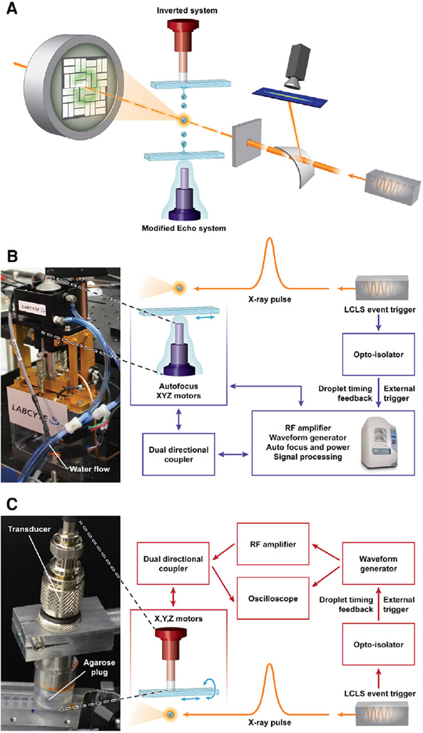
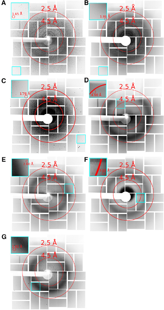
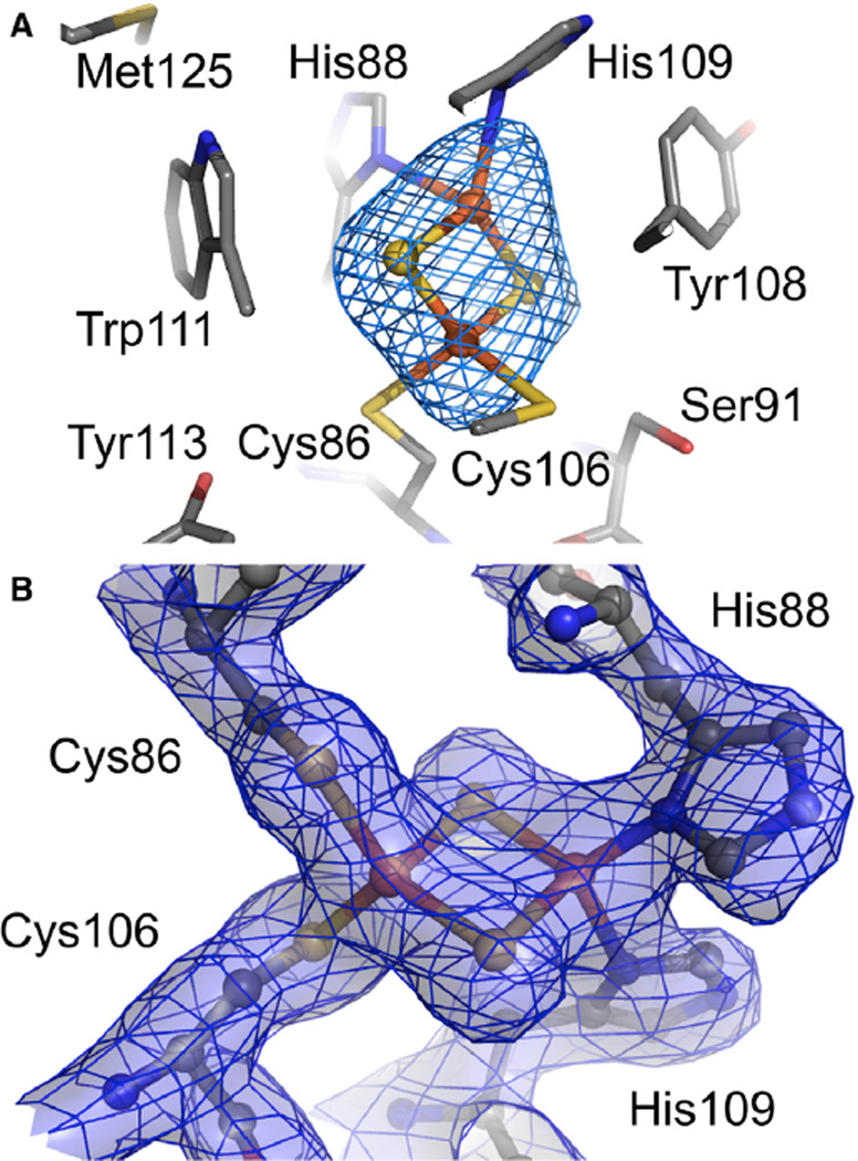
X-ray free-electron lasers (XFELs) provide very intense X-ray pulses suitable for macromolecular crystallography. Each X-ray pulse typically lasts for tens of femtoseconds and the interval between pulses is many orders of magnitude longer. Here we describe two novel acoustic injection systems that use focused sound waves to eject picoliter to nanoliter crystal-containing droplets out of microplates and into the X-ray pulse from which diffraction data are collected. The on-demand droplet delivery is synchronized to the XFEL pulse scheme, resulting in X-ray pulses intersecting up to 88% of the droplets. We tested several types of samples in a range of crystallization conditions, wherein the overall crystal hit ratio (e.g., fraction of images with observable diffraction patterns) is a function of the microcrystal slurry concentration. We report crystal structures from lysozyme, thermolysin, and stachydrine demethylase (Stc2). Additional samples were screened to demonstrate that these methods can be applied to rare samples.


Figures

Figure 1
Figure 1. Acoustic Injection for SFC
(A) Schematic overview image shows incoming X-ray pulses passing first through a bent transmissive silicon crystal, deflecting a portion of each pulse for spectrographic recording. The pulses arrive at the interaction region concurrent with a crystal-containing droplet. In the inverted system, the droplet is ejected downward out of a multiwell microplate. The modified Echo system is configured to eject droplets upwards out of a multiwell microplate. Diffraction patterns were recorded 108 mm downstream of the interaction point by a Cornell-SLAC pixel array detector. The three drops are meant to illustrate the repeatable nature of the ejection process and are not to scale. (B) Detail for the modified Echo system in which the acoustic injector (photo, left) was controlled through an “umbilical cord” with electrical and water connections to the Echo instrument. (C) Detail for the inverted system wherein an agarose plug maintained the acoustic connection between the transducer and the inverted microplate well (photo, left). Illustration courtesy Tiffany Bowman, BNL. See also Figure S1.
Figure 2
Figure 2. An Overlay, Composite, or Virtual Powder Pattern of the Diffraction Hits Observed for Acoustically Injected Samples
(A–G) Images show the maximum pixel values over selected runs merged by sample type. (A) Lysozyme (7,613 images), (B) Thermolysin (18,172 images), (C) Stc2 (26,365 images), (D) giant extracellular hemoglobin (554 images), (E) MauG-MADH (17,217 images), (F) PS-II in PEG 2000 (42,046 images), and (G) PS-II in PEG 5000 (18,197 images). Insets highlighted in cyan show individual application-specific integrated circuit tiles containing the highest-resolution reflections for each sample. Shadows appear on the top of the detector in some cases from positioning the sample holder within the cone of X-ray scatter. Some diffraction images contained multiple patterns per image. See also Figure S2.
Figure 3
Figure 3. An Example of the Quality of the Electron Density Maps around the Rieske Cluster in the Structure of Stachydrine Demethylase
(A) The 2Fe-2S Rieske cluster within Stc2 revealed by the 3-Å resolution Fo − Fc omit electron density maps contoured at +3 σ. (B) The 2 FoFc map to 2.2-Å resolution is contoured at 1.5 σ and displayed as mesh with semitransparent surfaces. Atoms are colored gray, red, blue, yellow, and orange for C, O, N, S, and Fe, respectively. See also Figure S3.

Comment in

Similar articles

Cited by

  • High-resolution crystal structures of transient intermediates in the phytochrome photocycle.
    Carrillo M, Pandey S, Sanchez J, Noda M, Poudyal I, Aldama L, Malla TN, Claesson E, Wahlgren WY, Feliz D, Šrajer V, Maj M, Castillon L, Iwata S, Nango E, Tanaka R, Tanaka T, Fangjia L, Tono K, Owada S, Westenhoff S, Stojković EA, Schmidt M. Carrillo M, et al. Structure. 2021 Jul 1;29(7):743-754.e4. doi: 10.1016/j.str.2021.03.004. Epub 2021 Mar 22. Structure. 2021. PMID: 33756101 Free PMC article.
  • Time-Resolved Macromolecular Crystallography at Pulsed X-ray Sources.
    Schmidt M. Schmidt M. Int J Mol Sci. 2019 Mar 20;20(6):1401. doi: 10.3390/ijms20061401. Int J Mol Sci. 2019. PMID: 30897736 Free PMC article. Review.
  • A multi-reservoir extruder for time-resolved serial protein crystallography and compound screening at X-ray free-electron lasers.
    Wranik M, Kepa MW, Beale EV, James D, Bertrand Q, Weinert T, Furrer A, Glover H, Gashi D, Carrillo M, Kondo Y, Stipp RT, Khusainov G, Nass K, Ozerov D, Cirelli C, Johnson PJM, Dworkowski F, Beale JH, Stubbs S, Zamofing T, Schneider M, Krauskopf K, Gao L, Thorn-Seshold O, Bostedt C, Bacellar C, Steinmetz MO, Milne C, Standfuss J. Wranik M, et al. Nat Commun. 2023 Dec 2;14(1):7956. doi: 10.1038/s41467-023-43523-5. Nat Commun. 2023. PMID: 38042952 Free PMC article.
  • Characterization of Biological Samples Using Ultra-Short and Ultra-Bright XFEL Pulses.
    Round A, Jungcheng E, Fortmann-Grote C, Giewekemeyer K, Graceffa R, Kim C, Kirkwood H, Mills G, Round E, Sato T, Pascarelli S, Mancuso A. Round A, et al. Adv Exp Med Biol. 2024;3234:141-162. doi: 10.1007/978-3-031-52193-5_10. Adv Exp Med Biol. 2024. PMID: 38507205
  • Drop-on-demand sample delivery for studying biocatalysts in action at X-ray free-electron lasers.
    Fuller FD, Gul S, Chatterjee R, Burgie ES, Young ID, Lebrette H, Srinivas V, Brewster AS, Michels-Clark T, Clinger JA, Andi B, Ibrahim M, Pastor E, de Lichtenberg C, Hussein R, Pollock CJ, Zhang M, Stan CA, Kroll T, Fransson T, Weninger C, Kubin M, Aller P, Lassalle L, Bräuer P, Miller MD, Amin M, Koroidov S, Roessler CG, Allaire M, Sierra RG, Docker PT, Glownia JM, Nelson S, Koglin JE, Zhu D, Chollet M, Song S, Lemke H, Liang M, Sokaras D, Alonso-Mori R, Zouni A, Messinger J, Bergmann U, Boal AK, Bollinger JM Jr, Krebs C, Högbom M, Phillips GN Jr, Vierstra RD, Sauter NK, Orville AM, Kern J, Yachandra VK, Yano J. Fuller FD, et al. Nat Methods. 2017 Apr;14(4):443-449. doi: 10.1038/nmeth.4195. Epub 2017 Feb 27. Nat Methods. 2017. PMID: 28250468 Free PMC article.

References

    1. Aerni H-R, Cornett DS, Caprioli RM. Automated acoustic matrix deposition for MALDI sample preparation. Anal. Chem. 2005;78:827–834. - PubMed
    1. Alonso-Mori R, Kern J, Gildea RJ, Sokaras D, Weng TC, Lassalle-Kaiser B, Tran R, Hattne J, Laksmono H, Hellmich J, et al. Energy-dispersive X-ray emission spectroscopy using an X-ray free-electron laser in a shot-by-shot mode. Proc. Natl. Acad. Sci. USA. 2012;109:19103–19107. - PMC - PubMed
    1. Aquila A, Hunter MS, Doak RB, Kirian RA, Fromme P, White TA, Andreasson J, Arnlund D, Bajt S, Barends TR, et al. Time-resolved protein nanocrystallography using an X-ray free-electron laser. Opt. Express. 2012;20:2706–2716. - PMC - PubMed
    1. Bachega JF, Bleicher L, Horjales ER, Santiago PS, Garratt RC, Tabak M. Crystallization and preliminary structural analysis of the giant haemoglobin from Glossoscolex paulistus at 3.2 A. J. Synchrotron Radiat. 2011;18:24–28. - PMC - PubMed
    1. Barends TRM, Foucar L, Botha S, Doak RB, Shoeman RL, Nass K, Koglin JE, Williams GJ, Boutet S, Messerschmidt M, et al. De novo protein crystal structure determination from X-ray free-electron laser data. Nature. 2014;505:244–247. - PubMed

Publication types