. 2016 Jan 1;6(11):8980-8991.
doi: 10.1039/C5RA26022A. Epub 2016 Jan 12.

Ultrahigh-throughput Generation and Characterization of Cellular Aggregates in Laser-ablated Microwells of Poly(dimethylsiloxane)

Affiliations

Ultrahigh-throughput Generation and Characterization of Cellular Aggregates in Laser-ablated Microwells of Poly(dimethylsiloxane)

Jacob L Albritton et al. RSC Adv. .

Abstract

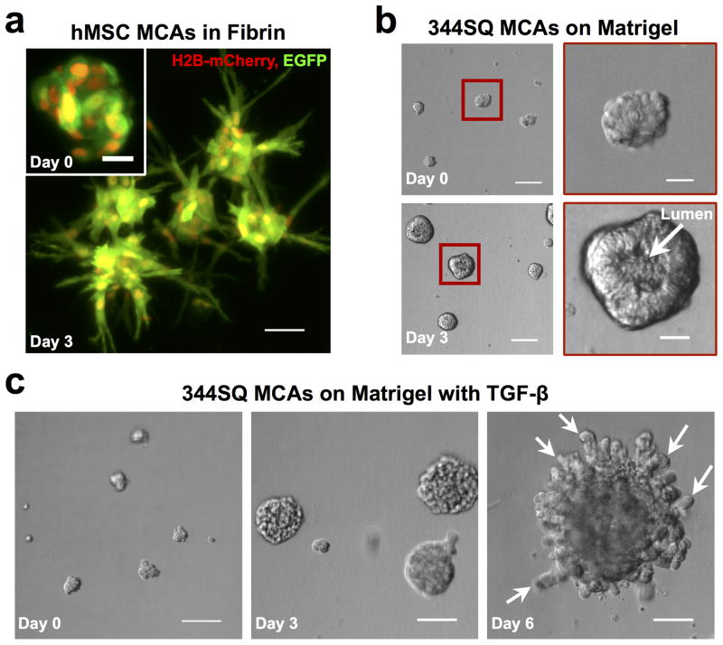
Aggregates of cells, also known as multicellular aggregates (MCAs), have been used as microscale tissues in the fields of cancer biology, regenerative medicine, and developmental biology for many decades. However, small MCAs (fewer than 100 cells per aggregate) have remained challenging to manufacture in large quantities at high uniformity. Forced aggregation into microwells offers a promising solution for forming consistent aggregates, but commercial sources of microwells are expensive, complicated to manufacture, or lack the surface packing densities that would significantly improve MCA production. To address these concerns, we custom-modified a commercial laser cutter to provide complete control over laser ablation and directly generate microwells in a poly(dimethylsiloxane) (PDMS) substrate. We achieved ultra rapid microwell production speeds (>50,000 microwells/hr) at high areal packing densities (1,800 microwells/cm2) and over large surface areas for cell culture (60 cm2). Variation of the PDMS substrate distance from the laser focal plane during ablation allowed for the generation of microwells with a variety of sizes, contours, and aspect ratios. Casting of high-fidelity microneedle masters in polyurethane allowed for non-ablative microwell reproduction through replica molding. MCAs of human bone marrow derived mesenchymal stem cells (hMSCs), murine 344SQ metastatic adenocarcinoma cells, and human C4-2 prostate cancer cells were generated in our system with high uniformity within 24 hours, and computer vision software aided in the ultra-high-throughput analysis of harvested aggregates. Moreover, MCAs maintained invasive capabilities in 3D migration assays. In particular, 344SQ MCAs demonstrated epithelial lumen formation on Matrigel, and underwent EMT and invasion in the presence of TGF-β. We expect this technique to find broad utility in the generation and cultivation of cancer cell aggregates, primary cell aggregates, and embryoid bodies.


Figures

Figure 1
Figure 1. High-throughput fabrication of conical microwells in poly(dimethylsiloxane) (PDMS)
(a) Schematic diagram (left) and photograph (right) of the selective laser ablation of a PDMS substrate to generate conical microwells. D = diameter, h = height (b) A single laser pulse generates a single microwell, and microwell height (h) can be controlled by changing either laser power (W) or laser pulse duration (ms) at each point of ablation; scalebar=500 μm. (c) To maximize packing density of microwells, custom software allowed the input of c, or center-to-center distance between adjacent microwells; scalebar=1,000 μm. (d) Vacuum removal of debris allowed large-scale microwell fabrication of a 63 cm2 area of PDMS (>100,000 microwells) in less than two hours. (e) PDMS microwell arrays can be optionally cast in polyurethane to create a master mold for an additional means of microwell reproduction; scalebar=2,000 μm (left), scalebar=500 μm (right).
Figure 2
Figure 2. Out-of-focus laser ablation generates distinct microwell shapes
Control over z-axial distance from the laser focal plane to the PDMS surface allows fabrication of microwells with distinct shapes. (a) Example z-axial distances, shown in the schematic diagram (left), generate microwells of various dimensions including cylinders (top row), sharp cones (middle row) and rounded cones (bottom row). Cross-sectional view of PDMS (left column), confocal microscopy of volume filling fluorescent dextran (middle column), and scanning electron microscopy of polyurethane negative casts (right column). (b) Custom control over laser ablation at each point allows for mixed, interleaved microwell shapes. Cross-sectional view (left); SEM (right). All scalebars=500 μm.
Figure 3
Figure 3. Cells aggregate inside multiwell plates with embedded PDMS microwell inserts
(a) 344SQ-GFP cell aggregates in a 12-well plate with 1.2 mm center-to-center spacing at ~80 microwells/cm2. Aggregates can be imaged directly inside microwells from below; scalebar=2,000 μm (left), scalebar=500 μm (middle). Aggregates can also be seen in a cross-sectional side view; scalebar=500 μm (right). (b) Labelled C4-2 cells in a 12-well plate with 0.25 mm center-to-center spacing at ~1,800 microwells/cm2; scalebar=2,000 μm (left), scalebar=200 μm (middle), scalebar=200 μm (right). (c) 344SQ-GFP aggregates in microwells from an Ommnitray (single-well plate with multiwell plate footprint) demonstrates feasibility of microwell production scale-up; scalebar=2,000 μm. (d) Labelled 344SQ MCAs isolated from microwells and the maximum intensity projection of a single MCA imaged with confocal microscopy (inset); scalebar=200 μm, scalebar=25 μm (inset). (e) MCA Maximum Feret Diameter histogram for microwells seeded with cells at 10, 25, and 75 cells/microwell and harvested after one day of aggregation. (f) MCAs from microwells seeded at 75 cells/microwell were filtered with a 40 μm cell strainer.
Figure 4
Figure 4. Cells aggregate in PDMS microwells made by replica molding
(a) Schematic of non-ablative PDMS microwell fabrication by replica molding. A polyurethane microneedle array (see Fig. 1e) can be cast from a pre-existing PDMS microwell template (Fig. 1d). The polyurethane cast can serve as master for successive casting of PDMS microwell arrays without the need for laser ablation. (b) Cross-sectional view of PDMS microwells made by replica molding; scalebar=200 μm. (c) Top view of 344SQ-GFP aggregates (Day 1) in PDMS microwells made by replica molding; scalebar=200 μm. (d) MCA Maximum Feret Diameter histogram for microwells seeded with cells at 10 cells/microwell and harvested after one day of aggregation.
Figure 5
Figure 5. hMSCs and 344SQ cells maintain mobility after aggregation inside PDMS microwells
(a) Human bone marrow derived mesenchymal stem cells grown in fibrin gels for 3 days. Cells are clearly sprouting away from the MCA center; scalebar=500 μm, scalebar=20 μm (inset). (b) 344SQ MCAs lumenize over time when cultured on Matrigel® (left); scalebar = 100 μm. Day 3 aggregates have lumen-like absence in the center compared with Day 0 aggregates (right); scalebar=25 μm. (c) hTGF-β (2ng/mL) induces 344SQ MCAs to sprout into Matrigel®; scalebar=100 μm.

References

    1. Bissell MJ, Radisky D. Nat Rev Cancer. 2001;1:46–54. - PMC - PubMed
    1. Fischbach C, Chen R, Matsumoto T, Schmelzle T, Brugge JS, Polverini PJ, Mooney DJ. Nat Methods. 2007;4:855–860. - PubMed
    1. Dreher MR, Liu W, Michelich CR, Dewhirst MW, Yuan F, Chilkoti A. J Natl Cancer Inst. 2006;98:335–344. - PubMed
    1. Nelson CM, Bissell MJ. Annu Rev Cell Dev Biol. 2006;22:287–309. - PMC - PubMed
    1. Griffith LG, Swartz MA. Nat Rev Mol Cell Biol. 2006;7:211–24. - PubMed

LinkOut - more resources