. 2016 Mar 21;11(3):e0151753.
doi: 10.1371/journal.pone.0151753. eCollection 2016.

The Circular RNA Cdr1as Promotes Myocardial Infarction by Mediating the Regulation of miR-7a on Its Target Genes Expression

Affiliations

The Circular RNA Cdr1as Promotes Myocardial Infarction by Mediating the Regulation of miR-7a on Its Target Genes Expression

Hai-Hua Geng et al. PLoS One. .

Abstract

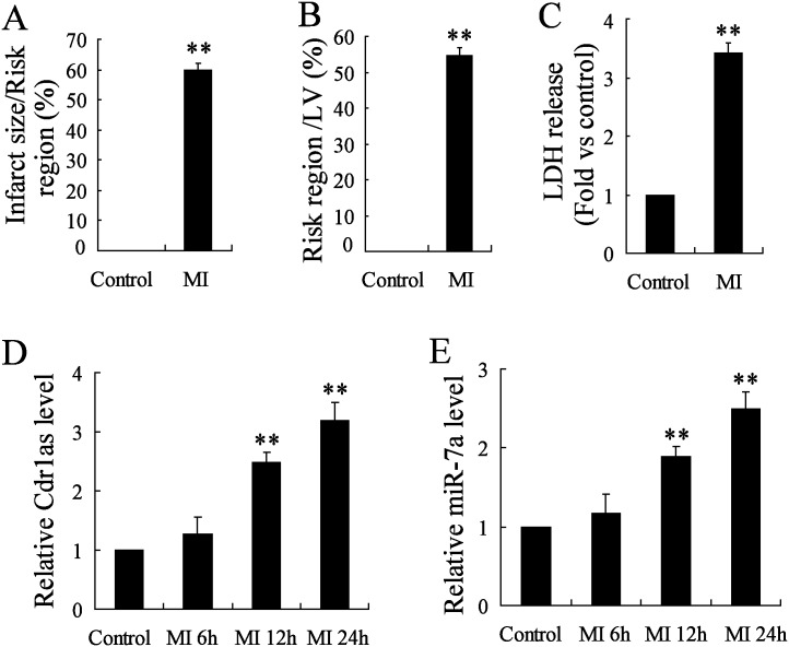
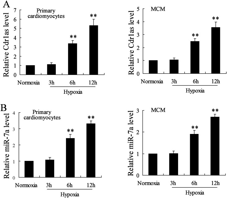
Objectives: Recent studies have demonstrated the role of Cdr1as (or CiRS-7), one of the well-identified circular RNAs (circRNAs), as a miR-7a/b sponge or inhibitor in brain tissues or islet cells. This study aimed to investigate the presence of Cdr1as/miR-7a pathway in cardiomyocytes, and explore the mechanism underlying the function of miR-7a in protecting against myocardial infarction (MI)-induced apoptosis.

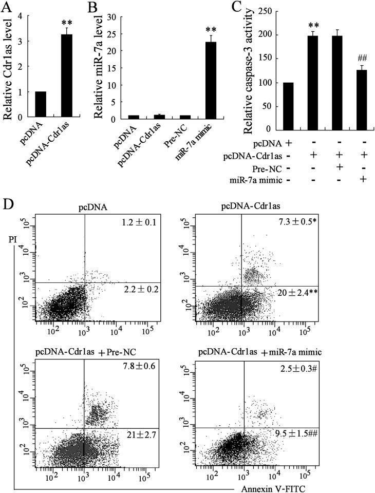
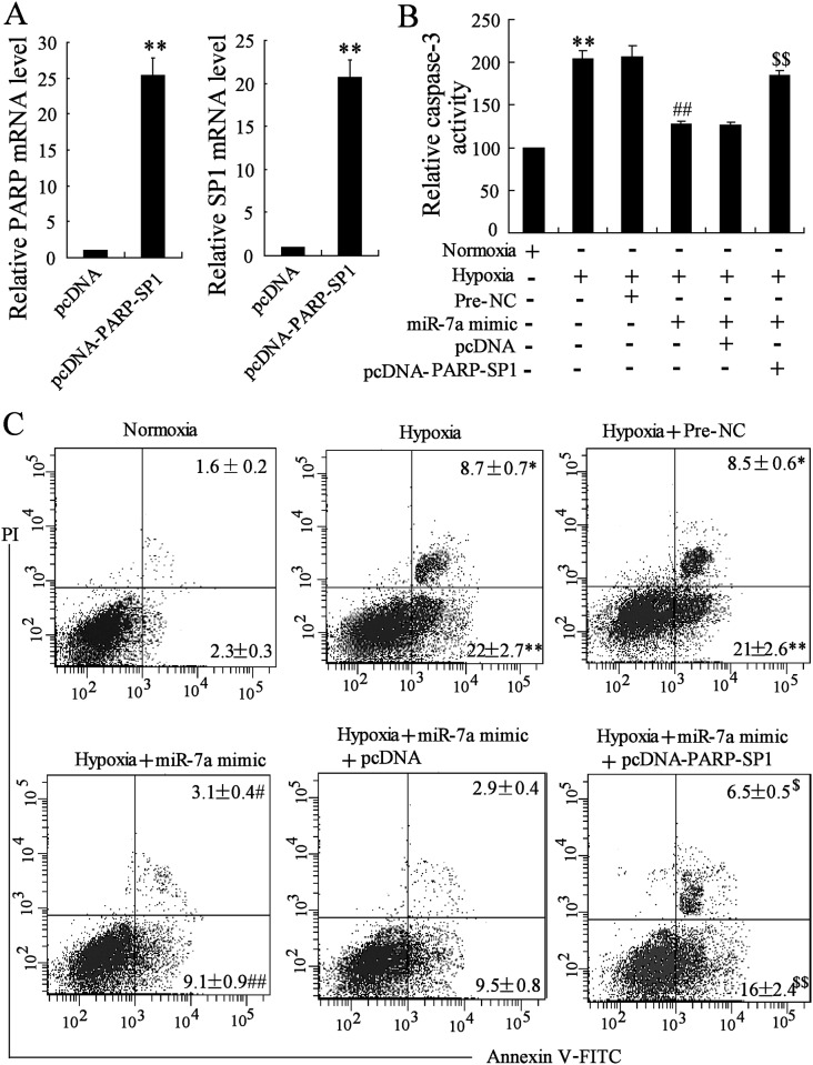
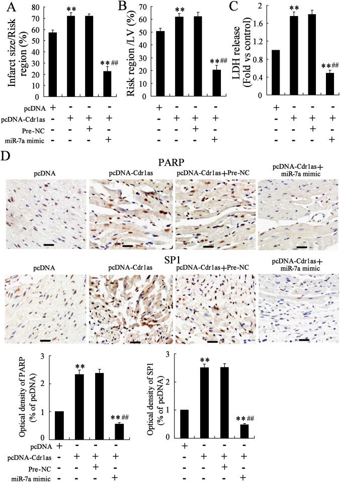
Methods: Mouse MI injury model was established and evaluated by infarct size determination. Real-time PCR was performed to quantify the expression of Cdr1as and miR-7a in cardiomyocytes. Cell apoptosis was determined by caspase-3 activity analysis and flow cytometry assays with Annexin V/PI staining. Transfection of Cdr1as overexpressing plasmid and miR-7a mimic were conducted for gain-of-function studies. Luciferase reporter assay and western blot analysis were performed to verity potential miR-7a targets.

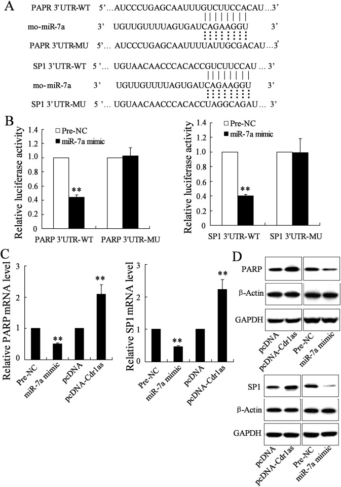
Results: Cdr1as and miR-7a were both upregulated in MI mice with increased cardiac infarct size, or cardiomyocytes under hypoxia treatment. Cdr1as overexpression in MCM cells promoted cell apoptosis, but was then reversed by miR-7a overexpression. The SP1 was identified as a new miR-7a target, in line with previously identified PARP, while miR-7a-induced decrease of cell apoptosis under hypoxia treatment was proven to be inhibited by PARP-SP1 overexpression. Moreover, Cdr1as overexpression in vivo increased cardiac infarct size with upregulated expression of PARP and SP1, while miR-7a overexpression reversed these changes.

Conclusions: Cdr1as also functioned as a powerful miR-7a sponge in myocardial cells, and showed regulation on the protective role of miR-7a in MI injury, involving the function of miR-7a targets, PARP and SP1.


Conflict of interest statement

Competing Interests: The authors have declared that no competing interests exist.

Figures

Fig 1
Fig 1. Upregulated expression of Cdr1as and miR-7a was detected in myocardial tissues from MI mice.
The infarct size (A), area at risk (B), and lactate dehydrogenase (LDH) release (C) were analyzed to examine the established mouse MI model, along with a sham group (control). And, myocardial tissues were disassociated at 6, 12, or 24 h after MI introduction, to detect the expression of Cdr1as (D) and miR-7a (E) by qRT-PCR analysis. n = 6, **P < 0.01 vs. control.
Fig 2
Fig 2. Cdr1as expression was elevated in hypoxia-treated cardiomyocytes with overexpressed miR-7a.
Primary cardiomyocytes, obtained from C57BL/6 mice, and mouse myocardia cell line MCM were subjected to hypoxia treatment for 3, 6, or 12 h for RNA expression analysis by qRT-PCR. (A) Expression of Cdr1as. (B) Expression of miR-7a. n = 3, **P < 0.01: compared to cells under normoxia condition.
Fig 3
Fig 3. MiR-7a overexpression reversed Cdr1as-induced apoptosis of MCM cells.
MCM cells were transfected with pcDNA-Cdr1as or miR-7a mimic for the overexpression of Cdr1as and miR-7a respectively, with pcDNA or Pre-NC as the respective negative control. After the transfection, Cdr1as or miR-7a expression level was confirmed by qRT-PCR. Analysis for caspase-3 activity and cell apoptosis were performed by the colorimetric method and Annexin V/PI staining respectively. A. Expression of Cdr1as. B. Expression of miR-7a. C. Relative caspase-3 activity under different conditions. D. Representative flow cytometry of apoptosis of MCM cells. The percentage of apoptotic cells with positive Annexin V signal were analyzed and compared accordingly. n = 3, *P < 0.05, **P<0.01: compared to cells transfected with pcDNA (A, C, D) or Pre-NC (B) as control; #P < 0.05, ##P < 0.01: compared to cells co-transfected with pcDNA-Cdr1as and Pre-NC.
Fig 4
Fig 4. Regulation of miR-7a on the expression of PARP and SP1 was inhibited by Cdr1as.
A. Conserved miR-7a binding sites in 3’-UTR of PARP and SP1. WT: wild type; MU: mutant type. B. Luciferase activity of the 3’-UTR of PARP and SP1 in miR-7a overexpressing cells, after normalized to that of Renilla. Cells transfected with Pre-NC were used as control. C. RT-PCR analysis of mRNA level of PARP and SP1 under miR-7a or Cdr1as overexpression. Cells transfected with Pre-NC (for miR-7a mimic) or pcDNA (for pcDNA-Cdr1as) were used as control D. Western blot analysis of protein level of PARP and SP1. n = 3, **P < 0.01 vs. control.
Fig 5
Fig 5. Hypoxia-induced apoptosis was mediated by the targeting of miR-7a on PARP and SP1.
MCM cells were transfected miR-7a mimic or co-transfected with miR-7a mimic and pcDNA-PARP-SP1, and then subjected to hypoxia treatment. A. Expression of PARP. B. Expression of SP1. C. Relative caspase-3 activity under different conditions. D. Representative flow cytometry of apoptosis of MCM cells. PI: propidium iodide. n = 3, *P < 0.05, **P < 0.01: compared to cells with pcDNA transfection (A, B) or under normoxia condition (C, D); #P < 0.05, ##P < 0.01: compared to cells with Pre-NC transfection under hypoxia condition; $P < 0.05, $ $P < 0.01: compared to cells co-transfected with miR-7a mimic and pcDNA under hypoxia condition.
Fig 6
Fig 6. MiR-7a overexpression reversed Cdr1as-induced bad effects in mouse MI model.
Three days after the transfection of pcDNA or pcDNA-Cdr1as, mice were subjected to MI introduction. Effect of Cdr1as overexpression on MI-induced infarct size (A), area at risk (B), lactate dehydrogenase (LDH) release (C), and the expression of PARP and SP1 (D; bar = 100μm) was analyzed, as well as miR-7a co-overexpression with Cdr1as through the delivery of miR-7a mimic three days before MI introduction. n = 6, **P < 0.01: compared to MI mice with pcDNA transfection; ##P < 0.01: compared to MI mice co-transfected with pcDNA-Cdr1as and Pre-NC.

References

    1. Mozaffarian D, Benjamin EJ, Go AS, Arnett DK, Blaha MJ, Cushman M, et al. Heart disease and stroke statistics-2015 update: a report from the american heart association. Circulation. 2015;131(4):e29 10.1161/CIR.0000000000000152 - DOI - PubMed
    1. Mackman N. Triggers, targets and treatments for thrombosis. Nature. 2008;451(7181):914–8. 10.1038/nature06797 - DOI - PMC - PubMed
    1. Swynghedauw B. Molecular mechanisms of myocardial remodeling. Physiological reviews. 1999;79(1):215–62. - PubMed
    1. Chavakis E, Koyanagi M, Dimmeler S. Enhancing the outcome of cell therapy for cardiac repair progress from bench to bedside and back. Circulation. 2010;121(2):325–35. 10.1161/CIRCULATIONAHA.109.901405 - DOI - PubMed
    1. Cho HJ, Lee HJ, Youn SW, Koh SJ, Won JY, Chung YJ, et al. Secondary sphere formation enhances the functionality of cardiac progenitor cells. Molecular therapy: the journal of the American Society of Gene Therapy. 2012;20(9):1750–66. 10.1038/mt.2012.109 - DOI - PMC - PubMed