. 2016 Mar 17;21(3):365.
doi: 10.3390/molecules21030365.

Silver Nanoparticles Exhibit the Dose-Dependent Anti-Proliferative Effect against Human Squamous Carcinoma Cells Attenuated in the Presence of Berberine

Affiliations

Silver Nanoparticles Exhibit the Dose-Dependent Anti-Proliferative Effect against Human Squamous Carcinoma Cells Attenuated in the Presence of Berberine

Arkadiusz Dziedzic et al. Molecules. .

Abstract

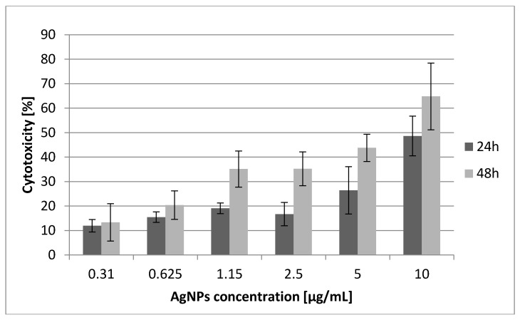
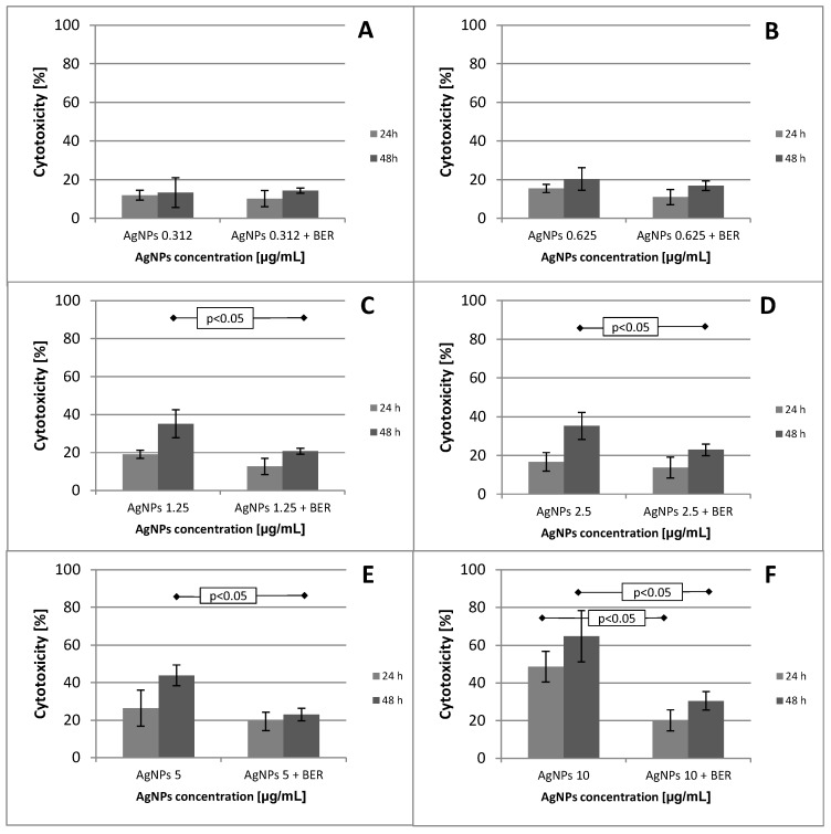
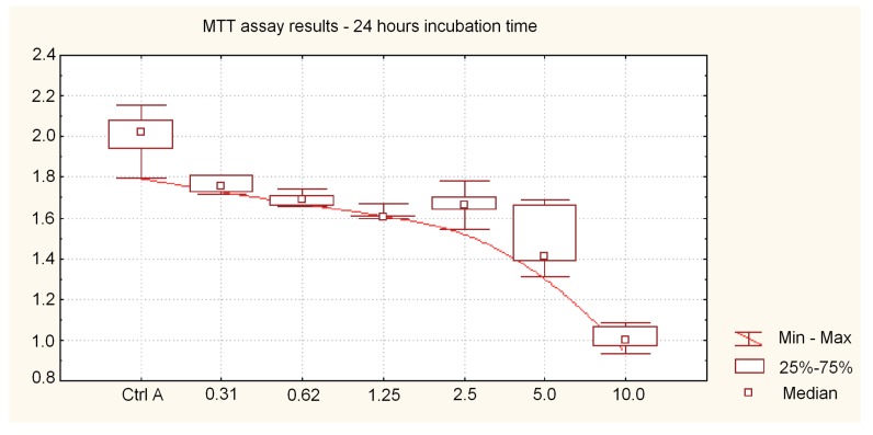
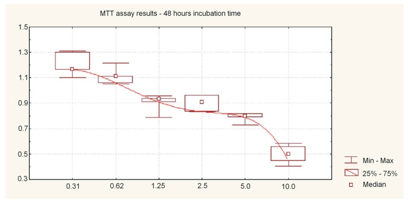
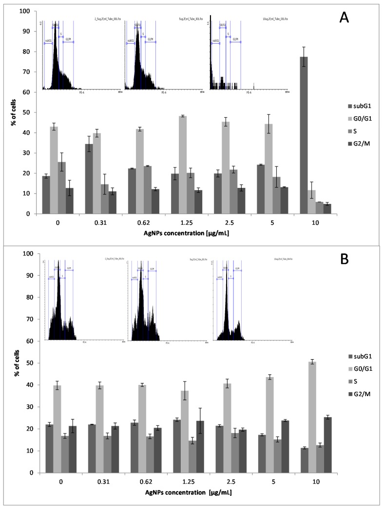
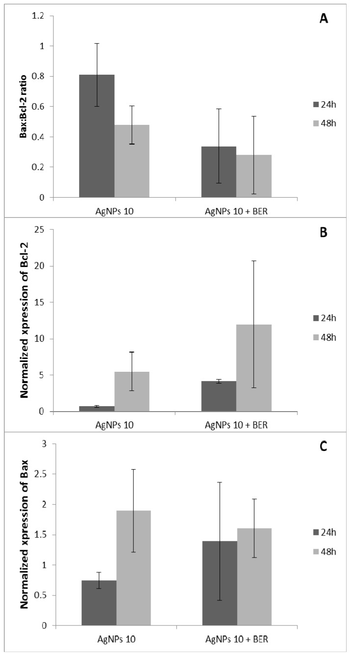
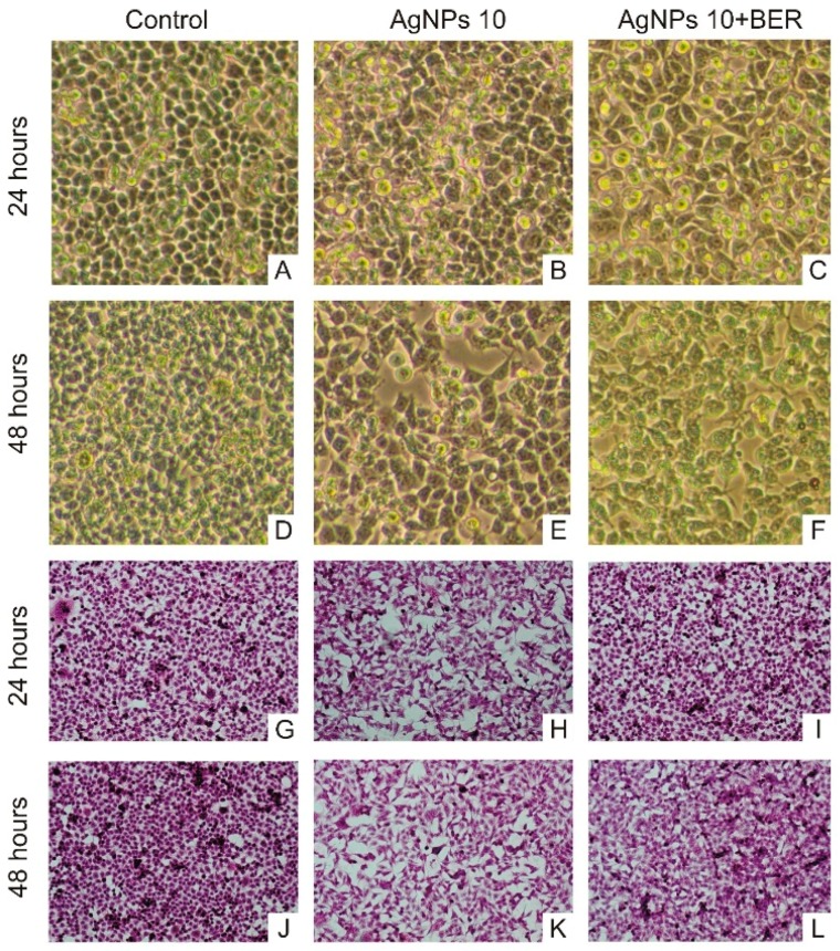
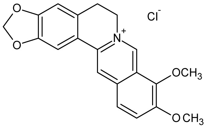
The biological activity of nanosize silver particles towards oral epithelium-derived carcinoma seems to be still underinvestigated. We evaluated the influence of low doses of nanosize scale silver particles on the proliferation and viability of malignant oral epithelial keratinocytes in vitro, alone and in conjunction with the plant alkaloid berberine. Cells of human tongue squamous carcinoma SCC-25 (ATCC CRL-1628), cultivated with the mixture of Dulbecco's modified Eagle's medium, were exposed to silver nanoparticles alone (AgNPs, concentrations from 0.31 to 10 μg/mL) and to a combination of AgNPs with berberine chloride (BER, 1/2 IC50 concentration) during 24 h and 48 h. The cytotoxic activity of AgNPs with diameters of 10 nm ± 4 nm was measured by 3-(4,5-dimethyl-2-thiazyl)-2,5-diphenyl-2H-tetrazolium bromide (MTT) assay. Cell cycle analysis was performed by treating cells with propidium iodide followed by flow-activated cell sorting. RT-QPCR reaction was used to assess expression of anti-apoptotic proteins Bcl-2 and pro-apoptotic protein Bcl-2-associated X protein Bax genes expression. Monodisperse silver nanoparticles at a concentration of 10 μg/mL arrested SCC-25 cells cycle after 48 h at the G0/G1 phase in a dose- and time-dependent manner through disruption G0/G1 checkpoint, with increase of Bax/Bcl-2 ratio gene expression. AgNPs exhibit cytotoxic effects on SCC-25 malignant oral epithelial keratinocytes, which is diminished when combined with BER. The AgNPs concentration required to inhibit the growth of carcinoma cells by 50% (IC50) after 48 h was estimated at 5.19 μg/mL. AgNPs combined with BER increased the expression of Bcl-2 while decreasing the ratio of Bax/Bcl-2 in SCC-25 cells. Silver particles at low doses therefore reduce the proliferation and viability of oral squamous cell carcinoma cells. SCC-25 cells are susceptible to damage from AgNPs-induced stress, which can be regulated by the natural alkaloid berberine, suggesting that nanoparticles may be potentially used in a chemoprevention/chemotherapy by augmentation of action of standard anti-cancer drugs.

Keywords: MTT assay; RT-QPCR; SCC-25; bax/bcl-2 gene expression; berberine; cell cycle arrest; oral carcinoma; silver nanoparticles.


Conflict of interest statement

The authors declare no conflict of interest.

Figures

Figure 1
Figure 1
Cytotoxic effects of silver nanoparticles (10 nm diameter, concentrations 0.31 μg/mL–10 μg/mL) on SCC-25 cancer cells. The percentage of cell death measured by MTT cytotoxicity assay. MTT values represent mean ± SD of three independent cytotoxicity experiments performed in quadruplicate (n = 12). The lower concentration of AgNPs (e.g., 0.625 μg/mL) after 48 h produced the same killing effect on SCC-25 cells (20%) as 3 μg/mL AgNP concentration after 24 h. Mean cytotoxicity between different AgNPs concentrations alone were highly significant above the concentration of 2.5 μg/mL (p < 0.01, ANOVA Friedman ANOVA test, Wilcoxon test).
Figure 2
Figure 2
MTT assay results after 24 and 48 h of cells culture incubation representing mean values for different concentrations of AgNPs alone and in combination with a fixed 1/2 IC50 (12.5 μg/mL) concentration of BER. AgNPs with BER inhibited significantly less the growth of SCC-25 in a dose-dependent manner which clearly demonstrates an attenuating effect of BER addition, particularly at 5 and 10 μg/mL concentrations (AF). Differences of mean cytotoxicity values between different AgNPs concentrations alone vs. AgNPs/BER were significant (p < 0.05 and p < 0.01, ANOVA Friedman ANOVA test).
Figure 3
Figure 3
Assessment of cell viability after treatment with various nanosilver particle concentrations during 24 h. Cell viability compared to the control (untreated cells) was plotted against the absorbance at 570 nm. MTT assay results after 24 h of incubation representing absorbance values (median, min-max) for 0.31 μg/mL to 10 μg/mL AgNP concentrations. AgNPs inhibited the cells’ growth in a dose-dependent manner. Differences of mean absorbance values between different AgNPs concentrations were highly significant (p < 0.01, ANOVA Friedman ANOVA test).
Figure 4
Figure 4
MTT assay results after 48 h of cell culture incubation representing absorbance values (median, min-max) for different dispersed Ag-NP concentrations (0.31 μg/mL to 10 μg/mL). Like after 24 h incubation time, AgNPs also inhibited the growth in a dose-dependent manner, with a significant variation of MTT absorbance values compared to the 24 h protocol. A “flattened” viability curve between the concentrations of 1.5 and 5 μg/mL of AgNP could be an effect of the experimental conditions. Differences of mean absorbance values between different AgNPs concentrations were highly significant (p < 0.01, ANOVA Friedman ANOVA test).
Figure 5
Figure 5
Representative flow cytometric histograms and bar graphs showing the cell cycle distribution when the SCC-25 cells were treated with AgNPs at 0.31, 0.62, 2.5, 5, 10 μg/mL for 24 h (A) and 48 h (B). Cells were stained with propidium iodide and subjected to flow cytometric analysis that collected 10,000 events. Alterations in the percentage of SCC-25 cells in subG1, G0/G1, S, and G2/M phases of cell cycle are presented as the mean ± SD of three independent experiments. The results show that AgNPs (10 µg/mL) has a significant effect on cell cycle arrest, contributing to their anticancer features. AgNPs treatment of SCC-25 with 10 µg/mL concentration for 24 h and 48 h resulted in a cell cycle (checkpoint) arrest within the subG1 and G0/G1 phase, respectively (p < 0.01 and p < 0.05, independent experiments).
Figure 6
Figure 6
Effect of AgNPs and combined action of AgNPs + BER on the expression levels of apoptosis-related genes Bax/Bcl-2 in SCC25 cells. The expression levels of Bax, Bcl-2 in SCC-25 cells were determined by RT-QPCR assay. Specificity of the reaction of amplification of all fragments of studied genes was assessed by means of polyacrylamide gel electrophoresis. Each sample was examined in two repetitions. (A) Representative bar graphs show the expression of relative level of Bax/Bcl-2 ratio, when the cells were treated with AgNPs at 10 μg/mL and combination of AgNPs/BER for 24 and 48 h (p < 0.05, ANOVA, AgNPs vs. AgNPs + BER); (B) Representative bar graphs show the expression of level of Bcl-2 gene, when the cells were treated with AgNPs at 10 μg/mL and combination of AgNPs/BER for 24 and 48 h (p < 0.05, ANOVA, AgNPs vs. AgNPs + BER); (C) Representative bar graphs show the expression of level of Bax gene, when the cells were treated with AgNPs at 10 μg/mL and combination of AgNPs/BER for 24 and 48 h.
Figure 7
Figure 7
Microscopic images (magnification 10 × 10) of untreated SCC-25 cells and ones treated with 10 μg/mL AgNPs and 10 μg/mL AgNPs with 12.5 μg/mL BER (1/2 IC50 BER concentration). (AF) Contrast-phase microscopy, (GL) hematoxylin & eosin staining. Morphological changes of SCC-25 exposed to AgNPs include decreased numbers of cells, with less regular, non-round shapes, and morphology variations.
Figure 8
Figure 8
Chemical structure of the berberine derivative—berberine chloride salt, containing a protoberberine skeleton and nitrogen cation ion. The positively charged berberine molecule possesses a great affinity to react rapidly with negatively charged anions, e.g., silver nanoparticles. This mechanism depends on the electrostatic interaction between the charged surfaces and cationic berberine. Besides the electrostatic forces, biding interactions may also play a significant role in the attenuation effect of BER vs. AgNPs. Hypothetically, the BER molecules structure turns into a less biologically active form after encapsulation by AgNPs, which is followed by further AgNP aggregation. The alteration of BER to encapsulated form and the interplay of AgNPs surface charge determine the interaction between the alkaloid and AgNPs.
Figure 9
Figure 9
SCC-25 squamous cell carcinoma cell morphology and spreading pattern: nuclear enlargement, highly irregular shaped cells, dense chromatin, prominent nucleoli, cells are connected to each other through short and slender extensions (A) nonstained culture, untreated SCC-25 cells, optical magnification 400×). Cytological features of untreated SCC-25 carcinoma cells exhibiting characteristic signs of cellular atypia: nuclear and pleomorphic cytoplasmic pattern, increase nucleus-cytoplasm ratio, irregular nuclear shapes, hyperchromasia (B) hematoxylin & eosin staining, magnification 400×). Cells were costained (blue) to label DNA nucleic acids. Hoechst staining shows that most cells have round and intact nuclei (C) immunofluorescent staining of untreated cells using Hoechst 33342 fluorescent dye).

References

    1. Marur S., Forastiere A.A. Head and neck cancer: changing epidemiology, diagnosis, and treatment. Mayo Clin. Proc. 2008;83:489–501. doi: 10.4065/83.4.489. - DOI - PubMed
    1. International Agency for Research on Cancer . GLOBOCAN 2012: Estimated Cancer Incidence, Mortality and Prevalence Worldwide in 2012. World Health Organization; Lyon, France: 2012.
    1. Howlader N., Noone A.M., Krapcho M., Garshell J., Miller D., Altekruse S.F., Kosary C.L., Yu M., Ruhl J., Tatalovich Z., et al. SEER Cancer Statistics Review, 1975–2011. In: Bethesda M.D., editor. National Cancer Institute; Bethesda, MD, USA: 2014. [(accessed on 14 March 2016)]. Available online: http://seer.cancer.gov/csr/1975_2011.
    1. Amir Z., Kwan S.Y., Landes D., Feber T., Williams S.A. Diagnostic delays in head and neck cancers. Eur. J. Cancer Care. 1999;8:198–203. doi: 10.1046/j.1365-2354.1999.00165.x. - DOI - PubMed
    1. Berrino F., Gatta G. Variation in survival of patients with head and neck cancer in Europe by the site of origin of the tumours. Eur. J. Cancer. 1998;34:2154–2161. doi: 10.1016/S0959-8049(98)00328-1. - DOI - PubMed

Publication types

MeSH terms

LinkOut - more resources