. 2016 Mar 21;11(3):e0151365.
doi: 10.1371/journal.pone.0151365. eCollection 2016.

Shape-Related Toxicity of Titanium Dioxide Nanofibres

Affiliations

Shape-Related Toxicity of Titanium Dioxide Nanofibres

Manfredi Allegri et al. PLoS One. .

Abstract

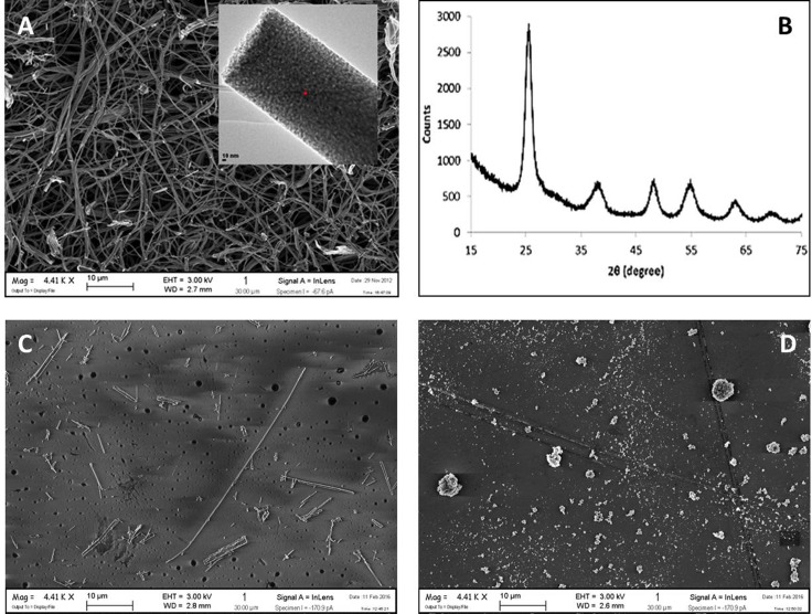
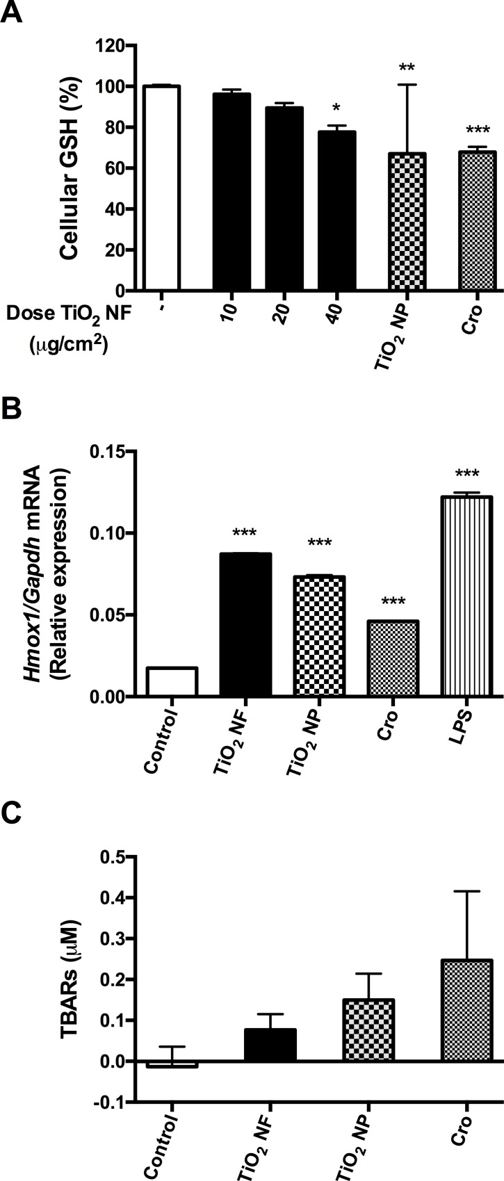
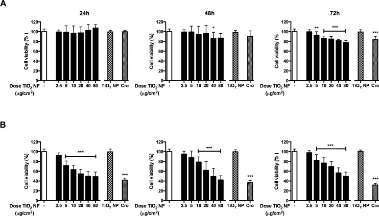
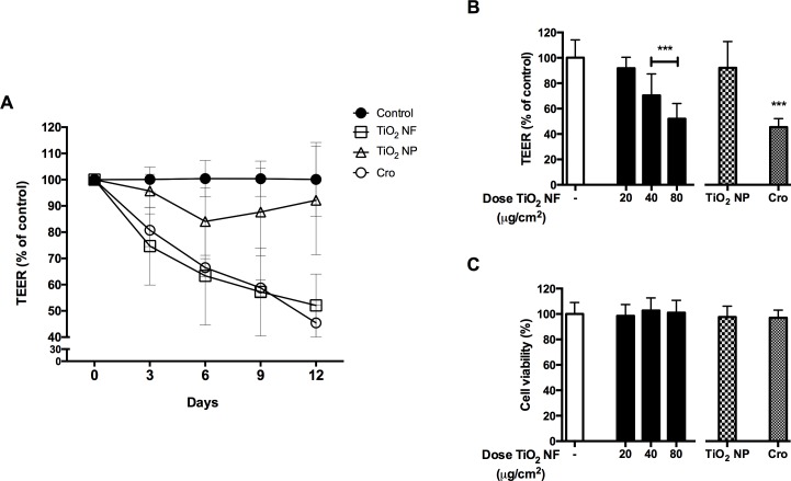
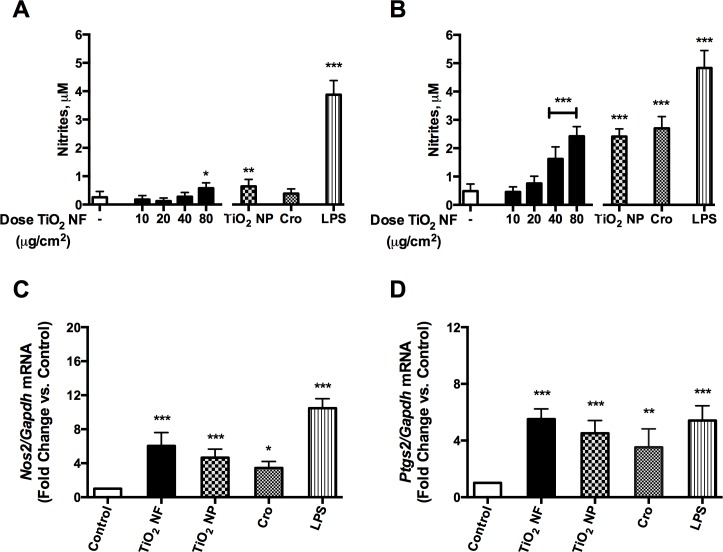
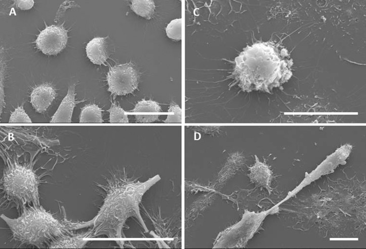
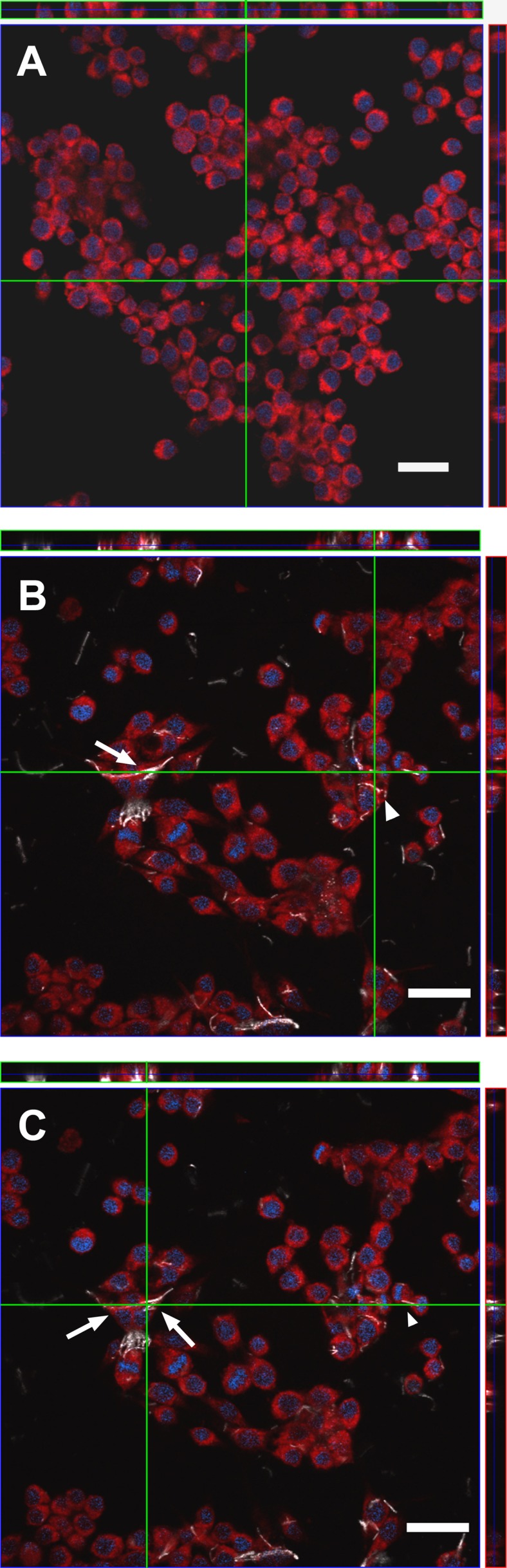
Titanium dioxide (TiO2) nanofibres are a novel fibrous nanomaterial with increasing applications in a variety of fields. While the biological effects of TiO2 nanoparticles have been extensively studied, the toxicological characterization of TiO2 nanofibres is far from being complete. In this study, we evaluated the toxicity of commercially available anatase TiO2 nanofibres using TiO2 nanoparticles (NP) and crocidolite asbestos as non-fibrous or fibrous benchmark materials. The evaluated endpoints were cell viability, haemolysis, macrophage activation, trans-epithelial electrical resistance (an indicator of the epithelial barrier competence), ROS production and oxidative stress as well as the morphology of exposed cells. The results showed that TiO2 nanofibres caused a cell-specific, dose-dependent decrease of cell viability, with larger effects on alveolar epithelial cells than on macrophages. The observed effects were comparable to those of crocidolite, while TiO2 NP did not decrease cell viability. TiO2 nanofibres were also found endowed with a marked haemolytic activity, at levels significantly higher than those observed with TiO2 nanoparticles or crocidolite. Moreover, TiO2 nanofibres and crocidolite, but not TiO2 nanoparticles, caused a significant decrease of the trans-epithelial electrical resistance of airway cell monolayers. SEM images demonstrated that the interaction with nanofibres and crocidolite caused cell shape perturbation with the longest fibres incompletely or not phagocytosed. The expression of several pro-inflammatory markers, such as NO production and the induction of Nos2 and Ptgs2, was significantly increased by TiO2 nanofibres, as well as by TiO2 nanoparticles and crocidolite. This study indicates that TiO2 nanofibres had significant toxic effects and, for most endpoints with the exception of pro-inflammatory changes, are more bio-active than TiO2 nanoparticles, showing the relevance of shape in determining the toxicity of nanomaterials. Given that several toxic effects of TiO2 nanofibres appear comparable to those observed with crocidolite, the possibility that they exert length dependent toxicity in vivo seems worthy of further investigation.


Conflict of interest statement

Competing Interests: The authors have declared that no competing interests exist.

Figures

Fig 1
Fig 1. FE-SEM image and XRD pattern of TiO2 nanofibres.
(A) The sample of TiO2 NF consists of discrete units of primary nano-particles, evident in the inset at higher magnification. (B) The XRD pattern of the material. (C) FE-SEM image of crocidolite. (D) FE-SEM image of TiO2 NP. For (A), (C) and (D). Bars, 10 μm.
Fig 2
Fig 2. Glutathione depletion, Hmox1 induction, and lipid peroxidation in macrophages.
Glutathione (GSH) content (A), the expression of Hmox1 (B) and the level of thiobarbituric acid reactive substances (TBARs) (C) were determined in Raw 264.7 cells after a 12-hour (B) or 24-hour (A-C) incubation with or without the indicated materials. For TiO2 NF, the doses were 10, 20, 40 μg/cm2 (A), 80 μg/cm2 (B), or 40 μg/cm2 (C); for crocidolite (Cro) and TiO2 NP, the doses were 40 μg/cm2 (A,C) or 80 μg/cm2 (B). Results are expressed as % of decrease (A) or relative expression (B) or fold-change (C) vs. control, untreated cultures. (A,C) Data are means ± SEM of 9 independent determinations. * p<0.05, ** p<0.01, *** p<0.001 vs. control. (B) Data are means ± SD of 4 values obtained in 2 separate experiments.
Fig 3
Fig 3. Effect of tested particles on cell viability.
Raw 264.7 (A) and A549 cells (B) were treated up to 72h with or without pristine TiO2 NF (range dose 2.5–80 μg/cm2), crocidolite (Cro; 80 μg/cm2) or TiO2 NP (80 μg/cm2), and cell viability was assessed with the resazurin assay. Data are means ± SD of twelve independent determinations obtained in three experiments. * p < 0.05, ** p < 0.01, *** p < 0.001 vs. control (untreated cultures).
Fig 4
Fig 4. Effect of tested materials on barrier integrity and cell viability of CaLu-3 cell monolayers.
The Trans-Epithelial Electrical Resistance (TEER), as a proxy of epithelial barrier competence, and cell viability were assessed in confluent monolayers of CaLu-3 cells after incubation with TiO2 NF, TiO2 NP or crocidolite (Cro) all used at 80 μg/cm2. (A) TEER (% of control) was recorded every 3d up to 12d. (B) TEER (% of control) measured at 12d. (C) Effect of TiO2 NF on cell viability, assessed by resazurin assay at the end of TEER experiments. Data are means ± SD of 8 independent determinations. *** p < 0.001 vs. control (untreated cultures).
Fig 5
Fig 5. Expression of pro-inflammatory markers in macrophages.
Macrophages were incubated up to 72h with TiO2 NF, at the indicated doses, TiO2 NP and crocidolite (Cro) at 80 μg/cm2, or with 10 ng/mL of LPS as a positive control. (A, B) Effects of TiO2 NF (dose range 10–80 μg/cm2) on NO production after 48h (A) or 72h (B). (C, D) Effects of the indicated materials (all used at 80 μg/cm2) on Nos2 (C) or Ptgs2 (D) gene expression in Raw 264.7 cells, assessed with RT-PCR after a 24h-exposure. (A, B) Data are means ± SD of 9 independent determinations obtained in three experiments. (C, D) Data are means ± SD of 4 independent determinations obtained in two experiments. For all the panels, * p < 0.05, ** p < 0.01 *** p < 0.001 vs. control (untreated cultures).
Fig 6
Fig 6. Characterization of the interaction between macrophages and materials by scanning electron microscopy.
Raw 264.7 cells were seeded on coverslips and incubated for 24h in the absence (A) or in the presence of TiO2 NF (B), TiO2 NP (C) or crocidolite asbestos (D) at a dose of 10 μg/cm2. Cells were then labelled, fixed, mounted for SEM analysis as described in Materials and methods. Bars, 20 μm.
Fig 7
Fig 7. Characterization of the interaction between macrophages and TiO2 NF by confocal microscopy.
Raw 264.7 macrophages were seeded on coverslides and incubated for 24h with TiO2 NF (10 μg/cm2). At the end of the experiment, cells were labelled and fixed as detailed in Materials and methods. (A) A single horizontal section of control, untreated cells is shown along with two orthogonal projections. (B, C) two single horizontal sections of NF-treated cells, taken at different planes, are shown along with two orthogonal projections. In (B) and (C), arrows point to pattern suggestive of cell fusion and arrowheads to an example of internalized material. In each panel, green lines highlight the planes where the orthogonal projections were taken. White, TiO2 NF; Blue, nucleus; Red, cytoplasm. Images report representative fields. Bars, 20 μm.

References

    1. Fubini B (1997) Surface reactivity in the pathogenic response to particulates. Environ Health Perspect 105 Suppl 5: 1013–1020. - PMC - PubMed
    1. Cho W-S, Duffin R, Poland CA, Duschl A, Oostingh GJ, MacNee W, et al. (2012) Differential pro-inflammatory effects of metal oxide nanoparticles and their soluble ions in vitro and in vivo; zinc and copper nanoparticles, but not their ions, recruit eosinophils to the lungs. Nanotoxicology 6: 22–35. 10.3109/17435390.2011.552810 - DOI - PubMed
    1. Donaldson K, Tran CL (2004) An introduction to the short-term toxicology of respirable industrial fibres. Mutation research 553: 5–9. - PubMed
    1. Donaldson K (2009) The inhalation toxicology of p-aramid fibrils. Crit RevToxicol 39: 487–500. - PubMed
    1. Donaldson K, Murphy F, Schinwald A, Duffin R, Poland CA (2011) Identifying the pulmonary hazard of high aspect ratio nanoparticles to enable their safety-by-design. Nanomedicine 6: 143–156. 10.2217/nnm.10.139 - DOI - PubMed

Publication types

MeSH terms