. 2016 Apr 1;126(4):1413-24.
doi: 10.1172/JCI82771. Epub 2016 Mar 21.

Alloantigen-specific regulatory T cells generated with a chimeric antigen receptor

Alloantigen-specific regulatory T cells generated with a chimeric antigen receptor

Katherine G MacDonald et al. J Clin Invest. .

Abstract

Adoptive immunotherapy with regulatory T cells (Tregs) is a promising treatment for allograft rejection and graft-versus-host disease (GVHD). Emerging data indicate that, compared with polyclonal Tregs, disease-relevant antigen-specific Tregs may have numerous advantages, such as a need for fewer cells and reduced risk of nonspecific immune suppression. Current methods to generate alloantigen-specific Tregs rely on expansion with allogeneic antigen-presenting cells, which requires access to donor and recipient cells and multiple MHC mismatches. The successful use of chimeric antigen receptors (CARs) for the generation of antigen-specific effector T cells suggests that a similar approach could be used to generate alloantigen-specific Tregs. Here, we have described the creation of an HLA-A2-specific CAR (A2-CAR) and its application in the generation of alloantigen-specific human Tregs. In vitro, A2-CAR-expressing Tregs maintained their expected phenotype and suppressive function before, during, and after A2-CAR-mediated stimulation. In mouse models, human A2-CAR-expressing Tregs were superior to Tregs expressing an irrelevant CAR at preventing xenogeneic GVHD caused by HLA-A2+ T cells. Together, our results demonstrate that use of CAR technology to generate potent, functional, and stable alloantigen-specific human Tregs markedly enhances their therapeutic potential in transplantation and sets the stage for using this approach for making antigen-specific Tregs for therapy of multiple diseases.


Figures

Figure 1
Figure 1. Construction, expression, and antigen specificity of an HLA-A2–specific CAR.
(A) Schematic of domains in the A2-CAR. Gly, glycine serine linker; Myc, Myc-tag; TM, transmembrane. (B) Schematic of bidirectional lentiviral vectors encoding the truncated nerve growth factor receptor (ΔNGFR) as a selectable marker under a minimal CMV promoter and CARs specific for HLA-A2 or HER2 under the EF1α promoter. (C) 293T cells were transfected with an empty vector (ΔNGFR), or a vector encoding a HER2-CAR, or the A2-CAR. Surface expression was measured by detection of the Myc-epitope tag by flow cytometry. UT, untransfected. (D) 293T cells expressing the HER2-CAR or A2-CAR were stained with HLA-A2 or HLA-A24 tetramers (tet). Data are representative of 2 independent experiments.
Figure 2
Figure 2. Generation of HLA-A2–CAR Tregs.
(A) Sorting strategy to isolate naive Tregs and Tconvs from CD25-enriched and CD25-depleted CD4+ cells, respectively. (B) Schematic diagram outlining the protocol to transduce naive human T cells with lentiviral vectors encoding CARs. (C) Extracellular expression of CARs was assessed by staining for the Myc-epitope tag. The average proportion of CAR+ cells in ΔNGFR-selected cells is summarized. The average CAR MFI was determined relative to ΔNGFR MFI on the same cell (n = 3). (D) Expression of FOXP3 was assessed at day 14; representative data are on the left and averaged data are on the right (n = 3). (E) TSDR methylation was determined by pyrosequencing of cells from males at day 14 (n = 4). Left panel shows data from each CpG residue, with averaged data combing all CpGs to the right. Significance determined by 2-way ANOVA. Data represent mean ± SEM. *P < 0.05.
Figure 3
Figure 3. A2-CAR–mediated stimulation activates Tregs.
(A) Signaling downstream of TCR or CAR stimulation was assessed in A2-CAR Tregs and Tconvs loaded with αCD3 (TCR) or αMyc (CAR) mAbs, which were then crosslinked by addition of αIgG. Cells were fixed at 0, 5, 10, and 20 minutes and stained for phosphorylated ZAP70 (n = 4). (BD) A2-CAR Tregs or Tconvs were stimulated with K562.64 cells loaded with αCD3/28 mAbs (TCR stim) or expressing HLA-A2 (CAR stim) (n = 4). After 1 day, T cells were assayed for expression of (B) CD69 and CD154, (C) CTLA-4, and (D) membrane-bound TGF-β (LAP) and GARP. Gates were set based on unstimulated controls. (E) After 48 hours, cytokine production upon A2-CAR stimulation was measured by cytometric bead array (n = 2). Averaged data represent mean ± SEM. Significance determined by 2-way ANOVA. *P < 0.05; **P < 0.01; ***P < 0.001.
Figure 4
Figure 4. A2-CAR–mediated stimulation and expansion maintains a Treg phenotype.
A2-CAR Tregs or Tconvs were stimulated with K562.64 cells loaded with αCD3/28 mAbs or expressing HLA-A2. (A) Expression of FOXP3, CTLA-4, CD25, and CD127 was assessed over 5 days on Tregs and Tconvs (n = 4). (B) Fold expansion based on cell counts at day 0 of restimulation and day 14, and viability of CAR- and TCR-expanded Tregs (n = 4). (C) Suppressive capacity of CAR-expanded A2-CAR Tregs when stimulated through endogenous TCR was assayed by titrated Treg ratios with HLA-A2 PBMCs stimulated with αCD3/28-coated beads (n = 3). (D) A2-CAR Treg phenotype after 2 weeks of expansion with TCR or CAR stimulation, both in the presence of IL-2. FOXP3 purity and CAR expression; representative plots on the left and summary plots on the right (n = 4). (E) Expression of CD25 and CTLA-4 on TCR-expanded or CAR-expanded Tregs (Tconv in gray, n = 4). Averaged data represent mean ± SEM. Significance determined by paired t test. *P < 0.05.
Figure 5
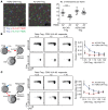
Figure 5. A2-CAR Tregs preferentially interact with HLA-A2+ PBMCs and are superior to polyclonal Tregs at suppressing alloantigen-stimulated proliferation in the presence of HLA-A2.
(A) A2-CAR or HER2-CAR Tregs were cultured in a 3D collagen gel with a mixture of HLA-A2+ and HLA-A2 PBMCs, each labeled with a unique dye. Colocalization of CAR Tregs (blue) with HLA-A2+ (red, interaction indicated by circles) and HLA-A2 (green, interaction indicated by squares) PBMCs was observed over time by fluorescence microscopy. Representative data are shown on the left, with a summary of the number of interactions over 4 hours to the right. Data represent mean ± SEM. Significance determined by 2-way ANOVA. (B and C) Suppressive capacity of transduced Tregs in allogeneic mixed leukocyte reactions was assayed by titrating the indicated ratios of A2-CAR or HER2-CAR Tregs into cultures with (B) HLA-A2+CD3+ responders (labeled with CPD) stimulated by HLA-A2 monocyte-derived DCs or (C) HLA-A2 CD3+ responders (labeled with CPD) stimulated by HLA-A2+ DCs. Division index of gated CD8+ cells was determined after 96 hours. Averaged data (mean ± SEM, n = 4) are shown. Significance determined by multiple t-tests with Holm-Šídák comparison. *P < 0.05; ***P < 0.001.
Figure 6
Figure 6. A2-CAR Tregs have minimal cytotoxic activity.
A2-CAR Tregs or Tconvs were cocultured with HLA-A2+ K562 cells, which were labeled with PKH26 at the indicated ratios for 24 hours. (A) Representative plots of intracellular staining for active caspase 3. (B) Averaged cell death (mean ± SEM, n = 4). (C) Similar data were obtained when HLA-A2+ PBMCs were used as targets (mean ± SEM, n = 4). Significance determined by multiple t tests with Holm-Šídák comparison. *P < 0.05; **P < 0.01; ****P < 0.0001.
Figure 7
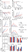
Figure 7. HLA-A2 CAR Tregs are superior to polyclonal Tregs at preventing xenogeneic GVHD.
Irradiated NSG mice were injected with PBS (n = 4) or 1 × 10⁷ HLA-A2+ PBMCs alone (n = 6) or with 1 × 10⁷ (1:1) or 0.5 × 10⁷ (1:2) A2-CAR or HER2-CAR Tregs (n = 4 for each group). Two independent experiments were performed using Tregs generated from independent donors. Data shown are the combined results for all mice in 2 experiments. Blood was monitored every 7 days, and spleen engraftment was analyzed at end point (flow cytometry). (A) Survival curve and (B) day of GVHD onset. Each symbol shape represents a unique human donor from whose blood Tregs were made. (C) Percentage of weight loss relative to the start of experiment. (D) GVHD score. (E) Engraftment in numbers and proportion of PBMCs (HLA-A2+) and (F) Tregs (HLA-A2). (G) Representative plots and summary data for proportions and per-cell expression of FOXP3 and the relevant CAR in blood of mice receiving A2-CAR or HER2-CAR Tregs (n = 4). Data represent mean ± SEM. Significance determined by log-rank test (A), 1-way ANOVA (B), and 2-way ANOVA (G). *P < 0.05; **P < 0.01.
Figure 8
Figure 8. A2-CAR–stimulated Tregs require exogenous IL-2 for long-term survival.
(A) A2-CAR Tregs or Tconvs were labeled with CPD, then stimulated with K562.64 cells loaded with αCD3/28 mAbs or expressing HLA-A2 in the absence or presence of 100 U/ml IL-2 for 5 days. Representative plots of FVDCD4+ cells (left) and percentage of proliferated compared with unstimulated controls are summarized (right, n = 4). (B) The proportion of viable CD4+ T cells at day 5 was determined as the proportion of cells in the lymphocyte gate that were FVDCD4+ (n = 4). (C) STAT5 phosphorylation was assessed in A2-CAR Tregs stimulated via the TCR (with αCD3), CAR (αMyc), or IL-2. Cells were fixed at 0, 5, 10, and 20 minutes, and the proportion of phospho-STAT5 positive cells was assessed (n = 3). Data represent mean ± SEM. Significance determined by 1-way ANOVA (B) or 2-way ANOVA (A). **P < 0.01; ***P < 0.001.

Comment in

Similar articles

Cited by

References

    1. Trzonkowski P, et al. First-in-man clinical results of the treatment of patients with graft versus host disease with human ex vivo expanded CD4+CD25+CD127– T regulatory cells. Clin Immunol. 2009;133(1):22–26. doi: 10.1016/j.clim.2009.06.001. - DOI - PubMed
    1. Di Ianni M, et al. Tregs prevent GVHD and promote immune reconstitution in HLA-haploidentical transplantation. Blood. 2011;117(14):3921–3928. doi: 10.1182/blood-2010-10-311894. - DOI - PubMed
    1. Brunstein CG, et al. Infusion of ex vivo expanded T regulatory cells in adults transplanted with umbilical cord blood: safety profile and detection kinetics. Blood. 2011;117(3):1061–1070. doi: 10.1182/blood-2010-07-293795. - DOI - PMC - PubMed
    1. Marek-Trzonkowska N, et al. Administration of CD4+CD25highCD127– regulatory T cells preserves beta-cell function in type 1 diabetes in children. Diabetes Care. 2012;35(9):1817–1820. doi: 10.2337/dc12-0038. - DOI - PMC - PubMed
    1. Bluestone JA, et al. Type 1 diabetes immunotherapy using polyclonal regulatory T cells. Sci Transl Med. 2015;7(315): doi: 10.1126/scitranslmed.aad4134. - DOI - PMC - PubMed

Publication types