. 2016 Mar 22:9:31.
doi: 10.1186/s13041-016-0212-8.

Valosin-containing protein is a key mediator between autophagic cell death and apoptosis in adult hippocampal neural stem cells following insulin withdrawal

Affiliations

Valosin-containing protein is a key mediator between autophagic cell death and apoptosis in adult hippocampal neural stem cells following insulin withdrawal

Bo Kyoung Yeo et al. Mol Brain. .

Abstract

Background: Programmed cell death (PCD) plays essential roles in the regulation of survival and function of neural stem cells (NSCs). Abnormal regulation of this process is associated with developmental and degenerative neuronal disorders. However, the mechanisms underlying the PCD of NSCs remain largely unknown. Understanding the mechanisms of PCD in NSCs is crucial for exploring therapeutic strategies for the treatment of neurodegenerative diseases.

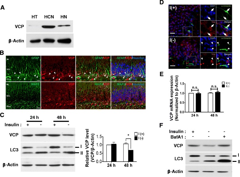
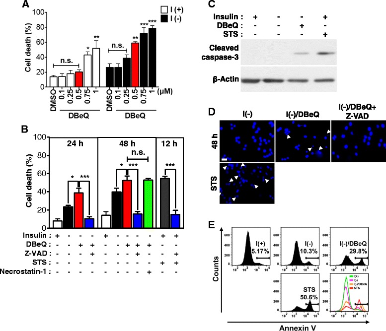
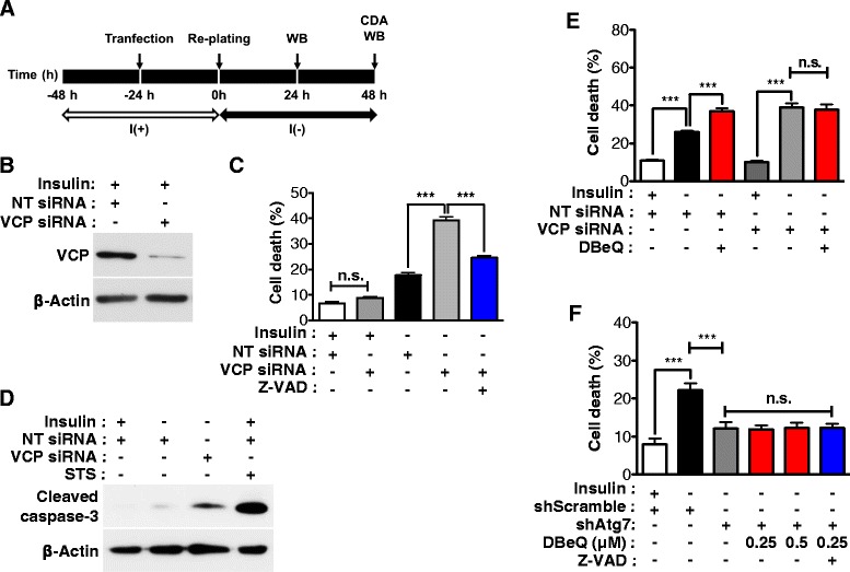
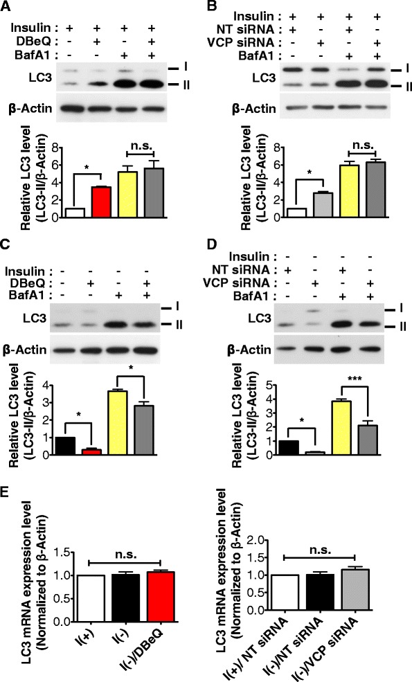
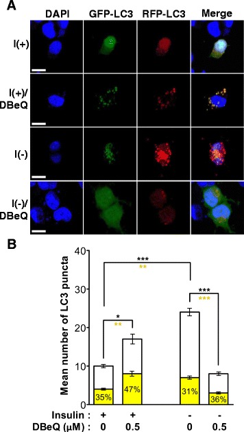
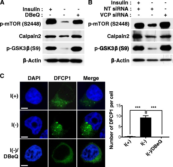
Result: We have previously reported that adult rat hippocampal neural stem (HCN) cells undergo autophagic cell death (ACD) following insulin withdrawal without apoptotic signs despite their normal apoptotic capabilities. It is unknown how interconnection between ACD and apoptosis is mediated in HCN cells. Valosin-containing protein (VCP) is known to be essential for autophagosome maturation in mammalian cells. VCP is abundantly expressed in HCN cells compared to hippocampal tissue and neurons. Pharmacological and genetic inhibition of VCP at basal state in the presence of insulin modestly impaired autophagic flux, consistent with its known role in autophagosome maturation. Of note, VCP inaction in insulin-deprived HCN cells significantly decreased ACD and down-regulated autophagy initiation signals with robust induction of apoptosis. Overall autophagy level was also substantially reduced, suggesting the novel roles of VCP at initial step of autophagy.

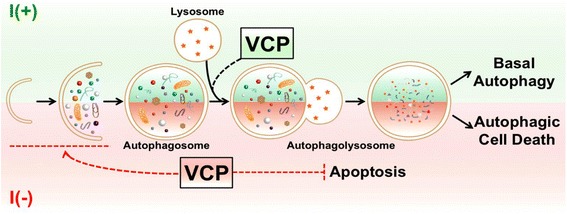
Conclusion: Taken together, these data demonstrate that VCP may play an essential role in the initiation of autophagy and mediation of crosstalk between ACD and apoptosis in HCN cells when autophagy level is high upon insulin withdrawal. This is the first report on the role of VCP in regulation of NSC cell death. Elucidating the mechanism by which VCP regulates the crosstalk of ACD and apoptosis will contribute to understanding the molecular mechanism of PCD in NSCs.

Keywords: Adult neural stem cells; Apoptosis; Autophagic cell death; Insulin withdrawal; Valosin-containing protein.


Figures

Fig. 1
Fig. 1
VCP is highly expressed in HCN cells and degraded through autophagy following insulin withdrawal. a VCP was abundantly expressed in HCN cells compared with hippocampal tissue prepared from 8-week-old rat and embryonic primary hippocampal neurons (9 days in vitro). b VCP was particularly localized in hippocampal neural stem cells in vivo, as indicated by arrowheads. GFAP and MAP2 were used as NSC and neuronal markers, respectively. Scale bar, 20 μm. c VCP protein level was significantly decreased at 48 h following insulin withdrawal. Quantitative analysis of VCP levels was normalized to β-actin. Quantitative data are represented at the mean ± SD (n = 3). Statistical significance was determined with an One-way ANOVA test by Tukey’s multiple comparison test. *p < 0.05. d VCP expression levels were decreased in ex vivo organotypic hippocampal slice culture following insulin withdrawal for 24 h. Arrow indicates the VCP-expressing HCN cells in I(+) condition, while arrowheads show the loss of VCP expression in I(-) condition. Scale bar, 20 μm. e Expression levels of VCP mRNA were not changed by insulin withdrawal. Quantitative data are represented at the mean ± SD (n = 3). n.s. not significant. f Degradation of VCP was prevented by Bafilomycin A1 treatment (BafA1, 30 nM for 3 h before harvest) following 48 h insulin withdrawal
Fig. 2
Fig. 2
Pharmacological inhibition of VCP switched ACD to apoptosis in insulin-deprived HCN cells. a A VCP inhibitor, DBeQ (0.5 μM) markedly increased cell death following insulin withdrawal for 24 h, but without significant effect on I(+) HCN cells. Quantitative data were determined using One-way ANOVA followed by Tukey’s multiple comparison test. *p < 0.05, **p < 0.01, ***p < 0.001. n.s., not significant. b Z-VAD-FMK significantly prevented cell death induced by DBeQ in I(-). Staurosporine (STS, 1 μM for 12 h) was treated in I(+) HCN cells as a positive control for apoptosis induction. However, addition of necrostatin-1 (10 μM) up to 48 h did not decrease cell death in DBeQ-treated I(-) HCN cells. Quantitative data are represented as the mean ± SD by One-way ANOVA using Tukey’s multiple comparison test (n = 3). **p < 0.01, ***p < 0.001. n.s., not significant. c Caspase 3 was activated in DBeQ-treated I(-) HCN cells. d Nuclear condensation was observed by Hoechst staining, as indicated by arrow. Nuclear condensation was detected in DBeQ-treated I(-) HCN cells, but prevented by Z-VAD-FMK. Scale bar is 20 μm. e Apoptosis induction was confirmed by Annexin-V staining and FACS analysis for detection of the exposure of phosphatidylserine
Fig. 3
Fig. 3
Genetic inhibition of VCP changed ACD to apoptosis in insulin-deprived HCN cells. a An experimental scheme for the knockdown of VCP. CDA, cell death assay; WB, Western blotting. b Knockdown of VCP was confirmed by Western blotting after 24 h treatment. NT, non-targeting. c VCP knockdown increased cell death and Z-VAD-FMK efficiently blocked an increase of cell death in I(-) HCN cells. Quantitative data are determined as the mean ± SD by One-way ANOVA followed by Tukey’s multiple comparison test (n = 3). ***p < 0.001. n.s., not significant. d Cleaved caspase 3 was detected in I(-) HCN cells following VCP knockdown at 48 h. e Cell death rate was not affected by DBeQ in I(-) HCN cells depleted of VCP. Quantitative data are determined as the mean ± SD by One-way ANOVA followed by Tukey’s multiple comparison test (n = 3). ***p < 0.001. n.s., not significant. f Atg7 knockdown decreased cell death in I(-) HCN cells. However, cell death rate was not altered by DBeQ or Z-VAD-FMK in I(-) HCN cells with stable knockdown of Atg7. Quantitative data are determined as the mean ± SD by One-way ANOVA followed by Tukey’s multiple comparison test (n = 3). ***p < 0.001. n.s., not significant
Fig. 4
Fig. 4
VCP inhibition reduced autophagic flux in insulin-deprived HCN cells. a-b DBeQ treatment (a) or VCP knockdown (b) induced a moderate increase in LC3-II in I(+) HCN cells. Blocking of autophagy using BafA1 gave rise to a significant increase of LC3-II with the same levels between BafA1 alone and BafA1/DBeQ or BafA1/VCP knockdown cells in I(+). Quantitative LC3-II levels were determined as the mean ± SD by One-way ANOVA followed by Tukey’s multiple comparison test (n = 3). **p < 0.01, ***p < 0.001. n.s., not significant. c-d DBeQ treatment (c) or VCP knockdown (d) led to a decrease in LC3-II in I(-) HCN cells. Blocking of autophagy using BafA1 gave rise to a significant increase of LC3-II, but the amount of accumulated LC3-II in BafA1/DBeQ or BafA1/VCP knockdown cells was significantly less than BafA1 alone in I(-). Quantitative LC3-II levels were determined as the mean ± SD by One-way ANOVA followed by Tukey’s multiple comparison test (n = 3). **p < 0.01, ***p < 0.001. n.s., not significant. e mRNA expression level of LC3 did not show different changes by VCP inactivation in I (-). Quantitative LC3 mRNA expression levels are represented as the mean ± SD (n = 3). Quantitative data was determined by Student’s t-test. *p < 0.05. n.s., not significant
Fig. 5
Fig. 5
Differential regulation of autophagy flux by VCP in I(+) and I(-) HCN cells. a mRFP-GFP-LC3 was used for autopahgic flux assay. DBeQ treatment in I(+) led to an increase in the percentage of yellow puncta with no marked increase in the number of the total LC3 puncta, suggesting impairment of autophagosome maturation. DBeQ treatment in I(-) greatly reduced the number of the total LC3 puncta, but the percentage of yellow puncta remained similar between I(-) and I(-)/DBeQ conditions. Scale bar, 10 μm. b Quantitative red and yellow puncta numbers were determined as the mean ± SD by One-way ANOVA followed by Tukey’s multiple comparison test (n = 16–35). Black and yellow asterisks indicate the comparison of the total and yellow puncta, respectively. *p < 0.05, **p < 0.01, ***p < 0.001
Fig. 6
Fig. 6
VCP is required for autophagy initiation signaling in HCN cells following insulin withdrawal. a-b Negative regulators of autophagy, mTOR and calpain 2 were suppressed in I(-), but reactivated by DBeQ treatment (a) or VCP knockdown (b). On the contrary, positive inducers of autophagy such as GSK-3β were activated following insulin withdrawal, but deactivated by DBeQ treatment (a) or VCP knockdown (b). c The high number of DFCP1 puncta observed in I(-) were greatly reduced by DBeQ treatment. The number of DFCP1 puncta was quantified with cut-off diameter of 0.5 μm. Scale bar, 5 μm. Quantitative DFCP1 puncta were determined as the mean ± SD by One-way ANOVA followed by Tukey’s multiple comparison test (n = 10). ***p < 0.001
Fig. 7
Fig. 7
A schematic diagram illustrating the novel function of VCP in ACD and apoptosis in HCN cells following insulin withdrawal. HCN cells undergo autophagic cell death following insulin withdrawal. In basal autophagy, VCP positively regulates autophagosome maturation. However, following insulin withdrawal, VCP regulates autophagy initiation and the crosstalk between ACD to apoptosis

References

    1. Fuchs Y, Steller H. Programmed cell death in animal development and disease. Cell. 2011;147(4):742–58. doi: 10.1016/j.cell.2011.10.033. - DOI - PMC - PubMed
    1. Clarke PG. Developmental cell death: morphological diversity and multiple mechanisms. Anat Embryol. 1990;181(3):195–213. doi: 10.1007/BF00174615. - DOI - PubMed
    1. Kerr JF, Wyllie AH, Currie AR. Apoptosis: a basic biological phenomenon with wide-ranging implications in tissue kinetics. Br J Cancer. 1972;26(4):239–57. doi: 10.1038/bjc.1972.33. - DOI - PMC - PubMed
    1. Shintani T, Klionsky DJ. Autophagy in health and disease: a double-edged sword. Science. 2004;306(5698):990–5. doi: 10.1126/science.1099993. - DOI - PMC - PubMed
    1. Wang CW, Klionsky DJ. The molecular mechanism of autophagy. Mol Med. 2003;9(3-4):65–76. - PMC - PubMed

Publication types