. 2016 Mar 22:6:23432.
doi: 10.1038/srep23432.

Integrating computational and chemical biology tools in the discovery of antiangiogenic small molecule ligands of FGF2 derived from endogenous inhibitors

Affiliations

Integrating computational and chemical biology tools in the discovery of antiangiogenic small molecule ligands of FGF2 derived from endogenous inhibitors

Chiara Foglieni et al. Sci Rep. .

Abstract

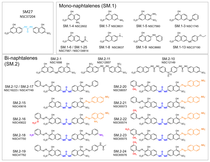
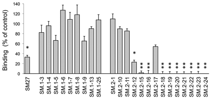
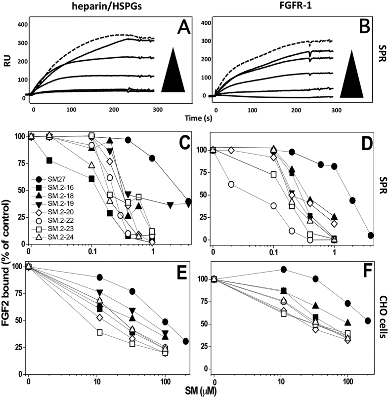
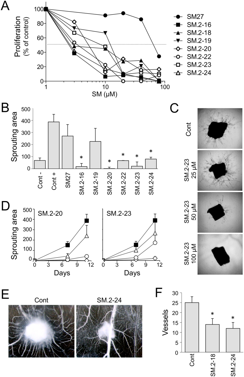
The FGFs/FGFRs system is a recognized actionable target for therapeutic approaches aimed at inhibiting tumor growth, angiogenesis, metastasis, and resistance to therapy. We previously identified a non-peptidic compound (SM27) that retains the structural and functional properties of the FGF2-binding sequence of thrombospondin-1 (TSP-1), a major endogenous inhibitor of angiogenesis. Here we identified new small molecule inhibitors of FGF2 based on the initial lead. A similarity-based screening of small molecule libraries, followed by docking calculations and experimental studies, allowed selecting 7 bi-naphthalenic compounds that bound FGF2 inhibiting its binding to both heparan sulfate proteoglycans and FGFR-1. The compounds inhibit FGF2 activity in in vitro and ex vivo models of angiogenesis, with improved potency over SM27. Comparative analysis of the selected hits, complemented by NMR and biochemical analysis of 4 newly synthesized functionalized phenylamino-substituted naphthalenes, allowed identifying the minimal stereochemical requirements to improve the design of naphthalene sulfonates as FGF2 inhibitors.


Figures

Figure 1
Figure 1. Structure of the lead compoud SM27 and the new identified hits, clustered according to structural features: mono-naphthalenes (SM.1 series) and bi-naphthalenes (SM.2 series).
For the most active SM.2 compounds, the main stereochemical requirements for binding and activity are colored: blue, urea bridge between the two napthalenes; orange, sulfonate-decorated additional naphtyl-azo group; purple, free amines on the two arylamine decorations in SM.2–18; red, substituents on the aryl-azo group.
Figure 2
Figure 2. FGF2-binding property of the selected hits, analyzed with an indirect assay, as ability to compete with the TSP-1 fragment E123CaG1 (comprising the type III repeats domain) for FGF2 binding, as described in Methods.
Binding of the biotinylated recombinant domain to FGF2 in the presence or absence of each molecule (5 μM). Data are the percentage of control binding, mean and SE of 3 experiments performed in triplicate. *p < 0.05 compared to control (ANOVA followed by Bonferroni test).
Figure 3
Figure 3
Inhibitory activity of the selected hits on the binding of FGF2 to heparin/HSPGs (A,C,E) or FGFR-1 (B,D,F). (A,B) Representative sensorgrams overlays derived by the injection of FGF2 (159 nM) in the absence (top hatched sensorgram) or in the presence of increasing concentrations of SM.2–22 on the heparin or FGFR-1 surfaces, respectively. (C,D) FGF2 (150 nM) was injected over heparin (C) or FGFR-1 (D) immobilized to a SPR sensorchip in the absence or in the presence of increasing concentrations of the indicated compounds and the amount of FGF2 bound to the surfaces in the different experimental conditions was measured. Each point is the mean of 3 experiments. (E,F) Binding of Eu-FGF2 (10 ng/ml) to CHO cell clones selectively expressing either HSPGs (E) or FGFR-1 (F) in the presence of increasing concentrations of the compounds. Each point is the mean of 2–3 experiments. Data are expressed as mean values of the percentage of bound FGF2 compared to control (in the absence of competing compounds).
Figure 4
Figure 4. Biological activity of the selected hits.
(A) Endothelial cell proliferation. BAEC were exposed to FGF2 (5 ng/ml) with increasing concentrations of molecules (3–80 μM). After 72 h, cells were stained and proliferation measured as absorbance. Data are the percentage of control proliferation (in absence of molecules), mean of value from 2 experiments performed in triplicate. (B–D) Aortic ring assay. Sections of murine aortas were embedded in Matrigel, in the presence of FGF2 (30 ng/ml) and the indicated small molecule. The formation of capillary structures sprouting from the rings was analyzed after 7 and 11 days as described in Methods, and the angiogenic response expressed as area covered by the sprouting structures (arbitrary units, mean and SE, n ≥ 6). (B) Antiangiogenic activity of the small molecules (100 μM). (C) Examples of time-dependent and dose-dependent effect of two compounds (SM.2–20 and SM.2–23), tested at 100 (diamond), 50 (circle) and 25 μM (triangle) compared to control (black squares). (D) Representative pictures of sprouting from control and SM.2–23 treated aortic sections. Original magnification, 20x. (E,F) Chorioallantoic membrane assay. FGF2 (200 ng) was administered in the absence or presence of the indicated compound (0.5 μg) on day 8 (n = 10). (E) Angiogenic response is evaluated 4 days later, and expressed as number of vessels entering the sponge (mean and SD). (F) Representative pictures are shown. Original magnification, 50x.
Figure 5
Figure 5. The best poses of SM.2–24, SM.2–19 and SM.2–18, resulting from the ensemble docking of the ligands into the heparin-binding pocket of FGF2.
In green: area of additional interactions compared to SM.2–19.
Figure 6
Figure 6
(A) Structure of the 4 phenylamino-substituted naphthalenes synthesized and analyzed in the present study. (B) Antiproliferative activity of the four anilino-naphthalenes was analyzed as in Fig. 4A. Data are the percentage of control proliferation (in absence of molecules). (C) Graphical representation of the combined 1HN and 15N FGF2 chemical shift perturbation determined for the various residues, according to formula image
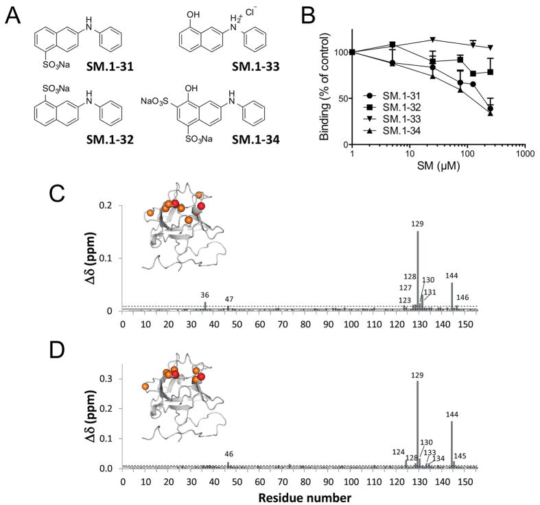
, following the addition of phenylamino-substituted naphthalenes. C) Chemical shift perturbation (CSP) induced by SM.1–34 addition in SM:FGF2 1:1 stoichiometric ratio D) CSP induced by SM.1–31 addition in SM:FGF2 1:3 stoichiometric ratio. The continuous and dashed lines represent the average and the average plus a standard deviation (SD) values, respectively. Residues affected by CSP > (<CSP>) + 1SD are mapped on the FGF2 structures as orange spheres. Residues Arg129 and Lys144, showing the highest CSP, are shown as red spheres.

References

    1. Wesche J., Haglund K. & Haugsten E. M. Fibroblast growth factors and their receptors in cancer. Biochem J 437, 199–213 (2011). - PubMed
    1. Turner N. & Grose R. Fibroblast growth factor signalling: from development to cancer. Nat Rev Cancer 10, 116–129 (2010). - PubMed
    1. Presta M. et al. Fibroblast growth factor/fibroblast growth factor receptor system in angiogenesis. Cytokine Growth Factor Rev 16, 159–178 (2005). - PubMed
    1. Cao Y. et al. Forty-year journey of angiogenesis translational research. Sci Transl Med 3, 114rv113 (2011). - PMC - PubMed
    1. Carmeliet P. & Jain R. K. Molecular mechanisms and clinical applications of angiogenesis. Nature 473, 298–307 (2011). - PMC - PubMed

Publication types

LinkOut - more resources