. 2016 Apr 20;44(7):2977-86.
doi: 10.1093/nar/gkw153. Epub 2016 Mar 21.

Identification of a mismatch-specific endonuclease in hyperthermophilic Archaea

Affiliations

Identification of a mismatch-specific endonuclease in hyperthermophilic Archaea

Sonoko Ishino et al. Nucleic Acids Res. .

Abstract

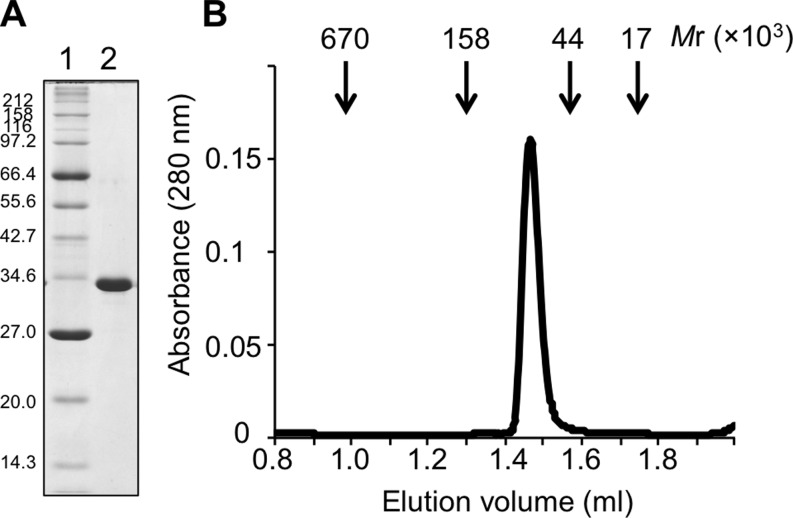
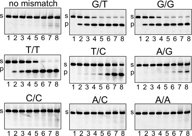
The common mismatch repair system processed by MutS and MutL and their homologs was identified in Bacteria and Eukarya. However, no evidence of a functional MutS/L homolog has been reported for archaeal organisms, and it is not known whether the mismatch repair system is conserved in Archaea. Here, we describe an endonuclease that cleaves double-stranded DNA containing a mismatched base pair, from the hyperthermophilic archaeon Pyrococcus furiosus The corresponding gene revealed that the activity originates from PF0012, and we named this enzyme Endonuclease MS (EndoMS) as the mismatch-specific Endonuclease. The sequence similarity suggested that EndoMS is the ortholog of NucS isolated from Pyrococcus abyssi, published previously. Biochemical characterizations of the EndoMS homolog from Thermococcus kodakarensis clearly showed that EndoMS specifically cleaves both strands of double-stranded DNA into 5'-protruding forms, with the mismatched base pair in the central position. EndoMS cleaves G/T, G/G, T/T, T/C and A/G mismatches, with a more preference for G/T, G/G and T/T, but has very little or no effect on C/C, A/C and A/A mismatches. The discovery of this endonuclease suggests the existence of a novel mismatch repair process, initiated by the double-strand break generated by the EndoMS endonuclease, in Archaea and some Bacteria.


Figures

Figure 1.
Figure 1.
Purified TK1898 protein. (A) Preparation of recombinant TK1898. The protein markers (lane 1) and the purified TK1898 (2 μg) (lane 2) were subjected to SDS-12% PAGE followed by staining with Coomassie Brilliant Blue (CBB). The sizes of the markers are shown on the left of the panel. (B) Gel filtration chromatography of the TK1898 protein. The arrowheads indicate the elution profile of the standard marker proteins, and the numbers indicate the relative molecular masses.
Figure 2.
Figure 2.
Cleavage of mismatch-containing DNA by TK1898. (A) The nucleotide sequences of the substrates (45 bp) containing the mismatched base pair. The position of the mismatched base pair is indicated by white letters on a black background. (B and C) The 5′-32P-labeled DNA substrates (5 nM) containing the mismatched base pair were incubated with various concentrations of TK1898 (0, 1.3, 2.5, 10 and 50 nM as a dimer)(lanes 1–5, 6–10, 11–15 and 16–20) at 55°C for 5 min. The 5′-32P-labeled ssDNA was incubated with no enzyme (lane 21) and 50 nM TK1898 (lane 22) at 55°C for 5 min. The products were analysed by native 10% PAGE followed by autoradiography. The base pairs at the 25th positions in the dsDNAs are indicted at the top of the panel, and the bases in the labeled strands are indicated by asterisks.
Figure 3.
Figure 3.
Cleavage pattern of mismatch-containing DNA by TkoEndoMS. (A) The nucleotide sequences of the substrate (45 bp) containing the mismatched base pair and the cleaved products are shown schematically. The mismatched base pair is indicated by white letters on a black background. The numbers indicate the lengths of the strands cleaved by TkoEndoMS. The 5′-32P-labeled upper strand (B), 5′-32P-labeled lower strand (C) and 3′-FITC-labeled upper strand (D) were used to make dsDNA substrates containing the mismatched base pair. The substrates (5 nM) were incubated with (+) or without (−) 50 nM TkoEndoMS (as a dimer) at 55°C for 5 min. The products were separated by 8 M urea-12% PAGE in TBE buffer. The size markers were loaded in lanes 1 and 12 in panel B, in lanes 1 and 10 in panel C and in lane 1 in panel D. The sizes are indicated on the side of each band.
Figure 4.
Figure 4.
Preference for base-pair mismatches of TkoEndoMS. Five nanomoles of Cy5-labeled dsDNA (45 bp), containing single base-pair mismatches (G/T, G/G, T/T, T/C, A/G, C/C, A/C, A/A), were incubated with various concentrations of TkoEndoMS (lanes 1, no protein; 2, 0.5 nM; 3, 1 nM; 4, 2.5 nM; 5, 5 nM; 6, 10 nM; 7, 25 nM; 8, 50 nM, as a dimer) at 55°C for 5 min. The base pairs are indicated on the top of each panel. The products were separated by 10% PAGE. The band assignments are indicated on the side of the panels, s, substrates; p, cleaved products.
Figure 5.
Figure 5.
Cleavage property of TkoEndoMS for DNAs containing insertions/deletions. The substrates were prepared with oligonucleotides. The numbers of the oligonucleotides correspond to those in Supplementary Table S2. The substrates (5 nM) were incubated with (+) or without (−) 20 nM TkoEndoMS (as a dimer) at 55°C for 30 min. The products were separated by 8 M urea-12% PAGE.
Figure 6.
Figure 6.
Cleavage of the structured DNA by TkoEndoMS. TkoEndoMS activities on the 3′-flapped duplex and the 3′-splayed duplex (A), the 5′-flapped duplex and the 5′-splayed duplex (B) and the normal duplex and the single strands (C) were analysed. Each panel included the products of the duplex containing a G/T mismatch, as a positive control. The inactivated mutant D165A (Mut) was used as a negative control. The substrates with the schematic illustrations are shown at the top of the figure. The 5′-Cy5-labeled strands are marked with asterisks. The numbers on the strands indicate the length of the oligonucleotides. The substrates (50 nM) were incubated with the indicated protein concentrations (as a dimer) at 55°C for 10 min. The products were separated by 8 M urea-12% PAGE in TBE buffer. The size markers were loaded in the first lane in panel A. The sizes are indicated on the left side of panel A.
Figure 7.
Figure 7.
DNA-binding properties of TkoEndoMS. EMSA of TkoEndoMS in the absence of MgCl2 with 2.5 mM EDTA (A) and in the presence of MgCl2 (B). Five nanomoles of Cy5-labeled ssDNA (15 nt) and dsDNA (15 bp) containing single base-pair mismatches were incubated with various concentrations of TkoEndoMS (lanes 1, no protein; 2, 0.5 nM; 3, 1 nM; 4, 2.5 nM; 5, 5 nM; 6, 10 nM as a dimer) at 37°C for 5 min. The base pairs are indicated on the top of each panel. The band assignments are indicated on the side of the panels.
Figure 8.
Figure 8.
Interaction of TkoEndoMS with TkoPCNA. (A) Purified recombinant proteins were analysed by SDS-12.5% PAGE. Three micrograms of each protein were loaded, and the gel was stained with CBB, lanes 1, marker; 2, WT; 3, ΔPIP. (B) Four nanomoler (as a dimer) of TkoEndoMS WT (upper panel) or ΔPIP (lower panel) were incubated with 5 nM DNA containing a G/T mismatch and TkoPCNA. The concentrations of TkoPCNA (as a trimer) are indicated above the gel. The products were separated by 10% native PAGE. The representative results are shown. The band assignments are indicated on the side of the panels, s, substrates; p, cleaved products. (C) The measurements of the products from four independently performed experiments are shown with the error bars (standard error of the mean).

Similar articles

Cited by

References

    1. Barnes D.E., Lindahl T. Repair and genetic consequences of endogenous DNA base damage in mammalian cells. Annu. Rev. Genet. 2004;38:445–476. - PubMed
    1. Reardon J.T., Sancar A. Nucleotide excision repair. Prog. Nucleic Acid Res. Mol. Biol. 2005;79:183–235. - PubMed
    1. Iyer R.R., Pluciennik A., Burdett V., Modrich P.L. DNA mismatch repair: functions and mechanisms. Chem. Rev. 2006;106:302–323. - PubMed
    1. Yasui A. Alternative excision repair pathways. Cold Spring Harb. Perspect. Biol. 2013;5:a012617. - PMC - PubMed
    1. Zhang J., Walter J.C. Mechanism and regulation of incisions during DNA interstrand cross-link repair. DNA Repair (Amst.) 2014;19:135–142. - PMC - PubMed

Publication types

MeSH terms