Comparative Study
. 2016 Apr;22(7-8):665-79.
doi: 10.1089/ten.TEA.2015.0546. Epub 2016 Apr 1.

Chondroinduction from Naturally Derived Cartilage Matrix: A Comparison Between Devitalized and Decellularized Cartilage Encapsulated in Hydrogel Pastes

Affiliations
Comparative Study

Chondroinduction from Naturally Derived Cartilage Matrix: A Comparison Between Devitalized and Decellularized Cartilage Encapsulated in Hydrogel Pastes

Emily C Beck et al. Tissue Eng Part A. 2016 Apr.

Abstract

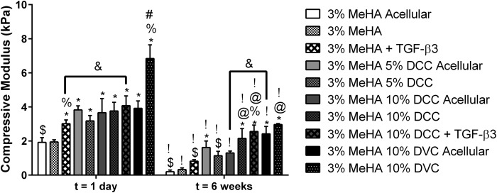
Hydrogel precursors are liquid solutions that are prone to leaking after surgical placement. This problem was overcome by incorporating either decellularized cartilage (DCC) or devitalized cartilage (DVC) microparticles into traditional photocrosslinkable hydrogel precursors in an effort to achieve a paste-like hydrogel precursor. DCC and DVC were selected specifically for their potential to induce chondrogenesis of stem cells, given that materials that are chondroinductive on their own without growth factors are a revolutionary goal in orthopedic medicine. We hypothesized that DVC, lacking the additional chemical processing steps in DCC to remove cell content, would lead to a more chondroinductive hydrogel with rat bone marrow-derived mesenchymal stem cells. Hydrogels composed of methacrylated hyaluronic acid (MeHA) and either DCC or DVC microparticles were tested with and without exposure to transforming growth factor (TGF)-β3 over a 6 week culture period, where swelling, mechanical analysis, and gene expression were observed. For collagen II, Sox-9, and aggrecan expression, MeHA precursors containing DVC consistently outperformed the DCC-containing groups, even when the DCC groups were exposed to TGF-β3. DVC consistently outperformed all TGF-β3-exposed groups in aggrecan and collagen II gene expression as well. In addition, when the same concentrations of MeHA with DCC or DVC microparticles were evaluated for yield stress, the yield stress with the DVC microparticles was 2.7 times greater. Furthermore, the only MeHA-containing group that exhibited shape retention was the group containing DVC microparticles. DVC appeared to be superior to DCC in both chondroinductivity and rheological performance of hydrogel precursors, and therefore DVC microparticles may hold translational potential for cartilage regeneration.


Figures

<b>FIG. 1.</b>
FIG. 1.
Biochemical contents and SEM images of hydrogel paste components. (A) PicoGreen content, (B) GAG content, and (C) Hydroxyproline content of DVC, DCC, and MeHA. After decellularization, there was a 44% reduction in DNA, a 23% reduction in GAG, and a 23% reduction in hydroxyproline content. Data are reported as mean + standard deviation (n = 5); ^below detectable limit; *significantly different from DVC (p < 0.05); #significantly different from DCC (p < 0.05). (D) SEM images of DCC and DVC microparticles under 500× and 12,000× magnifications. Under 500× magnification, the DCC microparticles were noted to have more smooth surfaces overall than the DVC microparticles, and under 12,000× magnification, the surfaces of the DCC microparticles were noted to have a grain-like appearance that was nonexistent in the DVC microparticles. Scale bars for the 500× and 12,000× magnifications are 100 and 5 μm, respectively. DCC, decellularized cartilage; DVC, devitalized cartilage; GAG, glycosaminoglycan; SEM, scanning electron microscope.
<b>FIG. 2.</b>
FIG. 2.
Macroscopic rheological evaluation of hydrogel precursors before and after crosslinking. All formulations were acellular unless noted. Non-Newtonian behavior was observed in solutions containing at least 5% DCC, whereas shape retention (indicated by the solution retaining extrusion orifice diameter) was only noted in 10% DCC and 3% MeHA + 10% DVC acellular formulations. All formulations containing MeHA retained their shape after crosslinking. Color images available online at www.liebertpub.com/tea
<b>FIG. 3.</b>
FIG. 3.
Yield stress (A) and storage modulus (B) of hydrogel precursor solutions. Only the 10% DCC, 3% MeHA + 10% DCC, and 3% MeHA + 10% DVC groups exhibited a measurable yield stress, whereas all groups had a measurable storage modulus. Data are reported as mean + standard deviation (n = 5); *significantly different from 3% MeHA acellular group (p < 0.05); #significantly different from 3% MeHA + 10% DCC acellular group (p < 0.05); $significantly different from 10% DCC group (p < 0.05); &significantly different from all other groups (p < 0.05).
<b>FIG. 4.</b>
FIG. 4.
Compressive moduli of crosslinked hydrogels after 1 day and 6 weeks of culture. Gels containing at least 10% DCC or DVC microparticles had significantly larger moduli than 3% MeHA gels alone. Data are reported as mean + standard deviation (n = 5); *significantly different from 3% MeHA at same time point (p < 0.05); %significantly different from acellular group of same formulation at same time point (p < 0.05); #significantly different from all other groups at same time point (p < 0.05); $significantly different from 3% MeHA + 10% DCC at same time point (p < 0.05); @significantly different from 3% MeHA + TGF-β3 and 3% MeHA + 5% DCC at same time point (p < 0.05); &p < 0.05 for specified comparison; !significantly different from same group at first time point (p < 0.05). TGF-β3, transforming growth factor-β3.
<b>FIG. 5.</b>
FIG. 5.
Swelling degree (A) and volume (B) of crosslinked hydrogels. The only gels with significantly smaller swelling degrees than the 3% MeHA gels were the 3% MeHA + 10% DCC acellular group and the 3% MeHA + 10% DVC acellular and cellular groups. At day 1, there were no significant differences between groups. However, the inclusion of DCC or DVC or exposure to TGF-β3 significantly reduced the volume at 6 weeks. Data are reported as mean + standard deviation (n = 5); *significantly different from 3% MeHA at same time point (p < 0.05); %significantly different from acellular group of same formulation at same time point (p < 0.05); $significantly different from 3% MeHA + 10% DCC at same time point (p < 0.05); !significantly different from same group at first time point (p < 0.05).
<b>FIG. 6.</b>
FIG. 6.
Biochemical content of gels over the 6 week culture period. (A) DNA content, (B) GAG content, and (C) hydroxyproline content. All gels contained significantly higher DNA content than their respective acellular groups at all time points, and all gels containing DCC or DVC had significant reductions in GAG over the 6 week culture period. Data are reported as mean + standard deviation (n = 5); ^below detectable limit; *significantly different from 3% MeHA at same time point (p < 0.05); %significantly different from acellular group of same formulation at same time point (p < 0.05); #significantly different from all other groups at same time point (p < 0.05); $significantly different from 3% MeHA + 10% DCC at same time point (p < 0.05); !significantly different from same group at first time point (p < 0.05); +significantly different from same group at previous time point (p < 0.05).
<b>FIG. 7.</b>
FIG. 7.
Relative gene expression of (A) collagen II, (B) collagen I, (C) Sox-9, and (D) aggrecan. The DVC group consistently outperformed the other groups in collagen II, Sox-9, and aggrecan expression, even when compared with TGF-β3 exposed groups. Data are reported as mean + standard deviation (n = 5); *significantly different from 3% MeHA at same time point (p < 0.05); #significantly different from 3% MeHA + TGF-β3 at same time point (p < 0.05); @significantly different from all DCC-containing groups at same time point (p < 0.05); %significantly higher than same group at previous time point (p < 0.05); $significantly higher than same group at first time point (p < 0.05), !significantly lower than same group at previous time point (p < 0.05); ^expression not detected.
<b>FIG. 8.</b>
FIG. 8.
Histological analysis of gels. All gels stained red/orange for GAGs, although no increase in the amount of staining was noted over the culture period. However, nodular Saf-O staining was noted in the 3% MeHA + TGF-β3 group. All DCC- and DVC-containing groups were stained for collagen II; however, no changes were noted in the location and intensity of collagen II staining over the culture period. Collagen I staining was noted again in all DCC- and DVC-containing groups. However, the intensity of collagen I staining decreased over the culture period for the 3% MeHA + 5% DCC and 3% MeHA + 10% DCC groups and appeared to increase slightly for the 3% MeHA + 10% DVC group. Aggrecan staining was noted in all DCC- and DVC-containing groups, where the aggrecan staining became more intense near the DCC and DVC microparticles in the 3% MeHA + 10% DCC + TGF-β3 and 3% MeHA + 10% DVC groups over the culture period. Scale bars are 200 μm. Color images available online at www.liebertpub.com/tea

References

    1. Rughani R.V., Branco M.C., Pochan D.J., and Schneider J.P. De novo design of a shear-thin recoverable peptide-based hydrogel capable of intrafibrillar photopolymerization. Macromolecules 43, 7924, 2010
    1. Todd R.H., and Daniel S.K. Hydrogels in drug delivery: progress and challenges. Polymer 49, 1993, 2008
    1. Beck E.C., Lohman B.L., Tabakh D.B., Kieweg S.L., Gehrke S.H., Berkland C.J., and Detamore M.S. Enabling surgical placement of hydrogels through achieving paste-like rheological behavior in hydrogel precursor solutions. Ann Biomed Eng 43, 2569, 2015 - PMC - PubMed
    1. Benders K., van Weeren P., Badylak S., Saris D., Dhert W., and Malda J. Extracellular matrix scaffolds for cartilage and bone regeneration. Trends Biotechnol 31, 169, 2013 - PubMed
    1. Burdick J.A., Mauck R.L., Gorman J.H., III, and Gorman R.C. Acellular biomaterials: an evolving alternative to cell-based therapies. Sci Transl Med 5, 176ps4, 2013 - PMC - PubMed

Publication types

MeSH terms