. 2016 Mar 22:6:23389.
doi: 10.1038/srep23389.

Improved dialytic removal of protein-bound uraemic toxins with use of albumin binding competitors: an in vitro human whole blood study

Affiliations

Improved dialytic removal of protein-bound uraemic toxins with use of albumin binding competitors: an in vitro human whole blood study

Xia Tao et al. Sci Rep. .

Abstract

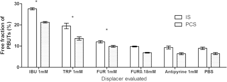
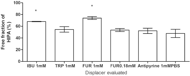
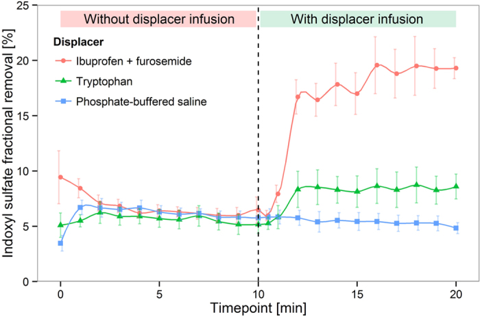
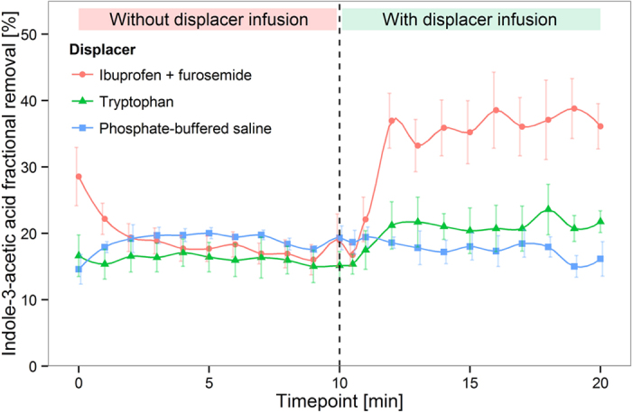
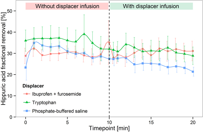
Protein-bound uraemic toxins (PBUTs) cause various deleterious effects in end-stage kidney disease patients, because their removal by conventional haemodialysis (HD) is severely limited by their low free fraction in plasma. Here we provide an experimental validation of the concept that the HD dialytic removal of PBUTs can be significantly increased by extracorporeal infusion of PBUT binding competitors. The binding properties of indoxyl sulfate (IS), indole-3-acetic acid (IAA) and hippuric acid (HIPA) and their binding competitors, ibuprofen (IBU), furosemide (FUR) and tryptophan (TRP) were studied in uraemic plasma. The effect of binding competitor infusion on fractional removal of PBUT was then quantified in an ex vivo single-pass HD model using uraemic human whole blood. The infusion of a combination of IBU and FUR increased the fractional removal of IS from 6.4 ± 0.1 to 18.3 ± 0.4%. IAA removal rose from 16.8 ± 0.3 to 34.5 ± 0.7%. TRP infusion increased the removal of IS and IAA to 10.5 ± 0.1% and 27.1 ± 0.3%, respectively. Moderate effects were observed on HIPA removal. Pre-dialyzer infusion of PBUT binding competitors into the blood stream can increase the HD removal of PBUTs. This approach can potentially be applied in current HD settings.


Conflict of interest statement

Xia Tao, Stephan Thijssen and Peter Kotanko are employees of Renal Research Institute, affiliated with Fresenius Medical Care North America (FMCNA). Peter Kotanko holds stock of FMCNA. Peter Kotanko is an inventor on a U.S. patent (assigned to FMCNA) on the method described in the paper. Chih-Hu Ho, Michael Henrie and Eric Stroup are employees of FMCNA. Garry Handelman declares no conflicts of interest.

Figures

Figure 1
Figure 1. IS and PCS displacement in uraemic plasma by furosemide, tryptophan and ibuprofen, determined in static RED assays.
Displacer concentration was 1 mmol/l, unless otherwise indicated. IS: indoxyl sulfate; PCS: p-cresol sulfate; IBU: ibuprofen; TRP: tryptophan; FUR: furosemide; PBS: phosphate buffered saline. Bars denote mean, error bars denote standard error of the mean (SEM), N = 3. *P < 0.05, compared to PBS.
Figure 2
Figure 2. HIPA displacement in uraemic plasma by furosemide, tryptophan and ibuprofen, determined in static RED assays.
Displacer concentration was 1 mmol/l, unless otherwise indicated. IBU: ibuprofen; TRP: tryptophan; FUR: furosemide; PBS: phosphate buffered saline. Mean ± SEM, N = 3. *P < 0.05, compared to PBS.
Figure 3
Figure 3. Dose-response relationships for IS displacement by ibuprofen, and synergistic effect of ibuprofen and furosemide in normal human plasma, determined in static RED assays.
Solid circle: ibuprofen with 180 μmol/l furosemide added; open circle: ibuprofen alone. Mean ± SEM, N = 3.
Figure 4
Figure 4. Indoxyl sulfate displacement in human whole blood HD model.
Mean ± SEM, N = 3.
Figure 5
Figure 5. Indoleacetic acid displacement in human whole blood HD model.
Mean ± SEM, N = 3.
Figure 6
Figure 6. Hippuric acid displacement in human whole blood HD model.
Mean ± SEM, N = 3.
Figure 7
Figure 7. Urea extraction ratio in human whole blood HD model.
Mean ± SEM, N = 3.
Figure 8
Figure 8. Setup of in vitro dialysis model using human whole blood.
(A) infusion site of displacers. (B) sample collection site for blood inlet. (C) sample collection site for dialysate outlet. (D) sample collection site for blood outlet.

References

    1. Vanholder R. et al. Review on uremic toxins: Classification, concentration, and interindividual variability. Kidney Int. 63, 1934–1943 (2003). - PubMed
    1. Liabeuf S., Drüeke T. B. & Massy Z. a. Protein-bound uremic toxins: New insight from clinical studies. Toxins (Basel). 3, 911–919 (2011). - PMC - PubMed
    1. Vanholder R., Glorieux G., De Smet R. & Lameire N. New insights in uremic toxins. Kidney Int. Suppl. 63, S6–S10 (2003). - PubMed
    1. Meijers B. K. I. et al. p-Cresyl sulfate and indoxyl sulfate in hemodialysis patients. Clin. J. Am. Soc. Nephrol. 4, 1932–8 (2009). - PMC - PubMed
    1. Sun C.-Y., Chang S.-C. & Wu M.-S. Uremic toxins induce kidney fibrosis by activating intrarenal renin-angiotensin-aldosterone system associated epithelial-to-mesenchymal transition. PLoS One 7, e34026 (2012). - PMC - PubMed

LinkOut - more resources