. 2016 Jun 20;44(11):5231-45.
doi: 10.1093/nar/gkw183. Epub 2016 Mar 21.

Involvement of budding yeast Rad5 in translesion DNA synthesis through physical interaction with Rev1

Affiliations

Involvement of budding yeast Rad5 in translesion DNA synthesis through physical interaction with Rev1

Xin Xu et al. Nucleic Acids Res. .

Abstract

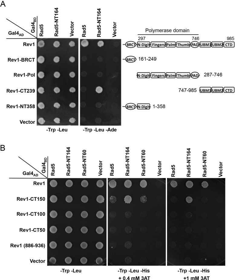
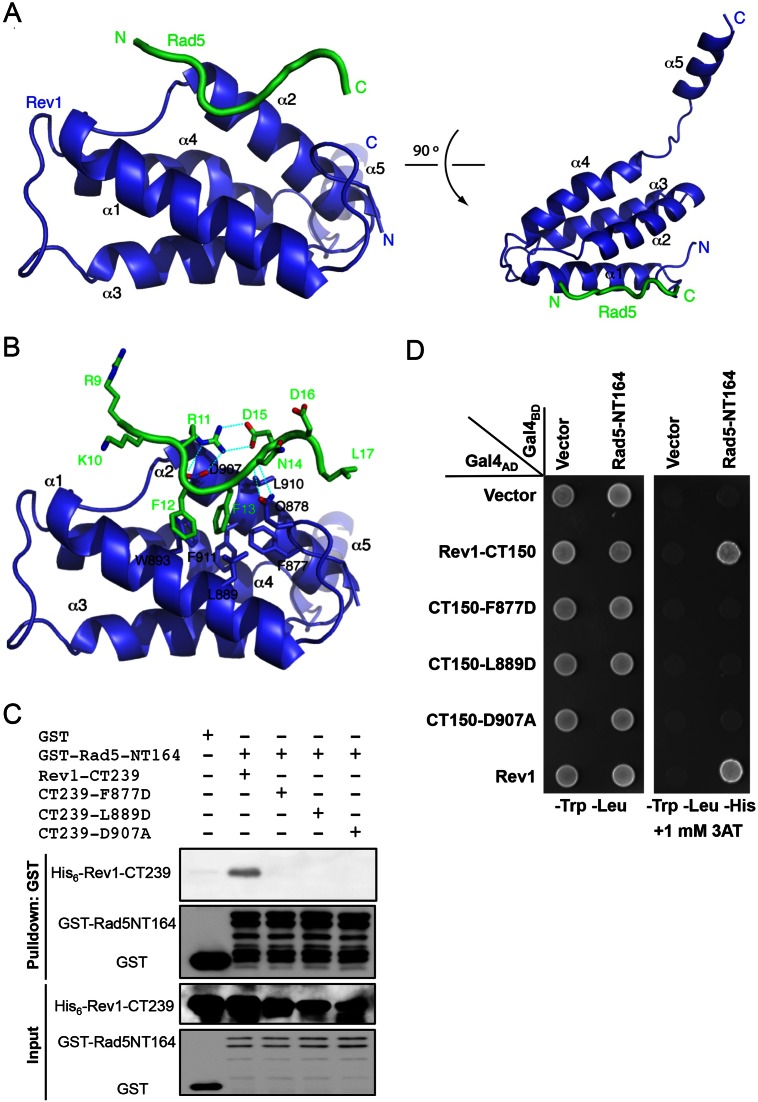
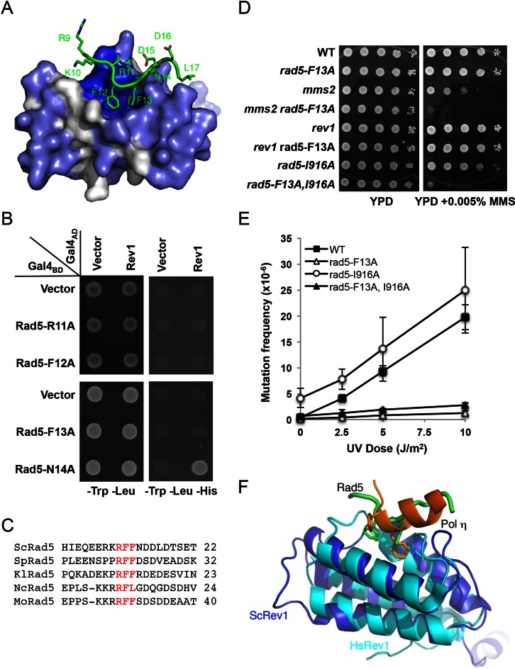
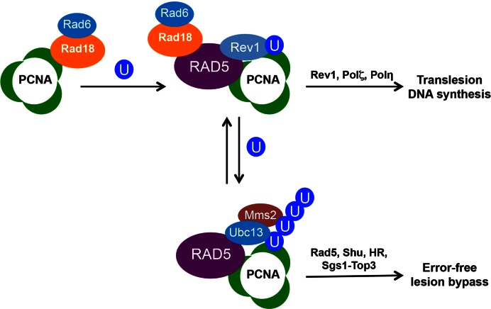
DNA damage tolerance (DDT) is responsible for genomic stability and cell viability by bypassing the replication block. In Saccharomyces cerevisiae DDT employs two parallel branch pathways to bypass the DNA lesion, namely translesion DNA synthesis (TLS) and error-free lesion bypass, which are mediated by sequential modifications of PCNA. Rad5 has been placed in the error-free branch of DDT because it contains an E3 ligase domain required for PCNA polyubiquitination. Rad5 is a multi-functional protein and may also play a role in TLS, since it interacts with the TLS polymerase Rev1. In this study we mapped the Rev1-interaction domain in Rad5 to the amino acid resolution and demonstrated that Rad5 is indeed involved in TLS possibly through recruitment of Rev1. Genetic analyses show that the dual functions of Rad5 can be separated and reconstituted. Crystal structure analysis of the Rad5-Rev1 interaction reveals a consensus RFF motif in the Rad5 N-terminus that binds to a hydrophobic pocket within the C-terminal domain of Rev1 that is highly conserved in eukaryotes. This study indicates that Rad5 plays a critical role in pathway choice between TLS and error-free DDT.


Figures

Figure 1.
Figure 1.
Mapping the Rev1-binding region in Rad5 by a yeast two-hybrid assay. (A) A diagram indicating Rad5 putative functional domains and the sites of truncation. (B) The Rev1-binding region is mapped to the N-terminal 223 amino acids of Rad5. (C) The Rev1-interacting domain is restricted to the N-terminal 60 amino acids of Rad5. (D) The N-terminal 30 amino acids of Rad5 are sufficient to interact with Rev1. Various combinations of Rad5 truncations and Rev1 as indicated were co-transformed into PJ69-4a. The transformants were spotted on control plates (SD-Leu-Trp) and selective plates (SD-Leu-Trp-His+3AT), which were incubated at 30°C for 4 days before photography. Numbers on the left panel indicate Rad5 amino acid sequences encoded by the pGBT plasmids. Only images from the control plates and selective plates containing 5 mM 3AT are presented.
Figure 2.
Figure 2.
Mapping amino acid residues in Rad5 required for the Rev1 interaction. (A) Multi-sequence alignment of budding yeast Rad5 N-terminus with Rad5 orthologs utilizing the ClustalW alignment tool on the Saccharomyces genome database. Residues with yellow highlight indicate complete conservation; those with pink highlight indicate high conservation; while those with green highlight indicate consensus. (B) The interaction region is limited to the conserved 16 amino acids. (C) Conserved residues were selected to make dual Ala point mutations in Rad5 and tested for their ability to interact with Rev1 in a Y2H assay. (D) A GST pull-down assay to examine physical interaction of purified Rad5-NT164 or its mutant form with His6-tagged Rev1-CT239. (E) Effects of deletion or point mutations within Rad5-(21-30) residues. The Y2H experimental conditions were as described in Materials and Methods and Figure 1.
Figure 3.
Figure 3.
Genetic interactions of the rad5-FN13,14AA point mutation with error-free DDT and TLS pathway mutations. (A) The rad5-FN13,14AA point mutation causes moderate sensitivity to MMS, as shown by a gradient plate assay. (B) Complementation of the rad5 ubc13 double mutant by single-copy plasmid carrying RAD5 or its rad5-FN13,14AA mutant form. (A and B) Overnight cultured yeast cells were imprinted onto the premade YPD or YPD + 0.015% MMS gradient plates and the plates were incubated at 30°C for 2 days before photography. Arrows indicate increasing MMS concentration. (C) Genetic interactions between rad5-FN13,14AA and mms2 or rev1 by a serial dilution assay. Overnight-cultured yeast cells were used to make a series of tenfold dilutions and then spotted to YPD or YPD plus various concentrations of MMS. The plates were incubated at 30°C for 2 days before photography. Only one representative MMS plate is shown. All strains are isogenic derivatives of HK578-10D.
Figure 4.
Figure 4.
Intragenic genetic interactions of RAD5 point mutations. (A) rad5-FN13,14AA and rad5-I916A mutations are synergistic with respect to MMS sensitivity. Experimental conditions were as described in Figure 3A and B. (B) Effects of rad5-FN13,14AA and rad5-I916A on spontaneous mutagenesis. (C) Effects of rad5-FN13,14AA and rad5-I916A on UV-induced mutagenesis. Strains in (A) are isogenic derivatives of HK578-10D and in (B and C) are isogenic derivatives of DBY747. Data in (B and C) are the average of at least three independent experiments with standard deviation.
Figure 5.
Figure 5.
The Rad5 interaction region is mapped to the C-terminus of Rev1 by yeast two-hybrid assays. (A) Physical interaction of Rev1 and its truncations with the full-length Rad5 or Rad5-NT164. Fragments remaining after Rev1 deletion are shown in the right panel relative to known functional domains. (B) Rev1-CT150 is the minimum region capable of interaction with Rad5. The Y2H conditions are as described in Figure 1.
Figure 6.
Figure 6.
Characterization of Rev1-CTD residues critical for the interaction with Rad5-NTD. (A) Two orthogonal views of the structure of Rev1-CTD bound with Rad5 fusion-peptide. Rev1 and Rad5 are colored blue and green, respectively. The N- and C-termini of the proteins are labeled. (B) Detailed interactions between Rev1 and Rad5 with labeled residues. (C) A GST pull-down assay to examine physical interaction between purified His6-tagged Rev1-CT239 or its mutant derivatives and GST-tagged Rad5-NT164. (D) A Y2H assay to examine physical interaction between Rev1-CT150 or its mutant derivatives and Rad5-NT164. The Y2H experimental conditions were as described in Figure 1.
Figure 7.
Figure 7.
Characterization of the Rad5-NTD RFF motif. (A) Rad5-NTD residues involved in binding to a conserved pocket of Rev1. Rev1 is shown as gray surface with conserved residues in blue and the Rad5 polypeptide is shown in green. (B) The ‘RFF’ motif of Rad5 is essential for the binding of Rad5 to Rev1, as judged by a Y2H assay. The experimental conditions were as described in Figure 1. (C) Sequence alignments around the ‘RFF’ motif among several fungal Rad5 homologs. The RFF motif is in red. Source of sequences: ScRad5, Saccharomyces cerevisiae, P32849.1; SpRad5, Schizosaccharomyces pombe, XP_001713034.1; KlRad5, Kluyveromyces lactis, XP_455865.1; NcRad5, Neurospora crassa, XP_958511.1; MoRad5, Magnaporthe oryzae, XP_003712540.1. (D) Inter- and intragenic interactions between rad5-F13A and DDT pathways mutations with respect to MMS-induced killing. (E) Effects of rad5-F13A and rad5-I916A on UV-induced mutagenesis. Strains are isogenic derivatives of DBY747. Data are the average of at least three independent experiments with standard deviation. (F) Structural alignment of Rev1 between budding yeast (blue) and human (cyan, PDB code 2LSK). hPolη (orange) binds to the same pocket of hRev1 as yRad5 (green) does with yRev1.
Figure 8.
Figure 8.
A revised working model of DNA-damage tolerance in budding yeast. Under DNA-damage conditions, the Rad6-Rad18 complex monoubiquitinates PCNA at the K164 residue. Rad5 interacts with both Rad18 (11) and PCNA (5,49), which is recruited to the damage site. Rad5-Rev1 physical interaction facilitates recruitment of Rev1 and other TLS polymerases for TLS, while Rad5-Ubc13-Mms2 promotes PCNA polyubiquitination and subsequent error-free lesion bypass, in which Rad5 serves as an E3 ligase and possibly a DNA helicase.

References

    1. Prakash L. Characterization of postreplication repair in Saccharomyces cerevisiae and effects of rad6, rad18, rev3 and rad52 mutations. Mol. Gen. Genet. 1981;184:471–478. - PubMed
    1. Broomfield S., Hryciw T., Xiao W. DNA postreplication repair and mutagenesis in Saccharomyces cerevisiae. Mutat. Res. 2001;486:167–184. - PubMed
    1. Lawrence C.W. Cellular functions of DNA polymerase zeta and Rev1 protein. Adv. Protein Chem. 2004;69:167–203. - PubMed
    1. Prakash L. The RAD6 gene and protein of Saccharomyces cerevisiae. Ann. N.Y. Acad. Sci. 1994;726:267–273. - PubMed
    1. Hoege C., Pfander B., Moldovan G.L., Pyrowolakis G., Jentsch S. RAD6-dependent DNA repair is linked to modification of PCNA by ubiquitin and SUMO. Nature. 2002;419:135–141. - PubMed

Publication types

Substances