. 2016 Apr 12;113(15):E2180-8.
doi: 10.1073/pnas.1515941113. Epub 2016 Mar 21.

Representation of spontaneous movement by dopaminergic neurons is cell-type selective and disrupted in parkinsonism

Affiliations

Representation of spontaneous movement by dopaminergic neurons is cell-type selective and disrupted in parkinsonism

Paul D Dodson et al. Proc Natl Acad Sci U S A. .

Abstract

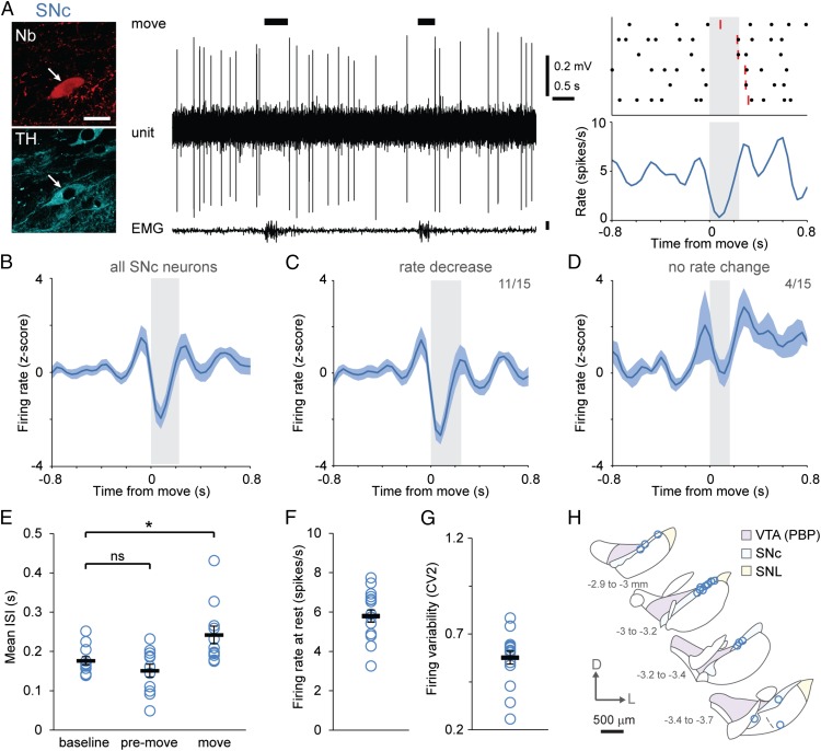
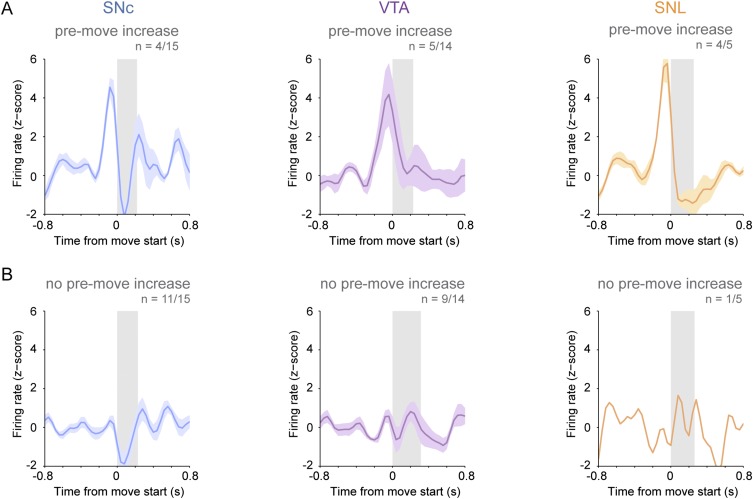
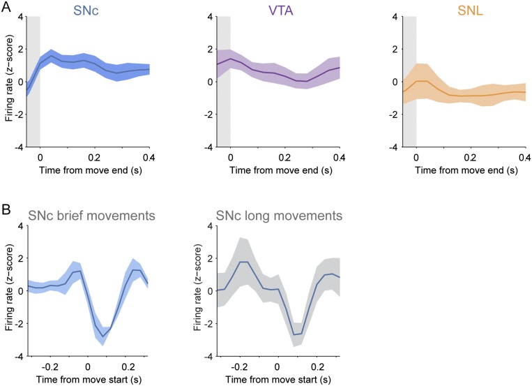
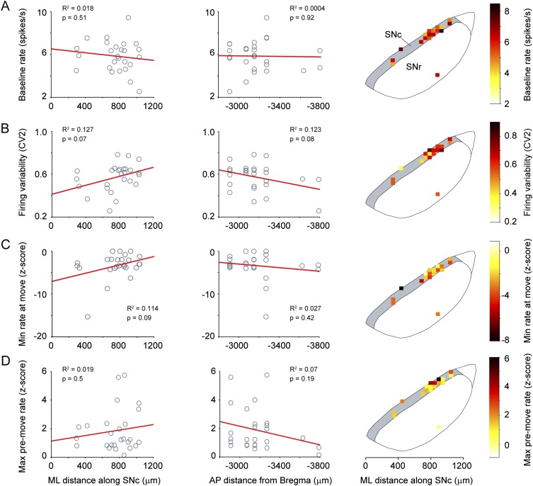
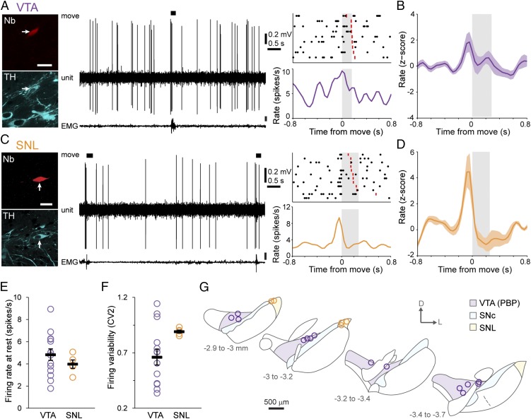
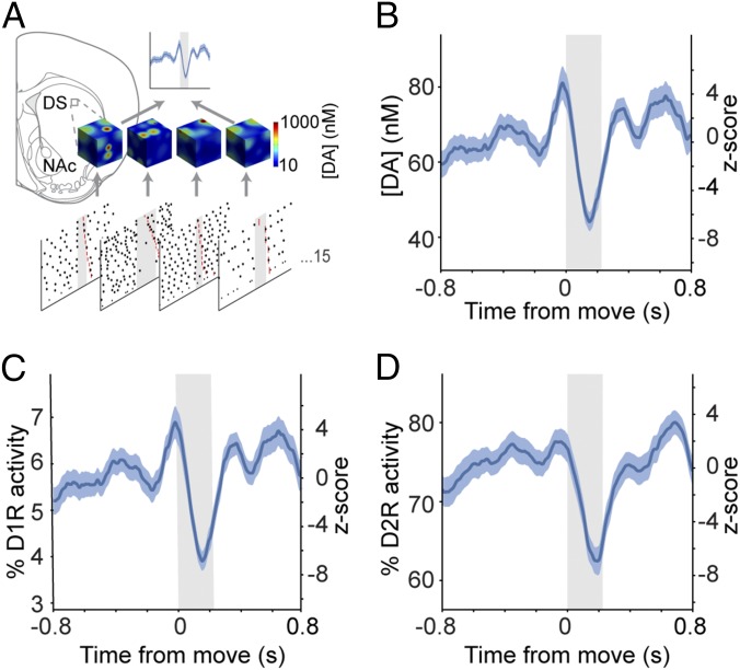
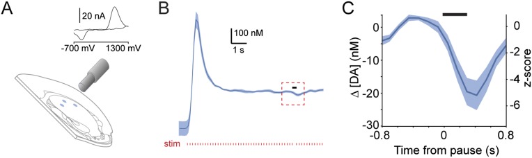
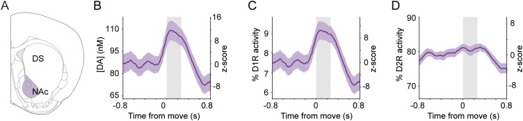
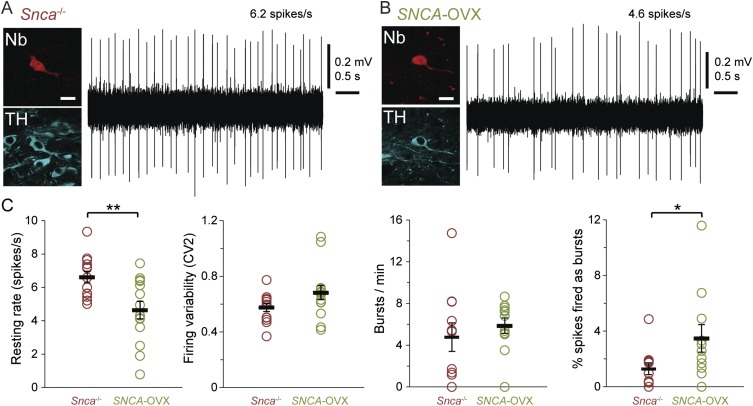
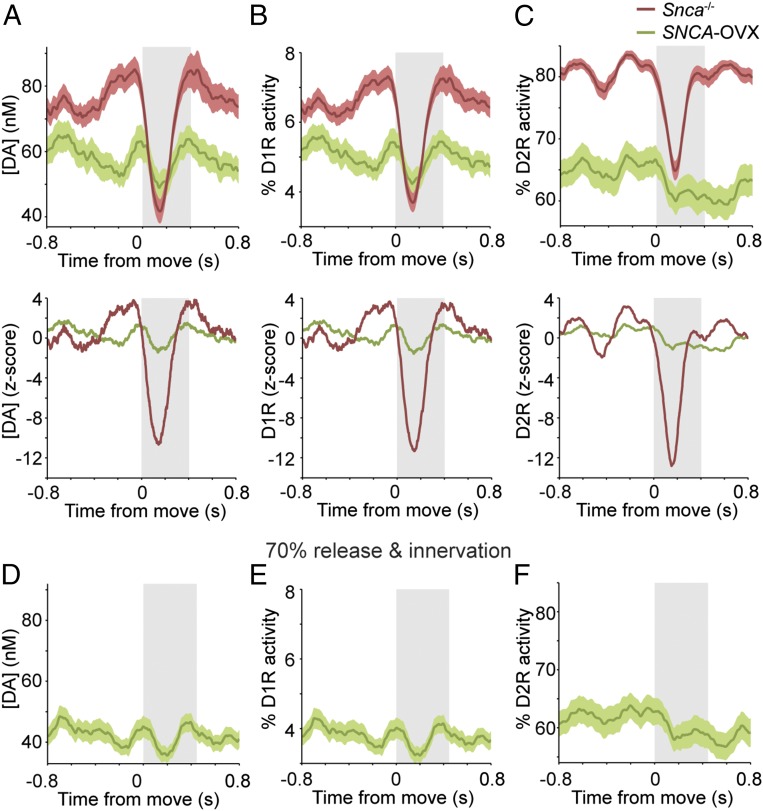
Midbrain dopaminergic neurons are essential for appropriate voluntary movement, as epitomized by the cardinal motor impairments arising in Parkinson's disease. Understanding the basis of such motor control requires understanding how the firing of different types of dopaminergic neuron relates to movement and how this activity is deciphered in target structures such as the striatum. By recording and labeling individual neurons in behaving mice, we show that the representation of brief spontaneous movements in the firing of identified midbrain dopaminergic neurons is cell-type selective. Most dopaminergic neurons in the substantia nigra pars compacta (SNc), but not in ventral tegmental area or substantia nigra pars lateralis, consistently represented the onset of spontaneous movements with a pause in their firing. Computational modeling revealed that the movement-related firing of these dopaminergic neurons can manifest as rapid and robust fluctuations in striatal dopamine concentration and receptor activity. The exact nature of the movement-related signaling in the striatum depended on the type of dopaminergic neuron providing inputs, the striatal region innervated, and the type of dopamine receptor expressed by striatal neurons. Importantly, in aged mice harboring a genetic burden relevant for human Parkinson's disease, the precise movement-related firing of SNc dopaminergic neurons and the resultant striatal dopamine signaling were lost. These data show that distinct dopaminergic cell types differentially encode spontaneous movement and elucidate how dysregulation of their firing in early Parkinsonism can impair their effector circuits.

Keywords: Parkinson's disease; alpha-synuclein; dopamine; substantia nigra; ventral tegmental area.


Conflict of interest statement

The authors declare no conflict of interest.

Figures

Fig. 1.
Fig. 1.
Dopaminergic SNc neurons exhibit a pause in firing during the onset of spontaneous movement. (A) Example of single-unit activity (Center) and a peri-event time histogram (PETH) (Lower Right) with the corresponding raster plot from an identified dopaminergic SNc neuron (Upper Right) during rest and spontaneous movement, the latter denoted by black bars, determined from video and EMG activity. The ends of individual movement epochs are denoted in rasters by red lines, and the mean duration of movement is indicated by gray shading. (Left) After recording, each neuron was juxtacellularly labeled with Neurobiotin (Nb) to identify its dopaminergic nature (by immunoreactivity to TH) and to confirm its location. (Scale bar, 20 μm.) (B–D) Mean normalized PETHs ± SEM. On average, SNc neurons (n = 15) transiently increased their activity just before movement and then paused their firing at the movement onset (B); 11 SNc neurons significantly decreased their rate during movement onset (C), and four did not change their rate significantly (D). (E) Mean interspike intervals (ISIs) during the baseline, premovement (ISIs ending in the 100 ms before movement), and movement (ISIs starting in the 100 ms preceding movement and ending after movement onset) periods. The ISI during movement onset was significantly longer than baseline ISIs [P < 0.01, n = 11 neurons that met analysis criteria (SI Materials and Methods), one-way repeated-measures ANOVA with Dunnett’s post hoc comparison]. (F and G) Firing rate (F) and variability quantified by CV2 (G) of all SNc neurons (n = 16) during alert rest. (H) Schematic coronal sections denoting locations within the SNc of all recorded and identified dopaminergic neurons. Adapted from ref. . The distance from bregma is shown on left. D, dorsal; L, lateral; PBP, parabrachial pigmented area of the VTA. Data are presented as mean ± SEM; *P < 0.05; ns, not significant.
Fig. S1.
Fig. S1.
A minority of dopaminergic neurons in the SNc and VTA and the majority of dopaminergic neurons in the SNL increase firing just before movement onset. Shown are mean normalized PETHs ± SEM of neurons exhibiting significant increases in firing rate (A) or no change in in firing rate (B) just before movement onset.
Fig. S2.
Fig. S2.
Dopaminergic neurons do not reliably represent the termination of brief movements, but SNc neurons represent the onset of longer movements. (A) Mean normalized PETHs ± SEM aligned to the end of each movement period (gray shading) for SNc, VTA, or SNL neurons show that, on average, these types of dopaminergic neurons do not alter their firing rates significantly at the end of movement (n = 15 SNc neurons, 14 VTA neurons, and 5 SNL neurons). (B) Mean normalized PETHs ± SEM aligned to the start of each movement for a set of SNc neurons (n = 5) that were recorded during both brief (Left) and longer (Right) movements. Note that the decreased firing of these neurons that occurred at the onset of brief movements also occurred at the onset of the long movements. Data for long-duration movements were pooled across neurons (13 movements, mean duration 35.87 ± 6.55 s).
Fig. S3.
Fig. S3.
Firing properties of SNc neurons are not related to their location within the nucleus. (AD, Left and Center) Firing properties [baseline firing rate (A), variability in firing rate (B), minimum firing rate at movement (C), and maximum firing rate premovement (D)] of SNc neurons from wild-type and Snca−/− mice were plotted according to their ML (Left) or AP (Center) positions in the SNc and were fit with a least-squares regression. R2 and P values from a Pearson product–moment correlation (n = 26 SNc neurons) are given. (AD, Right) Firing property heat maps were constructed by dividing the SNc into 50-µm2 segments. The color represents the mean value of all neurons within each segment. There were no significant relationships between ML or AP position or the mean firing rate of SNc dopamine neurons during the baseline period before movement (A), firing variability at rest (B), the minimum z-scored firing rate reached in the 160 ms following the start of movement onset (C), or the maximum z-scored firing rate during the 160 ms preceding movement (D).
Fig. 2.
Fig. 2.
The firing rate of VTA and SNL dopaminergic neurons does not change during movement onset. (A and C) Examples of single-unit activities and PETHs from identified dopaminergic neurons in the VTA (A) and SNL (C). (Scale bars, 20 µm.) (B and D) Mean normalized PETHs of all dopaminergic neurons in the VTA (B) and SNL (D). On average, neurons transiently increased their firing rates just before movement but did not significantly change firing during the movement period itself (gray shading). (E and F) Mean firing rate (E) and regularity of firing rate (F) of VTA and SNL dopaminergic neurons during alert rest (n = 14 VTA neurons and 5 SNL neurons). (G) Schematic coronal sections denoting the locations of all recorded and identified neurons in the VTA (purple) or SNL (orange). Data are presented as mean ± SEM.
Fig. 3.
Fig. 3.
Movement-related firing of SNc neurons significantly alters dopamine signaling in the dorsal striatum. (A) Schematic of the computational model of dorsal striatal dopamine signaling. Dopamine release and receptor activity in an ∼25-µm3 area of the dorsal striatum (DS) was modeled using movement-related activity from each recorded SNc neuron (n = 15) as exemplified by snapshot concentration plots. Single-neuron responses were then averaged to generate population-level estimates of dopamine concentration. NAc, nucleus accumbens. (B) Mean peri-movement dopamine concentrations. Note the decrease in dopamine timed with movement onset. (C and D) Peri-movement activity profiles of low-affinity D1 dopamine receptors (C) and high-affinity D2 dopamine receptors (D). Mean response ± SEM is plotted on the left axes; the z-score is plotted on the right axes.
Fig. S4.
Fig. S4.
Brief pauses significantly alter the extracellular dopamine concentration in dorsal striatum ex vivo. (A) Schematic of dopamine measurement in the dorsal striatum by FCV as used to determine the change in dopamine concentration associated with a brief pause in stimulation. (Inset) An example of a voltammogram. (B) Mean extracellular dopamine concentration (blue; SEM indicated by shading) elicited by a train of electrical stimuli (red ticks) delivered to dorsal striatum at 6 Hz (the approximate rate of firing of SNc neurons when animals were at rest). A brief pause in stimulation (denoted by black bar; 277-ms duration, mimicking the ISI of SNc neurons at movement onset) was introduced ∼7 s after the start of stimulation when the dopamine levels had reached a steady state. (C) Mean change in extracellular dopamine concentration in response to the 277-ms pause (black bar; see red box in B). The dopamine concentration was significantly lower in the 0.5 s following the initiation of the pause than during 0.5 s of baseline stimulation (P = 0.001, paired t test; n = 10 recording sites).
Fig. S5.
Fig. S5.
Computational modeling predicts that the movement-related firing of VTA dopaminergic neurons will lead to a peak in dopamine release in the nucleus accumbens during movement. (A) Schematic illustrating the region in which dopamine release and receptor activity were modeled using spike trains recorded from VTA neurons. DS, dorsal striatum; NAc, nucleus accumbens. (BD) Movement-related changes (mean PETH ± SEM) in dopamine concentration (B), in the activity of low-affinity D1 dopamine receptors (C), and in the activity of high-affinity D2 dopamine receptors (D) in the nucleus accumbens.
Fig. 4.
Fig. 4.
Dopaminergic SNc neurons in parkinsonian mice do not reliably represent movement onset in their firing rates. (A and C) Examples of single-unit activities and PETHs from identified dopaminergic SNc neurons in 2-year-old Snca−/− littermate controls (A) and SNCA-OVX parkinsonian mice (C). (B and D) Mean PETHs show that, on average, the firing rate of SNc neurons in Snca−/− mice (n = 11 neurons) decreased significantly at movement onset (B), but there was no significant change in the firing rate of SNc neurons in SNCA-OVX mice (n = 12 neurons) (D). (E) Mean ISIs of neurons in Snca−/− mice during the baseline, premovement, and movement periods (defined as in Fig. 1); ISIs were significantly longer during movement than at baseline (P < 0.001, n = 8 neurons, one-way repeated-measures ANOVA with Dunnett’s post hoc comparison). (F) ISIs were not significantly different in SNCA-OVX parkinsonian mice (P > 0.05, n = 6 neurons, one-way repeated-measures ANOVA). (G) Schematic coronal sections denoting locations of recorded and labeled neurons within the SNc [n = 13 neurons from Snca−/− mice (red) and 14 neurons from SNCA-OVX mice (green)]. Data are presented as mean ± SEM; ***P < 0.001; ns, not significant.
Fig. S6.
Fig. S6.
Dopaminergic SNc neurons have abnormally low firing rates in aged parkinsonian mice at rest. (A and B) Typical single-unit activities recorded from identified dopaminergic SNc neurons in 24-month-old Snca−/− littermate controls (A) or SNCA-OVX parkinsonian mice (B) during alert rest. After recording, individual neurons were juxtacellularly labeled with Neurobiotin (Nb); dopaminergic identity was verified by the expression of TH. (Scale bars, 20 µm.) (C) In vivo firing properties of SNc neurons in Snca−/− and SNCA-OVX mice during alert rest. The mean firing rate was significantly lower in SNCA-OVX mice (P < 0.01, t test), but firing variability was similar across the genotypes (P > 0.05, Mann–Whitney test). The number of bursts per minute fired by neurons was not significantly different across genotypes (P > 0.05, t test), but SNc neurons in SNCA-OVX mice fired a higher percentage of spikes as bursts (P < 0.05, Mann–Whitney test). Data are presented as mean ± SEM plotted (thick black bar and error bars); n = 13 neurons from Snca−/− mice and 14 neurons from SNCA-OVX mice. *P < 0.05, **P < 0.01.
Fig. 5.
Fig. 5.
Movement-related changes in striatal dopamine signaling are lost in parkinsonian mice. (A, Upper) Dorsal striatal dopamine concentrations (mean responses ± SEM) simulated using movement-related activity from SNc neurons recorded in Snca−/− littermate controls (red) or in parkinsonian SNCA-OVX mice (green). (Lower) Corresponding z-scores. (B and C, Upper) Activity of low-affinity D1 receptors (B) and high-affinity D2 dopamine receptors (C). (Lower) Corresponding z-scores. (DF) Dopamine concentration (D), D1 receptor activity (E), and D2 receptor activity (F) modeled with parameters adjusted to match deficits present in aged SNCA-OVX mice (the abnormally low firing rate of SNc neurons plus a 30% reduction in dopamine release and dopaminergic innervation).

References

    1. Montague PR, Dayan P, Sejnowski TJ. A framework for mesencephalic dopamine systems based on predictive Hebbian learning. J Neurosci. 1996;16(5):1936–1947. - PMC - PubMed
    1. Schultz W, Dayan P, Montague PR. A neural substrate of prediction and reward. Science. 1997;275(5306):1593–1599. - PubMed
    1. Schultz W. Behavioral dopamine signals. Trends Neurosci. 2007;30(5):203–210. - PubMed
    1. Matsumoto M, Hikosaka O. Two types of dopamine neuron distinctly convey positive and negative motivational signals. Nature. 2009;459(7248):837–841. - PMC - PubMed
    1. Lammel S, Lim BK, Malenka RC. Reward and aversion in a heterogeneous midbrain dopamine system. Neuropharmacology. 2014;76(Pt B):351–9. - PMC - PubMed

Publication types

LinkOut - more resources