. 2016 May 20;291(21):11055-63.
doi: 10.1074/jbc.M115.711333. Epub 2016 Mar 21.

Suppression of WHITE COLLAR-independent frequency Transcription by Histone H3 Lysine 36 Methyltransferase SET-2 Is Necessary for Clock Function in Neurospora

Affiliations

Suppression of WHITE COLLAR-independent frequency Transcription by Histone H3 Lysine 36 Methyltransferase SET-2 Is Necessary for Clock Function in Neurospora

Guangyan Sun et al. J Biol Chem. .

Abstract

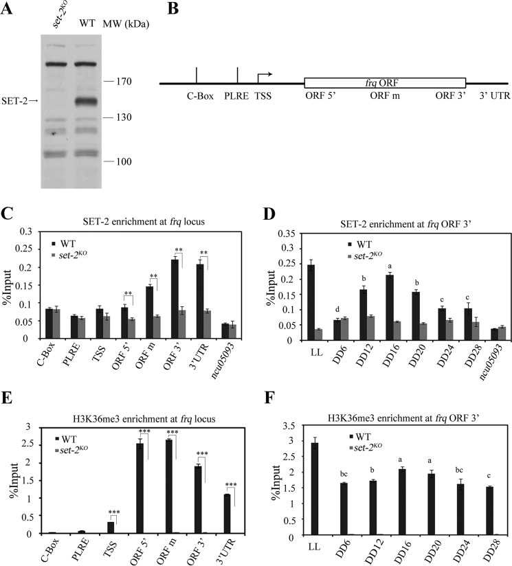
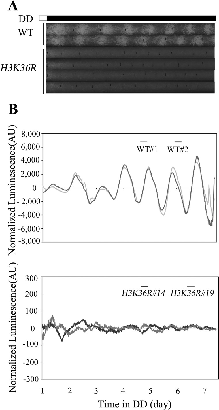
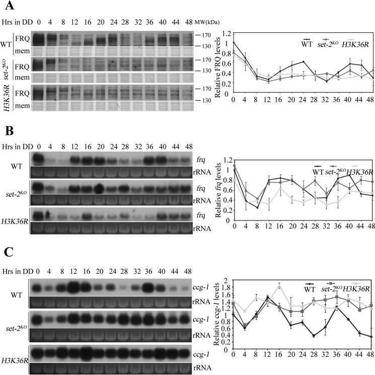
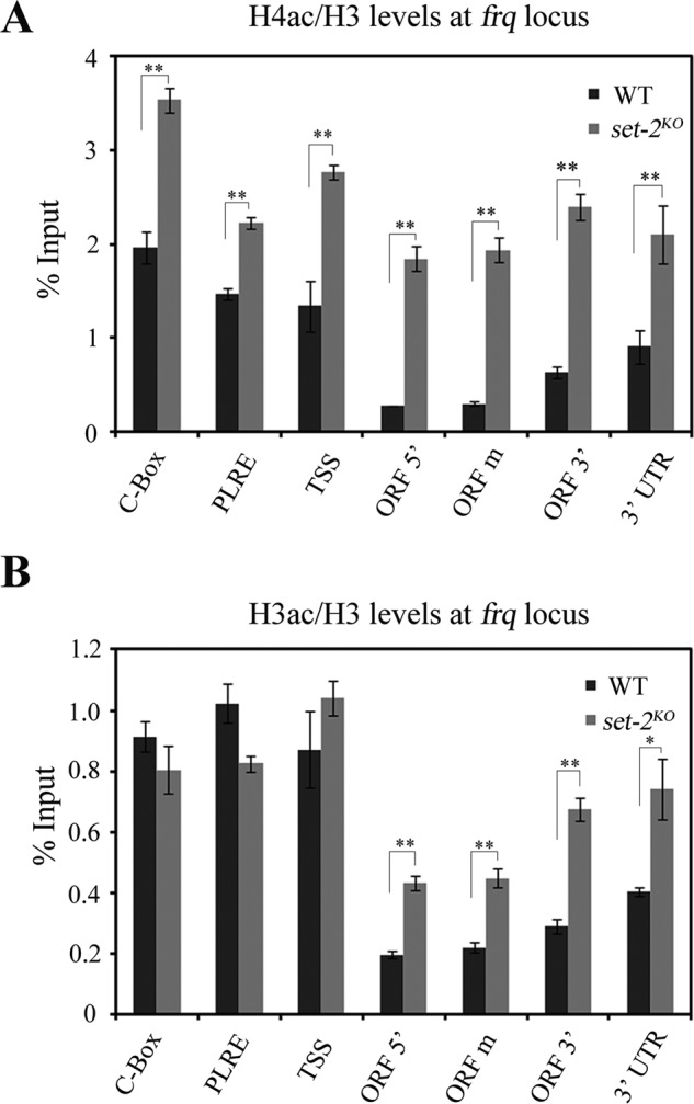
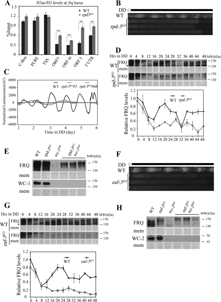
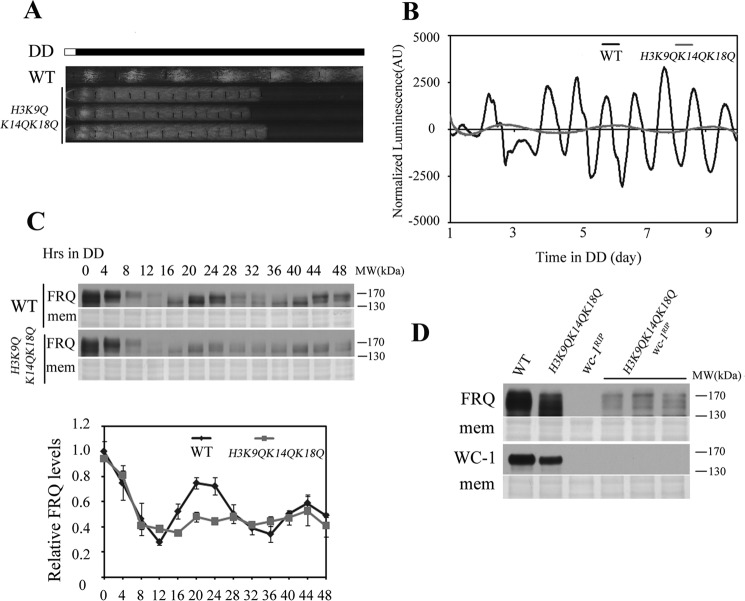
The circadian system in Neurospora is based on the transcriptional/translational feedback loops and rhythmic frequency (frq) transcription requires the WHITE COLLAR (WC) complex. Our previous paper has shown that frq could be transcribed in a WC-independent pathway in a strain lacking the histone H3K36 methyltransferase, SET-2 (su(var)3-9-enhancer-of-zeste-trithorax-2) (1), but the mechanism was unclear. Here we disclose that loss of histone H3K36 methylation, due to either deletion of SET-2 or H3K36R mutation, results in arrhythmic frq transcription and loss of overt rhythmicity. Histone acetylation at frq locus increases in set-2(KO) mutant. Consistent with these results, loss of H3K36 methylation readers, histone deacetylase RPD-3 (reduced potassium dependence 3) or EAF-3 (essential SAS-related acetyltransferase-associated factor 3), also leads to hyperacetylation of histone at frq locus and WC-independent frq expression, suggesting that proper chromatin modification at frq locus is required for circadian clock operation. Furthermore, a mutant strain with three amino acid substitutions (histone H3 lysine 9, 14, and 18 to glutamine) was generated to mimic the strain with hyperacetylation state of histone H3. H3K9QK14QK18Q mutant exhibits the same defective clock phenotype as rpd-3(KO) mutant. Our results support a scenario in which H3K36 methylation is required to establish a permissive chromatin state for circadian frq transcription by maintaining proper acetylation status at frq locus.

Keywords: Neurospora; SET-2 pathway; WC-independent frq; circadian rhythm; clock gene; gene transcription; histone modification.


Figures

FIGURE 1.
FIGURE 1.
SET-2 is recruited to frq gene and regulates its transcription by catalyzing H3K36me3. A, Western blot analysis showing a specific band present in the wild-type strain but not the set-2KO strain. The arrow points out the SET-2 protein band detected by our SET-2 antibody. WT, wild-type; MW, molecular weight. B, schematic depiction of the frq locus. C, ChIP analysis showing the enrichment of SET-2 at the frq locus in DD16. ChIP experiment performed on chromatin isolated at DD16 when frq expression is maximal by using the SET-2 antibody. The amount of DNA associated with SET-2 was determined by qPCR. D, rhythmic association of SET-2 at the 3′-end of frq gene in different time points. Samples were grown for the indicated hours in constant darkness (DD) prior to harvesting and processing for ChIP. LL, constant light. E, ChIP analysis shows that the H3K36me3 levels are increased at the 3′-end of frq in the wild-type strain. F, rhythmic changes of H3K36 trimethylation at the 3′-end of frq gene in different time points. The set-2KO strain was used as a negative control in the ChIP assay, no SET-2 binding at ncu05093 promoter in C and D also acts as a negative control. Error bars show the mean ± S.D. (n = 3). Significance in C and E was assessed by using a two-tailed t test. *, p < 0.05, **, p < 0.01; ***, p < 0.001. The statistical analysis was also performed on wild-type from DD6 to DD24 in D and F, bars with different letters in D and F are statistically significant, p < 0.05, one-way Anova and Tukey's HSD test.
FIGURE 2.
FIGURE 2.
H3K36 methylation is required for circadian rhythm. Race tube assay of the wild-type and H3K36R strains is shown in A, vertical black lines on race tubes mark daily growth fronts of the strains. The conidiation rhythm in H3K36R strains is lost compared with wild-type strains. Four independent H3K36R strains were shown in the figure. B, analyses of luciferase activity in wild-type, frq-luc, and H3K36R, frq-luc strains show that the luciferase activity rhythm is impaired in the H3K36R, frq-luc strains. Three independent H3K36R, frq-luc strains were used to test luciferase activity, and the result of two strains was shown in the figure. The low normalized luciferase signal levels in the H3K36R, frq-luc strain reflect the low-amplitude fluctuation of luciferase activity. Different strains' conidia suspension was placed on AFV medium and luminescence was recorded in real time using a LumiCycle. Raw data were normalized to subtract the baseline calculated by LumiCycle analysis software.
FIGURE 3.
FIGURE 3.
H3K36 methylation is essential for the Neurospora circadian clock. A, Western blot analysis shows that the circadian oscillation of FRQ in set-2KO and H3K36R is impaired when compared with wild-type strain. Samples were grown in constant darkness (DD) for indicated hours before harvest. The Coomassie Blue-stained membranes (mem) represent total protein in each sample and act as a loading control. Quantification of the FRQ protein is shown on the right side of Western blot. Northern blot analysis shows that frq (B) and ccg-1 (C) transcription are arrhythmic in set-2KO and H3K36R strains. Ribosome RNA (rRNA) bands stained by ethidium bromide shown below the Northern blot act as a loading control for each sample. Quantification of frq and ccg1 are shown on the right side of Northern blot. These experiments were performed at least three times, and one representative result is shown here.
FIGURE 4.
FIGURE 4.
SET-2 directs the histone acetylation state of frq ORF. ChIP analyses of H4ac (A) and H3ac (B) occurring at the frq locus. Both H4ac and H3ac levels at frq ORF region are increased in set-2KO strains compared with wild-type. ChIP experiment was performed on chromatin isolated at DD16.The amount of DNA associated with H4ac or H3ac was determined by qPCR. Significance was assessed by using a two-tailed t test. *, p < 0.05; **, p < 0.01. Error bars show the mean ± S.D. (n = 3).
FIGURE 5.
FIGURE 5.
Rpd3S-mediated deacetylation of frq coding region suppresses WC-independent frq expression. A, H3ac at the frq locus was examined by ChIP analysis in wild-type and rpd-3KO strains at DD16. The H3 acetylation levels at frq ORF region are higher in rpd-3KO strain than wild-type. Significance was assessed by using a two-tailed t test. *, p < 0.05; **, p < 0.01. Error bars show the mean ± S.D. (n = 3). B, conidiation rhythmicity is lost in rpd-3KO strains in the race tube assay. Vertical black lines on race tubes mark daily growth fronts of the strains. Three independent rpd-3KO strains run on race tube were shown in the figure. C, luciferase reporter assay showing the normalized frq promoter activity of wild-type, frq-luc and rpd-3KO, frq-luc strains. Three independent rpd-3KO, frq-luc strains were used to test luciferase activity and the result of two strains was shown in the figure. D, Western blot analysis shows the circadian oscillation of FRQ in rpd-3KO is impaired when compared with wild-type strain. Samples were grown in darkness (DD) for indicated hours before harvest. The Coomassie Blue-stained membranes (mem) represent total protein in each sample. Asterisks indicate nonspecific bands. The experiment was performed independently three times and one representative result was shown in the figure. E, Western blot analyses were performed using antibodies against FRQ or WC-1 in the wild-type, rpd-3KO, wc-1RIP, and rpd-3KO wc-1RIP strains. Samples were grown under constant light before harvest and three different rpd-3KO wc-1RIP double mutants were used. The Coomassie Blue-stained membranes (mem) represent total protein in each sample. F, race tube assay of wild-type and eaf-3KO strains. Three independent eaf-3KO strains were shown in the figure. G, Western blot analysis showing the circadian oscillation of FRQ in the wild-type and eaf-3KO strains. Both FRQ protein and phosphorylation rhythm in wild-type are abolished in the eaf-3KO strain. Samples were grown in constant darkness (DD) for indicated hours before harvest. The Coomassie Blue-stained membranes (mem) represent total protein in each sample. Asterisks indicate nonspecific bands. The experiment was performed independently three times and one representative result was shown in the figure. H, Western blot analyses were performed using antibodies against FRQ or WC-2 in the wild-type, eaf-3KO, wc-2RIP, and eaf-3KO wc-2RIP strains. Samples were grown under constant light before harvest and three different eaf-3KO wc-2RIPdouble mutants were used. The Coomassie Blue-stained membranes (mem) represent total protein in each sample.
FIGURE 6.
FIGURE 6.
The mimic acetylation states of histone H3 regulate frq transcription. A, H3K9QK14QK18Q strain lost the robust conidiation rhythm compared with WT in the race tube assay. Three independent H3K9QK14QK18Q strains were shown in the figure. B, analyses of luciferase activity in wild-type, frq-luc and H3K9QK14QK18Q, frq-luc strains. Three independent H3K9QK14QK18Q, frq-luc strains were used to test the luciferase activity and all strains lost the rhythmic luciferase activity compared with wild-type. One wild-type, frq-luc and one H3K9QK14QK18Q, frq-luc strain result was shown in the figure. C, Western blot analysis showing the circadian oscillation of FRQ in the wild-type and H3K9QK14QK18Q mutant strains. Samples were grown in constant darkness (DD) for indicated hours before harvest. The Coomassie Blue-stained membranes (mem) represent total protein in each sample. The experiment was performed independently at least three times, and one representative result was shown in the figure. D, Western blot analyses were performed using antibodies against FRQ or WC-1 in the wild-type, H3K9QK14QK18Q, wc-1RIP, and H3K9QK14QK18Q wc-1RIPstrains. Samples were grown on constant light before harvest and three different H3K9QK14QK18Q wc-1RIP strains were used. The Coomassie Blue-stained membranes (mem) represent total protein in each sample.

References

    1. Zhou Z., Liu X., Hu Q., Zhang N., Sun G., Cha J., Wang Y., Liu Y., and He Q. (2013) Suppression of WC-independent frequency transcription by RCO-1 is essential for Neurospora circadian clock. Proc. Natl. Acad. Sci. U.S.A. 110, E4867–E4874 - PMC - PubMed
    1. Baker C. L., Loros J. J., and Dunlap J. C. (2012) The circadian clock of Neurospora crassa. FEMS Microbiol. Rev. 36, 95–110 - PMC - PubMed
    1. Bell-Pedersen D., Cassone V. M., Earnest D. J., Golden S. S., Hardin P. E., Thomas T. L., and Zoran M. J. (2005) Circadian rhythms from multiple oscillators: lessons from diverse organisms. Nat. Rev. Genet. 6, 544–556 - PMC - PubMed
    1. Dunlap J. C. (1999) Molecular bases for circadian clocks. Cell 96, 271–290 - PubMed
    1. Dunlap J. C. (2006) Proteins in the Neurospora circadian clockworks. J. Biol. Chem. 281, 28489–28493 - PubMed

Publication types

MeSH terms

LinkOut - more resources