

Review
doi: 10.1038/cr.2016.35.
Epub 2016 Mar 22.
E2 enzymes: more than just middle men
Affiliations
- PMID: 27002219
- PMCID: PMC4822130
- DOI: 10.1038/cr.2016.35
Item in Clipboard
Review
E2 enzymes: more than just middle men
Cell Res.
2016 Apr.
Abstract
Ubiquitin-conjugating enzymes (E2s) are the central players in the trio of enzymes responsible for the attachment of ubiquitin (Ub) to cellular proteins. Humans have ∼40 E2s that are involved in the transfer of Ub or Ub-like (Ubl) proteins (e.g., SUMO and NEDD8). Although the majority of E2s are only twice the size of Ub, this remarkable family of enzymes performs a variety of functional roles. In this review, we summarize common functional and structural features that define unifying themes among E2s and highlight emerging concepts in the mechanism and regulation of E2s.
Figures
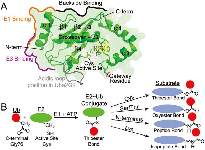
Overview of E2 structure and chemistry. (A) Important structural features and common binding surfaces described in the text are labeled on a representative UBC domain (Ube2D3; PDBID: 2FUH) shown in green. The position of the acidic loop in Ube2G2 (PDBID: 2CYX) when the UBC domain is aligned with Ube2D3 is shown in gray. (B) The C-terminal carboxylate of Ub is conjugated to the E2 active site cysteine in an E1-catalyzed, ATP-driven reaction. The E2∼Ub conjugate reacts with the side chain of a Cys, Ser, Thr, Lys, or N-terminus on a substrate to form the diverse ubiquitin linkages as shown.
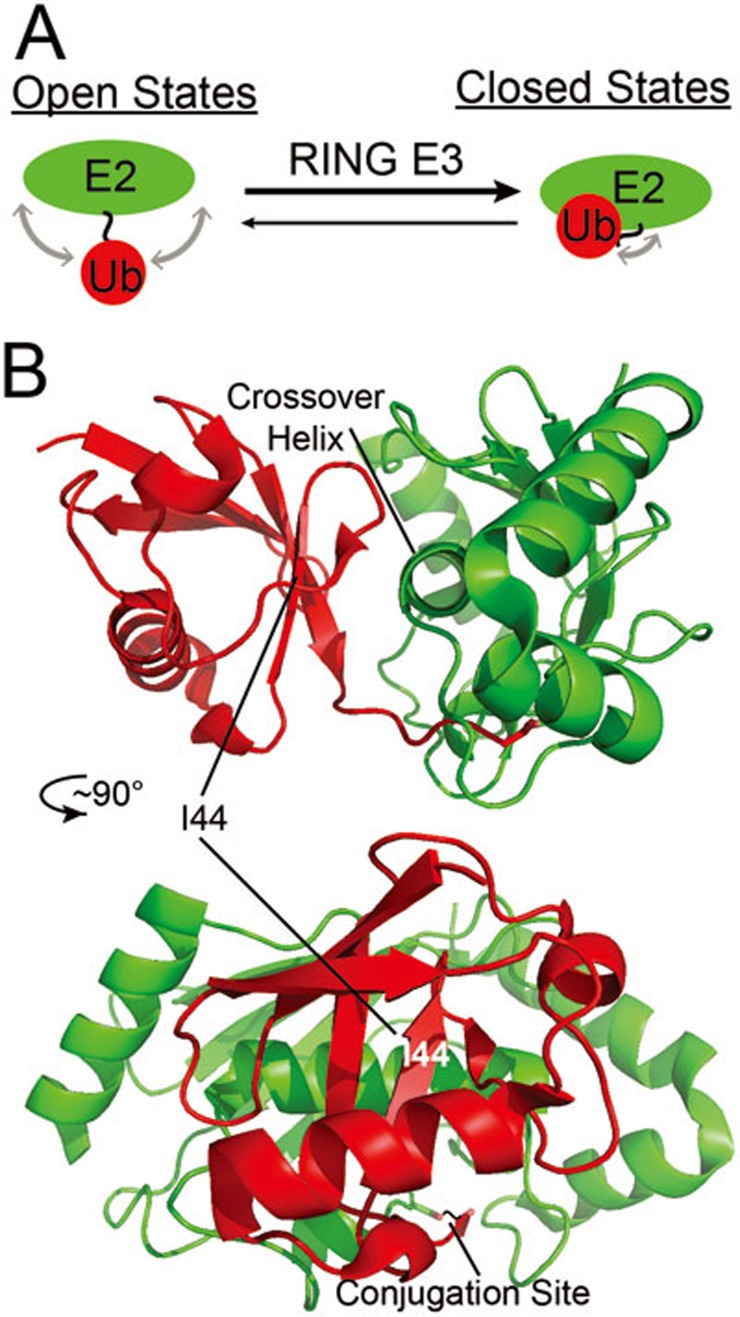
Ubiquitin positioning in the E2∼Ub conjugate. (A) A cartoon depiction of the dynamic “open states” in which Ub samples a range of various positions relative to the E2, but shifts towards population of more “closed states” upon binding of a RING-type E3. (B) Crystal structure of an E2∼Ub oxyester conjugate (Ube2D2) in a closed state showing the interface formed between the E2 (green) crossover helix and the I44 surface of Ub (red) upon binding a RING E3 (BIRC7 (not shown); PDBID: 4AUQ). The two representations are the same structure rotated ∼90° about the vertical axis. The E2 is in the same orientation as in Figure 1 in the bottom representation.
References
-
- Siepmann TJ. Protein interactions within the N-end rule ubiquitin ligation pathway. J Biol Chem 2003; 278:9448–9457. - PubMed
Publication types
MeSH terms
Substances
Grants and funding
LinkOut - more resources
Full Text Sources
Other Literature Sources
Miscellaneous
